

Page 1
Data
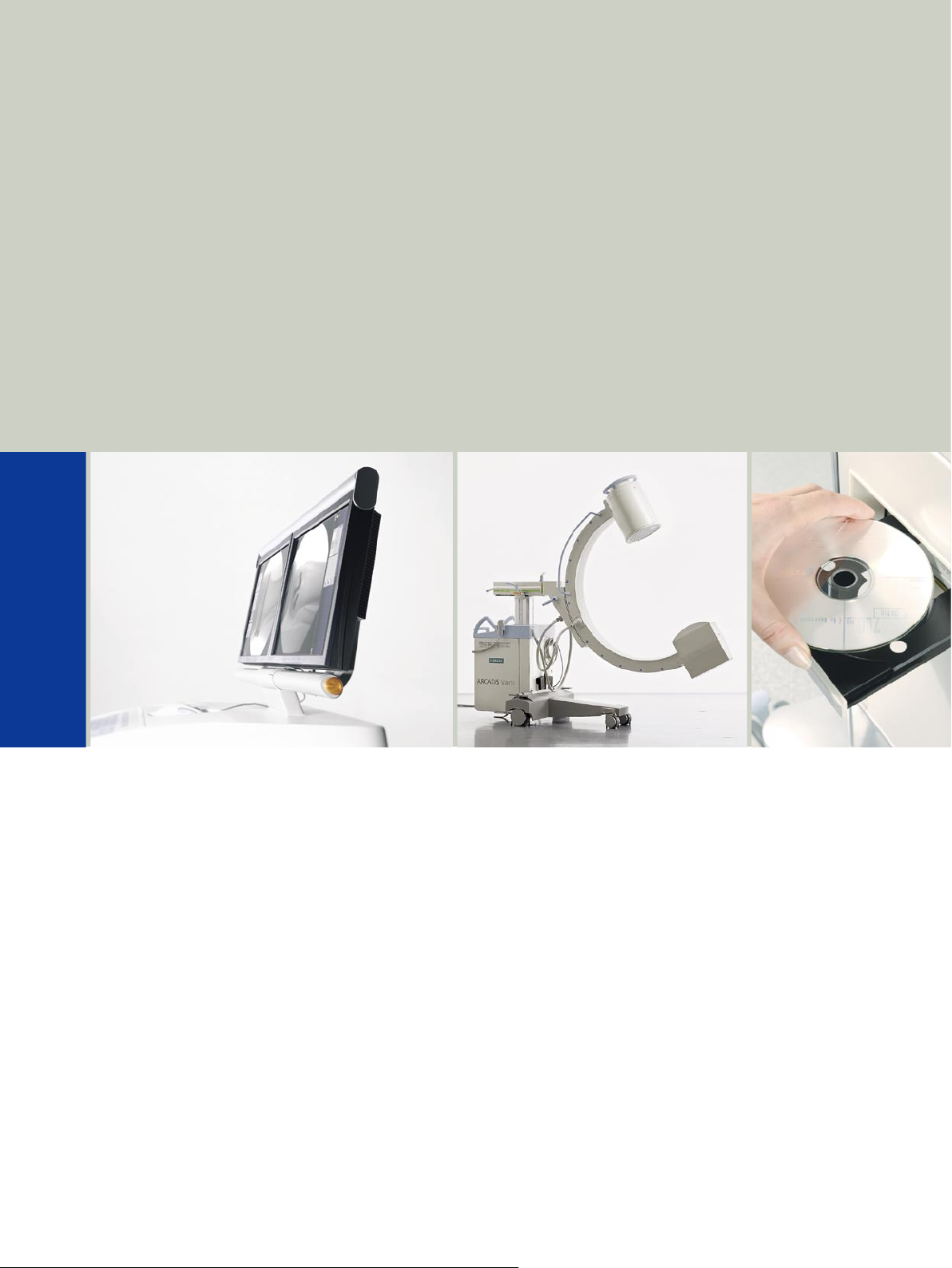
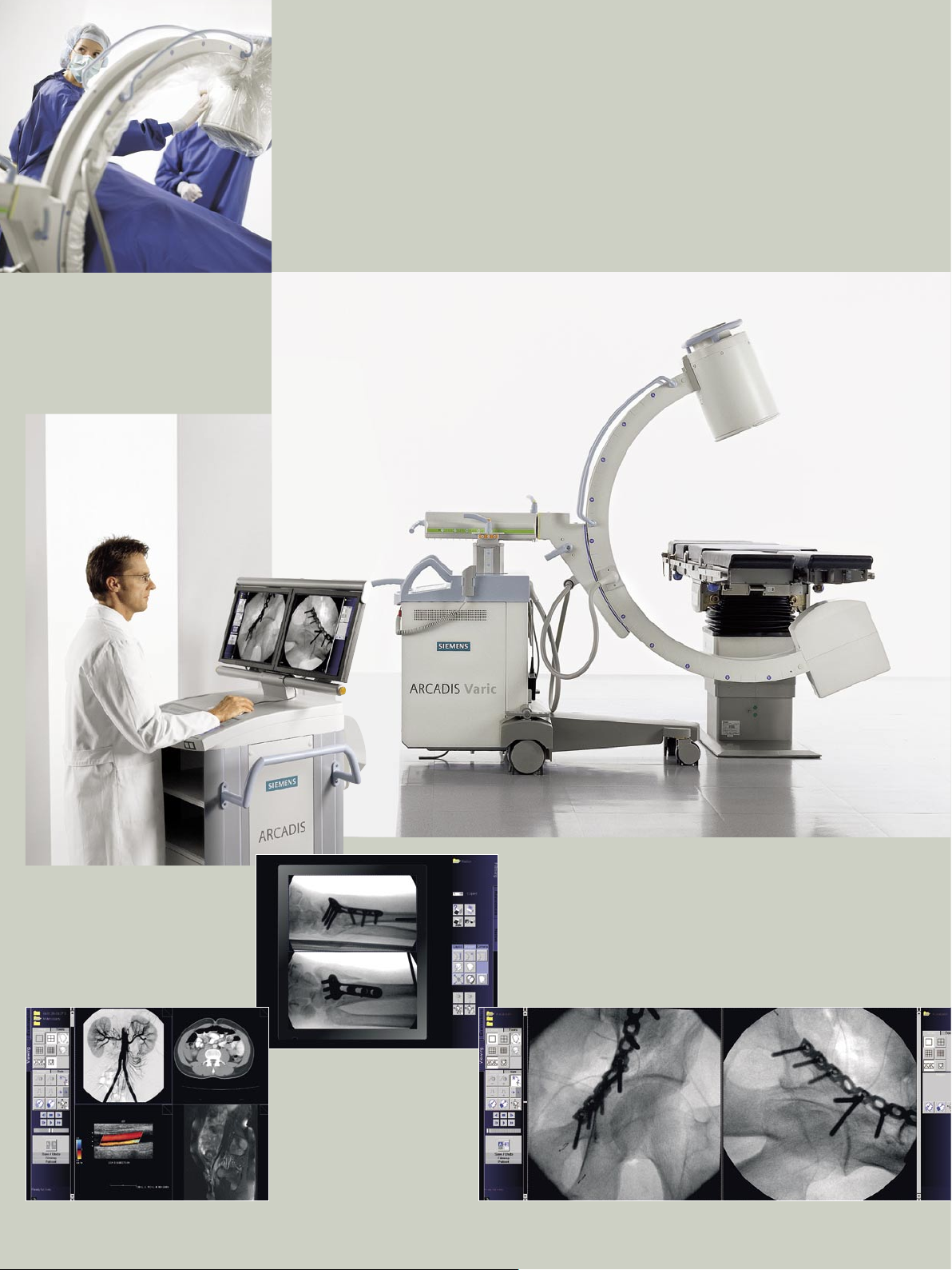
ARCADIS Varic
Maximize Efficiency in your OR
s
medical
Page 2

ARCADIS Varic
Maximize Efficiency in your OR
Siemens starts a new era in the OR with the syngo -based ARCADIS Varic.
syngo provides you with clinical patient information online, so you can optimize efficiency in your OR,
allowing entirely new possibilities.
ARCADIS Varic expands the boundaries of your OR by integrating modalities such as CT and MR,
as well as the entire clinical network into your OR work process.
ARCADIS Varic sets new standards for efficiency in the OR
Increased efficiency through syngo
• Uniform, intuitive user interface for system operation, image postprocessing and networking
• Workflow-oriented task card structure
• Comprehensive connectivity with other modalities and clinical networks
Increased efficiency through optimized clinical workflow
• Maximum flexibility in patient registration
• Easy, intuitive selection of application-specific user programs with VPA (Virtual Patient Anatomy)
• Real multimodality viewing before, during, and after the procedure
• Virtually unlimited possibilities for documentation and archiving
• Fluoro loop* / LSH* (Last Scene Hold)
Increased efficiency with brilliant 1K2 image quality
• Optimally matched, continuous 1K2 imaging chain, from image acquisition to viewing and archiving
• Large power reserves through 2.3 kW / 23 mA generator power and Power Mode
• High-luminance TFT displays
Increased efficiency through comprehensive connectivity and specialized interfaces
• Supports virtually all DICOM 3 functionalities
• Integrated, digital 1K2 navigation interface NaviLink 2D* with automatic image transfer
• syngo fastView for convenient selection and viewing of clinical images on the PC
Increased efficiency through user-friendly operation
• Excellent maneuverability due to compact and lightweight design
• Intelligent color coding for fast and precise positioning and operation
ARCADIS Varic – an intelligent investment for today and tomorrow!
* Option
2
Page 3
Page 4

Technical data
5
Image display/processing
Data transfer and documentation
Options
ARCADIS Varic
syngo
Patient data administration
Image acquisition
C-arm
Orbital movement 130° (– 40° to + 90°)
Angulation ± 190°
Horizontal movement 20 cm (7.9”)
Immersion depth 73 cm (28.7”)
Swivel range ± 12.5°
Vertical travel 45 cm (17.7”), motorized
Source-I.I. distance 100 cm (39.4”)
Free space 78 cm (30.7”)
X-ray generator / tube
Max. pulsed output 2.3 kW
Converter control frequency 15 kHz to 30 kHz
kV range 40 kV to 110 kV
Fluoroscopy 0.2 mA to 15.2 mA (max. 1000 W)
Digital Radiography 0.2 mA to 23 mA (max. 1000 W)
Cassette exposures max. 20 mA
Pulsed Fluoroscopy up to 23 mA
Pulse width min. 7 ms
Pulse rate up to 8 p/s, up to 15 p/s*
Power Mode enables temporary max. output in continuous fluoroscopy and
pulsed fluoroscopy
Single tank with stationary anode
Focal spot nominal value (IEC 336) 0.6
Nominal voltage (IEC 613) 110 kV
Optical anode angle (IEC 788) 9°
Inherent filtration ≥ 3 mm Al equivalent
Collimator system
Iris diaphragm for concentric, radiation-free collimation
Semi-transparent slot diaphragm for symmetric, radiation-free collimation, with unlimited rotation
* Option
4
Page 5

Image display/processing
Data transfer and documentation
Options
X-ray TV system
High-resolution X-ray TV system in maintenance-free CCD technology with 1024 x 1024 (1K2) full-size CCD sensor
Constant image brightness due to automatic gain control
High contrast and high spatial resolution
TV matrix 1K
2
Digital image rotation ± 360°
X-ray image intensier
Metal-enamel technology with Mu-metal shielding
Precision electron optics with minimal image distortion and consistent high resolution across the entire image field
Cesium-iodide input screen for minimum quantum noise and excellent resolution
Anti- glare output screen with scattered light trap for high contrast dynamics and prevention of scattered light effects
High-transparency input window
Nominal diameter 23 cm (9”)
Zoom format 15 cm (6”)
Grid PB 17/70, f0 100
Cassette holder*
Format 24 cm x 30 cm (9.5“ x 12“)
Grid Pb r17 N70, f0 85
Displays
18” TFT monochrome display
High-contrast display with high luminance
Screen size 18” (46 cm)
Image matrix 1280 x 1024
Maximum brightness, typical 600 cd/m
2
Horizontal / vertical viewing angle 170° / 170°
19“ TFT color display
Screen size 19" (48 cm)
Image matrix 1280 x 1024
Maximum brightness, typical 280 cd/m2
Horizontal / vertical viewing angle 170° / 170°
5
Page 6

Technical data
7
Image display/processing
Data transfer and documentation
Options
ARCADIS Varic
syngo
Patient data administration
Image acquisition
syngo
Fully digital, syngo-based online imaging system with continuous 1K2 imaging chain (image acquisition, image
processing, storage, archiving and documentation). Integrated, uninterruptible power supply (UPS) ensures that image
and patient data are secure in the event of a power outage
syngo -based applications Intuitive menu guidance with function-oriented task cards
Dedicated, application-related user programs VPA (Virtual Patient Anatomy)
with anatomic assignment and selection
Direct dose level selection for individual adaptation of the radiation dose to
the patient’s anatomy. Ensures lowest possible dose at high image quality
Dynamic and static reference image display
Simultaneous display of subtracted and native images (with subtraction option)
Patient data administration
Patient data administration Emergency registration
Pre-registration
Manual patient registration
Registration via database query (Patient Browser)
Registration via DICOM Worklist*
Image acquisition
Operating modes
Selection of 8 application-specific fluoroscopy and radiography curves for the individual operating modes
Digital Fluoroscopy (FL) Continuous fluoroscopy with 30 f/s (1K2/12-bit matrix)
Digital filtration
Manual/automatic image storage
Sliding weighted averaging for low-noise image display with minimum dose
Digital Radiography (DR) Digital filtration (1K2/12-bit matrix)
Sliding weighted averaging for low-noise image display with minimum dose
Digital Pulsed Fluoroscopy (PFC) Variable frame rate 0.5 to 8 f/s, (0.5 to 15 f/s*)
Digital Subtraction Angio- Variable frame rate 0.5 to 8 f/s, (0.5 to 15 f/s*)
graphy* / Roadmap*
simultaneous storage of fill image
Simultaneous dual-channel output for image acquisition and postprocessing,
* Option
6
Page 7

Image display/processing
Data transfer and documentation
Options
Image acquisition
Dose optimization Integrated dose measuring chamber* with automatic entry of the
accumulated dose in a radiation report
Dose level selection
I.I. laser aimer for attachment to image intensifier*
System-integrated I.I. laser aimer*
System-integrated laser targeting device on tube side*
Image display/processing
Image display Split screen (1, 4, 9, 16 on 1)
Digital zoom, fixed zoom, roaming
Image intensifier zoom (optical zoom)
Digital image rotation
Movie function for playback of scenes
Digital shutters
Left/right and top/bottom image reversal
Positive/negative image inversion
Fluoro loop* / LSH*
Image processing Application-specific lookup tables (LUTs) for optimum contrast and brightness
Spatial frequency filtration for edge-enhanced image display
Pixelshift, Remask, Landmark (with subtraction option)
Edge enhancement
Noise reduction
Motion detection with active noise reduction
Text / graphic functions Marking, image annotation and comment
Measuring* of angles and distances
7
Page 8

Technical data
9
Image display/processing
Data transfer and documentation
Options
ARCADIS Varic
syngo
Patient data administration
Image acquisition
Data transfer and documentation
DICOM Basic* DICOM Storage Send/Receive
DICOM interface for image data communication in a clinical network (PACS)
based on the DICOM 3 standard
Sending, receiving and storing of images
DICOM Storage Commitment
Archiving confirmation from the image archive
DICOM Print
For printing within the network, on a DICOM-compatible camera or DICOM-
compatible printer
syngo Filming Task Card
Enables dedicated image selection prior to printing;
preview and grouping of images on the virtual film sheet
DICOM Advanced* DICOM Advanced contains all the functions of DICOM Basic, plus:
DICOM Query/Retrieve
Retrieval of studies from a digital archive, a workstation, or other imaging
systems; e.g., MR, CT
Multimodality viewing
DICOM Worklist Management
For importing patient/examination data from an independent HIS/RIS system,
including HIS/RIS queries via special search criteria
MPPS (Modality Performed Procedure Step)
For importing / exporting examination data from / to an independent
HIS/RIS system
All DICOM Advanced functions are also available as individual items in
combination with DICOM Basic
* Option
8
Page 9

Image display/processing
Data transfer and documentation
Options
Data transfer and documentation
Image memory 40,000 images on hard disk in 1K
Dual network interface* Network hub for simultaneous connection of the ARCADIS C-arm system with
a navigation system (stand alone) and an IT network
NaviLink 2D* Integrated 2D navigation interface for digital, lossless transfer of 2D image
information to the navigation system
DICOM Offline Media (CD r/w) For documenting images on CD in DICOM and BMP format
DICOM Viewer for viewing patient images on the PC (single images only);
the DICOM viewer can be written to CD
External monitor connections* Monitor Out Live (L):
For connecting up to 2 external live monitors
Monitor Out Live and Reference (L + R):
For connecting up to 2 external live and reference monitors each
SXGA (1280 x 1024); 75 Hz; 5x BNC (galvanic separation)
Video splitter* Live monitor (L):
Video splitter output for connecting an external live monitor
Reference monitor (R):
Video splitter output for connecting an external reference monitor
VGA interface (splitter), 1 x 15 pin VGA (no galvanic separation)
Printer* Digital printer for printing on paper
Digital high-end printer for printing on film or paper
HIPAA* Security and Privacy (Health Insurance Portability and Accountability Act)
2
matrix
9
Page 10

Technical data
11
Image display/processing
Data transfer and documentation
Options
ARCADIS Varic
syngo
Patient data administration
Image acquisition
Options
Image acquisition Digital Subtraction Angiography / Roadmap
Pulsed fluoroscopy, pulse rate up to 15 f/s
I.I. laser aimer for attachment to image intensifier
System-integrated I.I. laser aimer
System-integrated laser targeting device on tube side
Integrated dose measuring chamber
Cassette holder
Image display/processing Fluoro loop / LSH
Measuring of angles and distances
Data transfer and documentation DICOM Basic
DICOM Advanced
Dual network interface
NaviLink 2D
External monitor connections
Video splitter
Printer
HIPAA
Accessories Sterile covers for C-arm, X-ray tube and image intensifier
NaviVision Fully integrated optical navigation platform from BrainLAB (BrainLAB AG,
D-Heimstetten) for 2D navigation with ARCADIS Varic and ARCADIS Orbic
NaviVision is not commercially available in the United States (U.S.A.)
Operating data
Power requirements 100 V, 110 V, 120 V, 127 V, 200 V, 230 V, 240 V (± 10%); 50/60 Hz (± 1 Hz)
Environmental conditions (operation)
Temperature range + 15°C to + 35°C
Relative humidity 15% to 75%, non-condensing
Barometric pressure 700 hPa to 1060 hPa
Weight
C-arm chassis 236 kg (520 lbs)
Monitor trolley with integrated UPS 190 kg (418 lbs)
10
Page 11

Dimensions in cm (inches)
Image display/processing
Data transfer and documentation
Options
11
Page 12

CAUTION
LASER RADIATION
DO NOT STARE
INTO BEAM
PEAK POWER < 1 mW
WAVE LENGTH 540 - 700 mm
CLASS II LASER PRODUCT
(For U.S. and Canada)
The information in this document contains general
descriptions of the technical options available
and may not always apply in individual cases.
The required features should therefore be specified
in each individual case at completion of contract.
Siemens reserves the right to modify the design
and specifications contained herein without prior
notice. Please contact your local Siemens sales
representative for the most current information.
Original images always lose a certain amount of
detail when reproduced.
In the interest of complying with legal requirements
concerning the environmental compatibility of our
products (protection of natural resources, waste
conservation), we recycle certain components.
Using the same extensive quality assurance measures
as for new components, we guarantee the quality
of these recycled components.
Siemens AG
Wittelsbacher Platz 2
D-80333 Muenchen
Germany
Headquarters
Siemens AG, Medical Solutions
Henkestr. 127, D-91052 Erlangen
Germany
Telephone ++49 9131 84-0
www.siemens.com/medical
Contact addresses: A
In the USA
Siemens Medical Solutions USA, Inc.
51 Valley Stream Parkway
Malvern, PA 19355
Telephone: +01 610 448 4500
Telefax: +01 610 448 1620
In Germany
Siemens AG, Medical Solutions
Special Systems
Allee am Röthelheimpark 2
D-91052 Erlangen
Germany
Telephone ++49 9131 84-0
siemens.com/medical
Siemens Medical
Solutions that help
© 04.2006 Siemens AG
Order No. A91001-M1310-G932-3-7600
Printed in Germany
SP PLM DA 04064
 Loading...