

Page 1
Digital Camera Digital Sight Series
Digital Camera System for Microscopy
The best choice in digital camera
systems for microscopes
Page 2
Nikon has developed an integrated imaging system for
microscopy by creating both microscopes and digital cameras
that work seamlessly together and with peripheral equipment.
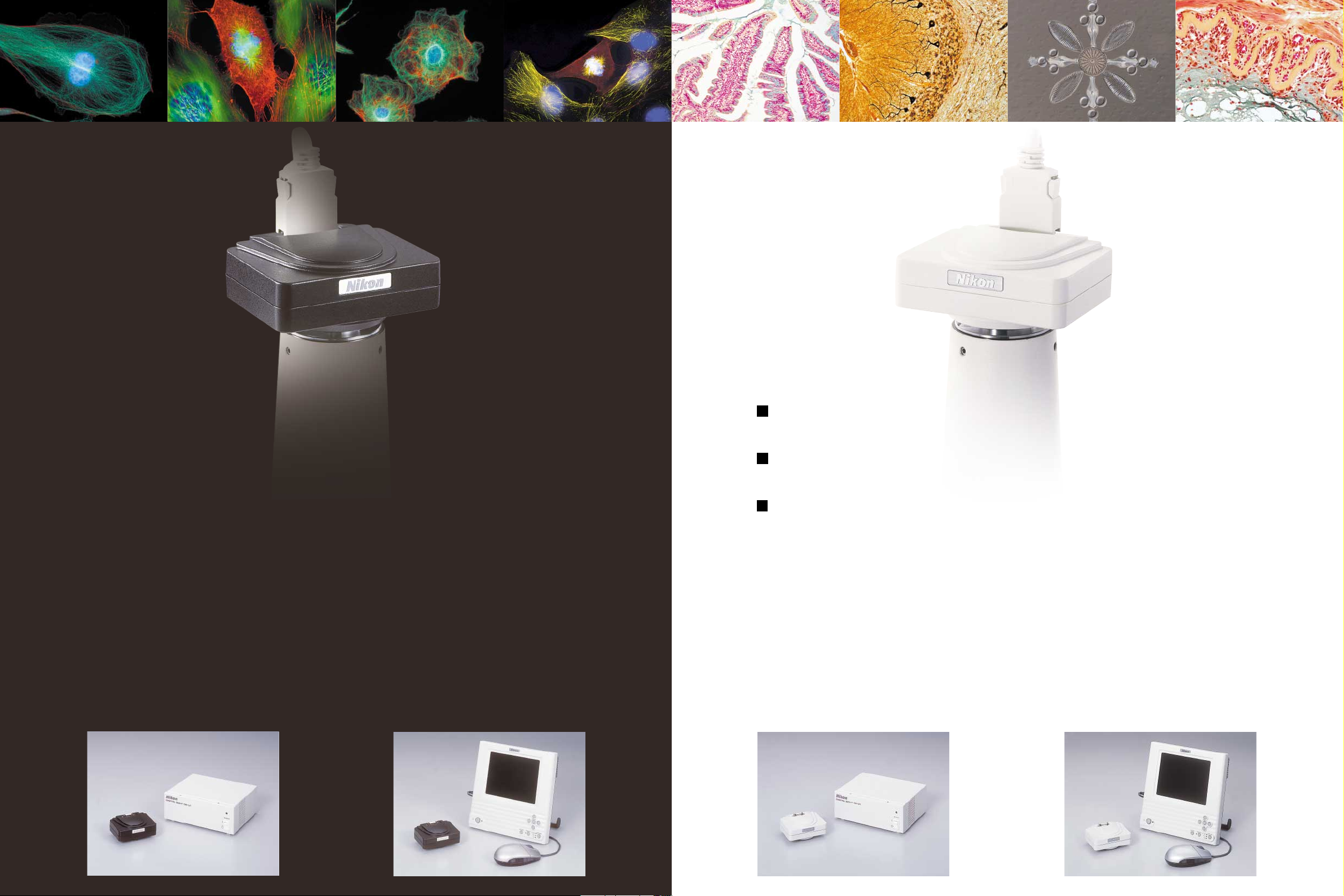
The compact camera head houses a 5.0-megapixel CCD.
Either color cooled type or standard type is selectable.
The color cooled camera head minimizes thermal noise
and creates ideal
conditions for taking fluorescence images.
Now you can get the best combination of cameras and
controllers to meet your observation needs.
DS-5Mc-U1
Cooled camera with USB interface controller
Even faint fluorescence and darkfield images are captured with high definition and low noise. Efficiently
capture and manage images from your PC with the
dedicated camera controlling software developed for
microscopes. Ideal for research applications involving image analysis and processing.
DS-5Mc-L1
Cooled camera with stand-alone controller
The controller, which has a large, built-in 6.3-inch
monitor, allows easy acquisition of clear, noiseless
fluorescence images without having to connect to
a PC. It's networking capabilities allow you to easily expand your system according to your research
needs.
A stand-alone
controller for capturing images without a PC, or a USB
interface controller for controlling the camera from a PC, can be selected.
DS-5M-U1
Standard camera with USB interface controller
Transferring large volumes of photographed
images to a PC is easy and fast thanks to the USB
interface. Image analysis and database creation
are also made easy with exclusive software for
microscopic image observation.
Standard camera with stand-alone controller
Optimum photography is realized with a single
click by virtue of menus that itemize photographic
conditions according to the observation method.
The camera's stand-alone design allows photos to
be taken without a PC.
DS-5M-L1
2 3
Page 3
Camera Heads
Its compact body is equipped with a high-definition, 5.0-megapixel 2/3-inch CCD. This standardtype camera head realizes high-resolution photomicrography with a maximum of 2,560 x 1,920
pixels. In all observation methods, including
brightfield, phase difference, and differentiation
interference, acquiring beautiful pictures faithful
to the real specimen is possible.
DS-5M
Standard CCD Camera
An optimum high-definition,
low-noise cooled camera head
for fluorescence image photography
The DS-5Mc's 5.0-megapixel 2/3-inch CCD attains high-definition, high-resolution pictures with a maximum of 2,560 x 1,920
pixels. And a Peltier cooling mechanism that maintains the
temperature of the CCD at -20˚C below ambient room temperature reduces the effects of thermal noise and the generation of hot pixels. Even when photographing weak fluores-
DS-5Mc
Cooled CCD Camera
4 5
cence or darkfield specimens requiring prolonged exposures,
it is able to acquire clear, low-noise images.
With 5.0 megapixels of high resolution,
the DS-5M captures beautiful microscope
images that are true to reality
Page 4

Control Units
DS-U1
PC Control type
One-touch connection to a PC via USB interface
This compact control unit can be connected quickly to any PC via its
USB2.0 interface without the need for a dedicated board.
Controlling the camera with a PC expands the user's system
and allows the user to perform all work, including capturing, analyzing,
and processing images, with only one piece of software (Act-2U).
Both DS-5Mc and DS-5M camera heads are connectable.
High-speed image transfer to a PC
With a high frame-per-second image transfer rate via the USB2.0 interface,
VGA (640 x 480 pixels) image data (live images) can be viewed on a monitor at a speed of 15 frames-per-second, enabling users to perform focus
adjustments and other tasks without strain.
Automatic detection of imaging status
In configuration with the ECLIPSE 80i microscope and DIH-M digital imaging head, DS-U1 automatically detects status data for objectives, zooming
magnification, optical ports, or fluorescence filters and saves the data
along with captured image files. It facilitates easy managing of the histories of images.
DS-5Mc
Camera Head
Exclusive camera cable (3m)
DS-5M
Camera Head
C-mount
adapter
Microscope
USB
Act -2U
Imaging Software
Camera Control Unit DS-U1
USB
Windows 2000/XP
Application software for imaging, processing, and analyzing
The software developed specifically for capturing microscope images offers a rich
array of functions for capturing large numbers of images easily and efficiently. It also
provides various functions for managing image data to create image databases.
This software features various image analyzing and image processing functions.
Users can do all this and more with only a single piece of software.
1
2
3
4
Easy-to-use interface
1. Main frame
2. Main toolbar
5
3. Toolbox
4. Annotation toolbox
5. Process view window
Shows the flow of image processing.
You can easily alter the process while working.
6
7
And you can set various modules,
and insert or delete them as well.
6. Property view window
7. Capture control window
8. Thumbnail window
8
Main features
Shading compensation
Time lapse capturing
Annotation
Histogram
ROI (Region of Interest) analysis
Image filtering:
Laplace (edge sharpner), Kirsh (gradient enhancer),
Sobel (edge enhancer), Median (pixel balancer)
Resolution changing
Image cropping
Image rotation
Configuration with the ECLIPSE 80i microscope and DS-5Mc camera head
Mirror effect
6 7
Page 5

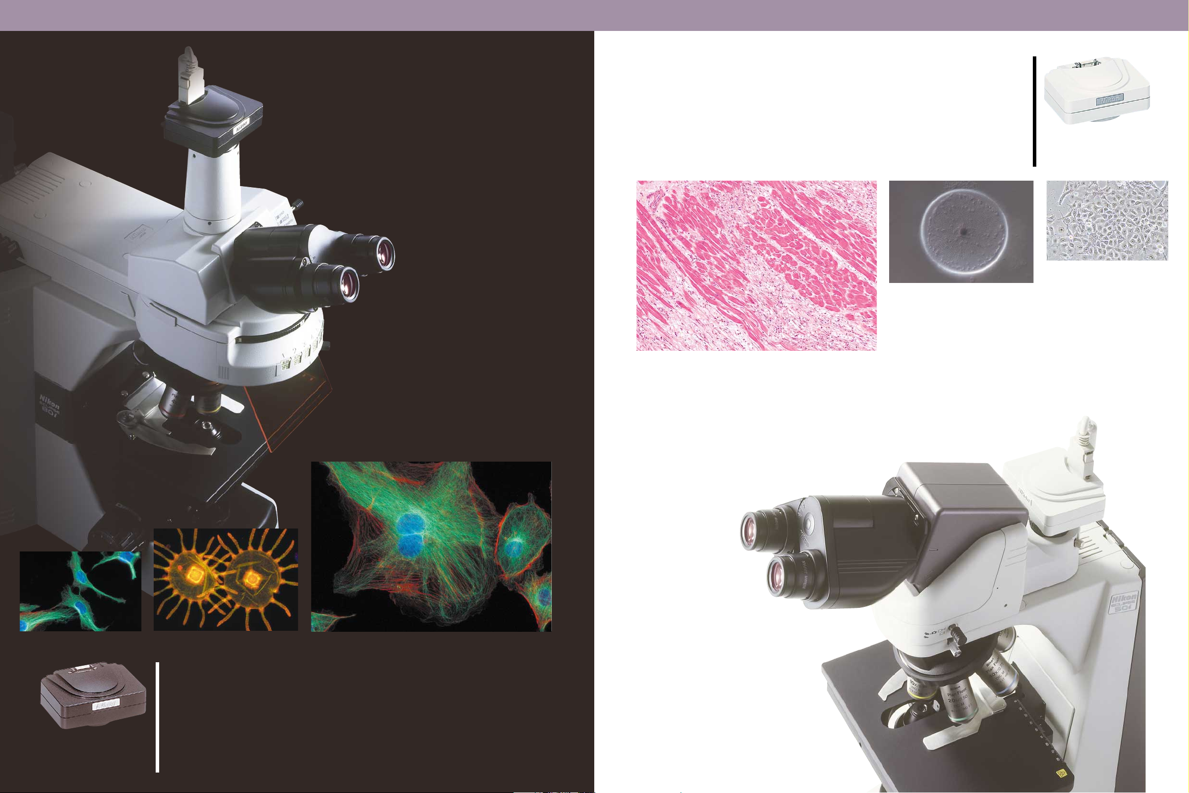
Control Units
DS-L1
Stand-alone type
Darkfield/
Fluorescence
image
Brightfield
image
Easy capturing of digital images without a PC
Observation, photography, and networking are all possible with this
single unit. There's no need to connect to a PC or external monitor.
By equipping a large, 6.3-type LCD monitor in a compact body,
focusing images on the monitor becomes possible. Both DS-5Mc
and DS-5M camera heads are connectable.
DS-5Mc
Camera Head
Microscope
C-mount
adapter
RGB output
PC monitor
DS-5M
Camera Head
Exclusive camera cable (3m)
100Base-TX
Ethernet
Universal-type
AC power adapter
USB
USB mass storage class
(Windows 2000/XP)
DS-L1 Camera Control Unit
with 6.3-inch LCD monitor
USB
USB HUB
TM
Compact Flash
Exclusive remote controller
USB mouse
USB keyboard
card
Phase contrast
DIC/
image
Scene function for one-click optimum
photography
Based on Nikon's experience manufacturing microscopes, optimal pre-programmed imaging modes are provided in the menu,
to allow the appropriate camera settings to be selected according to the desired observation method (bright, fluorescence, and
DIC/phase contrast). Optimal images can be captured with a simple click. The user can also customize settings and save up to
seven for quick retrieval.
Networking capabilities
Sharing images with PCs is possible with its networking function.
Distance measurement
Easily measures the distance between
any two points specified by the user.
(not available during digital zoom)
XY Scale Display
Independently movable scales with
an X-axis and a Y-axis are included for
measuring the size of samples, just
like you would with a ruler.
Count marking function
Up to 99 serial numbers can be
marked to provide a convenient way
to confirm the number of notes onscreen. They can also be easily saved
and printed.
Screen Pattern
Grid or concentric circle patterns can
be displayed. The center point can be
moved, and dot/solid, line/center, and
through/colors
patterns
(seven colors are available)
can be selected.
Two-screen split display
A frozen image can be displayed
alongside the live image for easy comparison.
Wide variety of other tools
Text and pen input: Input any character on the screen via a mouse or a
USB-connected keyboard. Also, lines
and figures can be drawn by hand
using the pen tool.
Thumbnail display: Images stored on
a CompactFlash
played together on-screen. File names
and photographic information can also
be displayed.
Superimpose function: Saved images
can be superimposed over live images,
for easy comparison.
TM
card can be dis-
Automatic detection of imaging status
In configuration with the ECLIPSE 80i microscope and DIH-M digital
imaging head, DS-L1 automatically detects status data for objectives,
zooming magnification, optical ports, or fluorescence filters and saves
the data along with captured image files. It facilitates easy managing of
the histories of images.
Configuration with the TS100 microscope
8 9
and DS-5M camera head
Page 6

Flexibly fits your specific needs
System Lineup
DS-5Mc-U1
Comparing confocal images with fluorescence images
The DS-5Mc cooled camera is ideal for research that compares confocal and epi-fluorescence images, as it is capable of capturing clear,
noiseless images even with weak fluorescent specimens. When configured with the ECLIPSE 80i microscope and DIH-M digital imaging
head, DS-U1 automatically detects objectives or fluorescence filters
and saves the data together with the image file.
DS-5Mc-L1
Capturing fluorescence images without a PC
Clear, noiseless fluorescence images can be taken with the
simple GUI, without connecting to a PC or external monitor.
With its networking capabilities, users can share images with
PCs. Objectives or fluorescence filter data is automatically
detected and can be attached to the image file.
Configuration with the ECLIPSE 80i microscope
and DIH-M digital imaging head
DS-5Mc-U1
Capturing and analyzing fluorescence images
The high-speed image transfer capabilities of the USB
interface enable users to easily focus images from their
PCs. It is possible to capture noiseless fluorescence
images and conduct a wide array of image analyses with
the same software.
Configuration with the ECLIPSE 80i microscope
DS-5M-L1
Handy for capturing digital images
Easily capture high-definition images without a PC
while saving space. You can capture and focus
images on the large monitor built into the controller. Optimal camera settings for each observation method can be selected from the menu.
10 11
The Digital Sight series is flexible and can be combined
and freely incorporated into your observation system to
optimize various applications.
Configuration with the ECLIPSE 80i microscope,
DIH-M digital imaging head, and confocal C1 imaging attachment
Configuration with the ECLIPSE TE2000-E microscope
DS-5M-U1
Creating a database of images
Capture high-definition, 5.0-megapixel images. The USB interface is useful for connecting your camera to a PC with just onetouch. It is suitable for researchers who need to take a large
number of images to create a database.
Configuration with the ECLIPSE 50i microscope
Specifications
(mm)
DS-5Mc/DS-5M
40.9
90.6
75.3
DS-U1
68.4
144.5
180.5
DS-L1
203
77
204
Camera Heads
CCD 2/3 in. high-density CCD: Total number of pixels: 5.24 million (effective 5.07 million)
CCD cooling device
Sensitivity
A/D conversion 12-bit
Lens mount C-mount
Exposure time 1/1000 to 600 sec 1/1000 to 60 sec
Dimensions Camera head: 90.6 (W) x 40.9 (H) x 75.3 (D) mm
Weight Camera head: approx. 290g Camera head: approx. 230g
System composition Camera Cable (3m)
Optional accessories 0.7x Relay lens (C-mount)
DS-5Mc (Cooled CCD Camera) DS-5M (Standard CCD Camera)
Peltier Device: Ambient temperature -20ºC
2400 lx, F5.6 or greater; equivalent to ISO 260
DS-U1 Camera Control Unit (PC Control type)
Exposure control Program AE, Shutter-priority AE, Focus AE, Manual with AE lock function
Exposure correction Correction range: ±2.0EV, Step: 1/3EV
Digital zoom 5 to 2400%
Interval shooting 5 sec. - 12 hr. intervals
Exposure metering Average metering, Peak hold metering
Exposure metering range
White balance Set method, Color balance adjustable
Compensation Brightness, Contrast, Gamma correction, Rotation, Flip-flop, Crop, Shading correction,
Storable image size 2560 x 1920 pixels, 1280 x 960 pixels, 640 x 480 pixels
Storage format BMP, TIFF, JPEG, JPEG2000
Live display mode
Interface
Power supply AC100-240V 50/60Hz
Power consumption 43VA
Dimensions Control unit: 180.5 (W) x 68.4 (H) x 144.5 (D) mm
Weight Control unit: approx. 1000g
Operating environment
System composition Power cord
3 selectable sizes
Monochrome, Nega/posi
Center scan mode (15 frames/sec. max.), 1.3M interlace mode (6.8 frames/sec. max. 2x2 binning mode)
1.3M progressive mode (7.5 frames/sec. max), 5M interlace mode (3.75 frames/sec. max)
USB2.0 device port (computer control connector), USB1.1 host port (microscope connector)
0-40˚C, 85% RH max. (without condensation)
Act-2U Imaging Software System Requirements
Computer type DOS PC supporting USB2.0
CPU Pentium 4, 1.7GHz or faster (Pentium 4, 2.4GHz or faster recommended)
RAM 512MB or more (1GB or more recommended)
USB2.0 2 ports
Hard disk 100MB to install, 300MB or more free space to run (on launch disk)
Operating system Windows 2000 Professional (SP4 or later, English or Japanese),
Graphics
Others CD-ROM drive (to install), Microsoft USB2.0 driver
The above system requirements list does not constitute a guarantee that all computers and systems meeting these criteria will be able to run the software.
Windows XP Professional (English or Japanese), pre-installed versions only
1280 x 1024 pixels or more, 16-bit color or more (24-bit color recommended), DirectX 9.0b support
DS-L1 Camera Control Unit (Stand-alone type)
Exposure control Program AE, Shutter-priority AE, Focus AE, Manual with AE lock function
Exposure correction Correction range: ±2.0EV, Step: 1/3EV
Digital zoom Up to 16x (8 steps)
Interval shooting 10 sec. - 6 hr. intervals
Exposure metering Average metering, Peak hold metering
Exposure metering range
White balance Set method, Color balance adjustable
Compensation
Image size 2560 x 1920 pixels, 1280 x 960 pixels, 640 x 480 pixels
Storage format BMP, JPEG (4-step compression)
Live display mode 4 selectable modes : 5M interlace mode (3.75 frames/sec. max.)
Interface USB1.1 host port (USB mouse, USB keyboard connection)
Power supply AC100-240V 50/60Hz
Power consumption 138VA
Dimensions Control unit: 203 (W) x 204 (H) x 77(D) mm
Weight Camera control unit: approx. 1300g, AC adapter: approx. 350g
Operating environment
System composition AC adapter, Power cord, CompactFlash card (64MB), Mouse
Networking Ethernet (10/100Base-TX), DHCP compatible, HTTP, TELENET or FTP server, FTP client
LCD monitor 6.3-in. TFT color LCD XGA (1024 x 768, 60Hz)
External monitor output
Storage media CompactFlash card (Type 1, Type II)
Optional accessories Exclusive remote control unit
3 selectable sizes
Gamma Compensation (9 types), Shading compensation (Auto: 5 step, Custom setting: 2 types) ,
Color/Monochrome, Color enhancement, Hue rotation, Black level adjustment,
Vertical and horizontal rotation
1.3M progressive mode (7.5 frames/sec. max.) , 1.3M interlace mode (6.8 frames/sec. max. 2x2 binning mode)
Center scan mode (15 frames/sec. max. , displays 1/2 in center of V direction)
USB1.1/2.0 device port (Mass Storage Class support)
0-40ºC, 85% RH max. (without condensation)
Analog RGB: SXGA (1280 x 1024, 60Hz), XGA (1024 x 768, 60Hz)
Page 7

TO ENSURE CORRECT USAGE, READ THE
CORRESPONDING MANUALS CAREFULLY
BEFORE USING THE EQUIPMENT.
WARNING
NIKON CORPORATION
Instruments Company
NIKON CORPORATION
Yokohama Plant
NIKON INSTECH CO., LTD.
Parale Mitsui Bldg.,8, Higashida-cho, Kawasaki-ku,
Kawasaki, Kanagawa 210-0005, Japan
phone: +81-44-223-2167 fax: +81-44-223-2182
www.nikon-instruments.jp/eng/
NIKON INSTRUMENTS (SHANGHAI) CO., LTD.
CHINA phone: +86-21-5058-5055 fax: +86-21-5058-5060
NIKON SINGAPORE PTE LTD
SINGAPORE phone: +65-6559-3618 fax: +65-6559-3668
NIKON MALAYSIA SDN. BHD.
MALAYSIA phone: +60-3-78763887 fax: +60-3-78763387
NIKON INSTRUMENTS EUROPE B.V.
Schipholweg 321, 1171PL Badhoevedorp, NL
phone: +31-20-44-96-222 fax: +31-20-44-96-298
www.nikon-instruments.com/
NIKON FRANCE S.A.
FRANCE phone: +33-1-45-16-45-16 fax: +33-1-45-16-00-33
NIKON GMBH
GERMANY phone: +49-211-9414-0 fax: +49-211-9414-322
NIKON INSTRUMENTS S.p.A.
ITALY phone: +39-55-3009601 fax: +39-55-300993
NIKON AG
SWITZERLAND phone: +41-43-277-2860 fax: +41-43-277-2861
NIKON UK LTD.
UNITED KINGDOM phone: +44-20-8541-4440 fax: +44-20-8541-4584
NIKON INSTRUMENTS INC.
1300 Walt Whitman Road, Melville, N.Y. 11747-3064, U.S.A.
phone: +1-631-547-8500; +1-800-52-NIKON (within the U.S.A. only) fax: +1-631-547-0306
www.nikonusa.com/
NIKON CANADA INC.
CANADA phone: +1-905-625-9910 fax: +1-905-625-0103
Code No. 2CE-MSPH-1
Printed in Japan (0404-12) Am/M
Company and product names included in this brochure are the
registered trademarks of the respective companies. Specifications
and equipment are subject to change without any notice or obligation
on the part of the manufacturer. April 2004.
'2004 NIKON CORPORATION
NIKON CORPORATION
Fuji Bldg., 2-3, Marunouchi 3-chome, Chiyoda-ku,
Tokyo 100-8331, Japan
www.nikon.com/
Highly flexible digital camera systems
for microscopy can be selected
for the optimum combination of units.
Digital Camera System for Microscopy
En
 Loading...