

Page 1
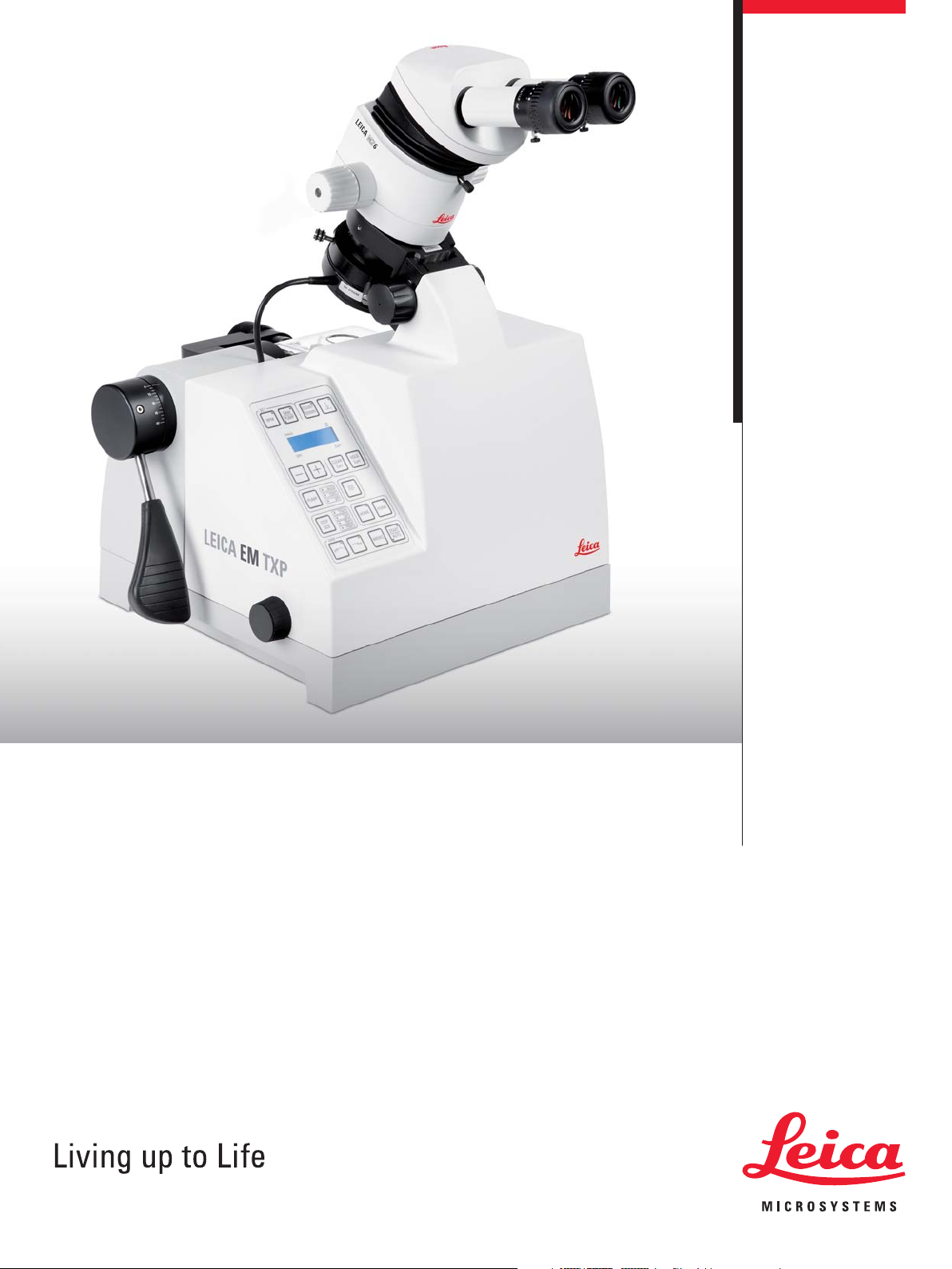
Leica EM TXP
Target Surfacing System
Page 2
2
Leica EM TXP – Target Surfacing
90° rotation view
Processing observation
Front face observation
The Leica EM TXP is a unique target preparation device especially
developed for cutting and polishing samples prior to examina tion by
SEM, TEM and LM techniques. It excels with challenging speci mens where pinpointing and preparing barely visible targets
becomes easy. Before the Leica EM TXP, sawing, milling, grinding
and polishing exactly to the target was often a very time-consuming
and difficult procedure as points of interest were easily missed and
specimens often difficult to handle due to their small size. With the
Leica EM TXP such samples can easily be prepared. Furthermore,
due to its versatility, the EM TXP is a very efficient tool for sample
pre-preparation prior to ion beam milling and ultramicrotomy.
Leica Design by W. Hölbl
Page 3
3
System
Stereomicroscopic target observation during the working process
With the specimen pivot arm the sample can be observed during
preparation at an angle between 0° and 60°, directly onto the front
face, or 90° to the front face for distance determination with an
eyepiece graticule. The Leica EM TXP features brilliant ring LED
top light and optimized backlight illumination for excellent viewing.
I Accurate location and preparation of microtargets
I
In-situ
observation with a stereomicroscope
I Multifunctional machine processing
I Automatic process control
I to produce a mirror-like surface quality
I Brightness control and segment selection
I of LED-ring illumination
Integrated Viewing System
Cross section of watch assembly
(processed without embedding)
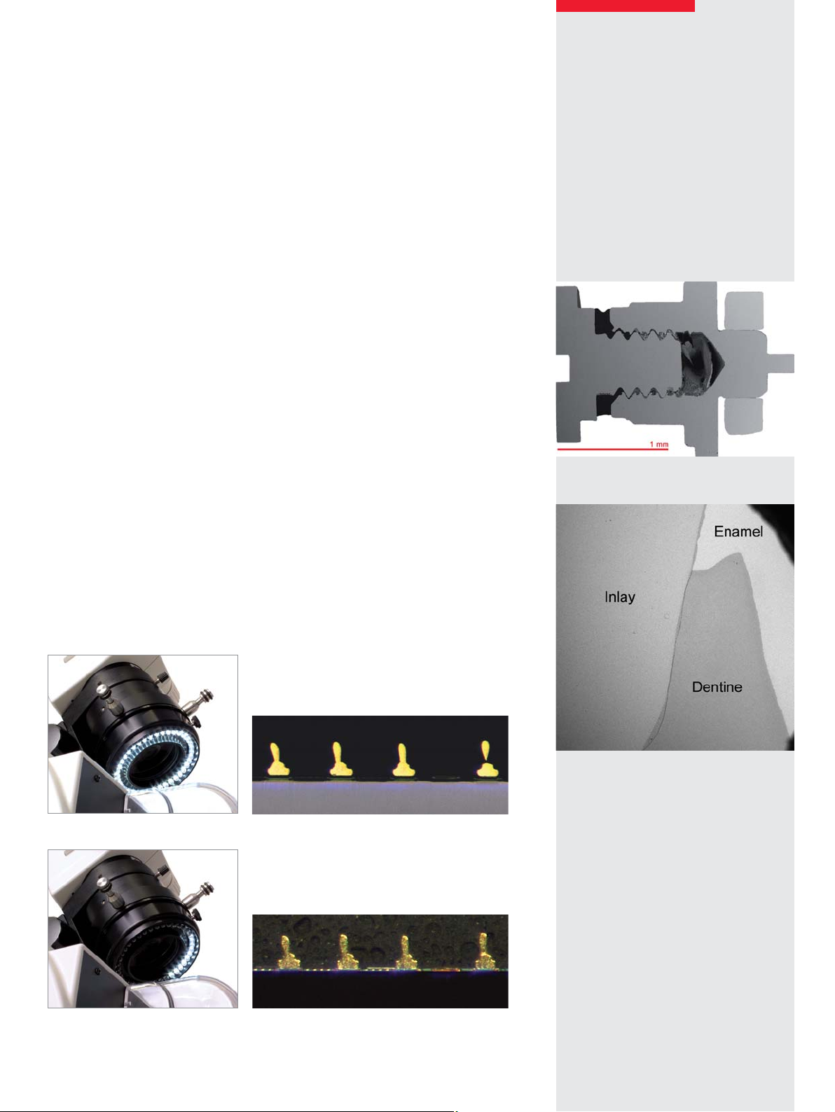
Viewing with quarter ring illumination
Cross section of a tooth
(processed without embedding)
Viewing with full ring illumination
Page 4

4
Once the sample is clamped into the specimen holder and inserted in the pivot arm, the specimen can be:
– milled
– sawn
– drilled
– ground
– and polished
without removing the sample from the Leica EM TXP and simply
changing the tools while observing the process directly through
the stereomicroscope. The tool and sample are enclosed within a
protective chamber with a transparent cover for safety. This prevents access to moving parts and avoids particulate matter
escaping. During milling a low-noise extraction and filtration unit
with a Hepa filter (optional) provides a safe, dust-free environment.
Process Possibilities
Diamond and tungsten carbide millers
Diamond disc cutter
Lapping inserts 15, 9, 6, 3, 1, 0.5 µm
Page 5

5
Let the Leica EM TXP do the job
The Leica EM TXP automatic process control mechanism saves
you from time-consuming routine sample preparation:
– with the automatic E-W guiding mechanism
– with the force-regulated feed control
– with the distance or time countdown function
– with force-controlled auto-advance for core-drilling
and level sensor for the integrated lubricant cooling system
Integrated Automatic Process Control
– Pivot arm lever
– Hand wheel for manual feed in steps of
0.5, 1, 10 and 100 µm
– Control panel for manual operation and setting
of all parameters for automatic preparation
E-W guiding mechanism
(manual or automatic)
Peristaltic
cooling system
SM LED gold wire bond
PCB cross section with soldered pin
Page 6

6
Observation via stereo microscope of the Leica EM TXPSawing
After the sample is fixed onto the stub a 3 mm disc is cut out using the
core-drill in conjunction with the force-regulated auto-advance.
The first side is then finished using the grinding and polishing foils.
The surface of the stub is milled with the tungsten carbide miller
so it is parallel to the grinding and the polishing plane for processing
the second side of the sample.
Target preparation on surfaced sample for incident light LM and SEM
All processing steps are carried out consecutively on the Leica EM TXP without removing the sample for pinpointing the area of
interest via another microscope or for making any adjustments. Preparing specimens observed during operation with the integrated stereomicroscope avoids the time consuming interruption of locating the target with a stand alone microscope and then
re-aligning the sample in the polishing instrument.
Sample thinning for transmitted light LM or prior to ion thinning for TEM
Specimen thinning with the Leica EM TXP offers the advantage of observing the complete process during treatment and distance
monitoring at each process step without the need to remove the sample for checking in another instrument.
The Leica EM TXP is a unique target surfacing system developed for cutting and polishing samples. Alignment of sample details
can be rapidly performed using the alignment accessory. Thus, the EM TXP is the instrument of choice for pre-preparation of the
sample prior to ion beam slope cutting with e.g. the Leica EM TIC020.
Sample holder attached on the alignment accessory.
The micro-targets can be aligned during the process
while observing with the stereo microscope.
Within a few minutes all sample details are aligned. LM-image of gold
wire bondings processed with the cut-off wheel and ground with 6 µm
diamond foil during alignment. The surface finish is good enough for
subsequent finishing with the ion beam slope cutter, EM TIC020.
Page 7

7
The area of interest via the stereomicroscope of the Leica EM TXP
The prepared first side is fixed onto the surface of the stub.
The second side is then cut, ground and polished. The thickness of
the sample can be determined with the advance counter display.
In such a manner, even hard and brittle materials can be thinned to be
subsequently prepared for TEM investigation e.g. for ion thinning with
the Leica EM RES101.
Grinding/polishing
The Leica EM TIC020 features three ion guns in one assembly
enabling > 4 mm cutting width and > 1 mm cutting depth.
Gold-wire bondings of IC-package during processing with the
Leica EM TIC020. The dark area above the gold wires has already been
polished by the ion beam.
Page 8

8
Gold-wire bonding of IC-package prepared
with the Leica EM TXP (LM image).
Leica EM RES101 for ion beam processing
of TEM and SEM samples.
Same gold wire bonding subsequently processed for
half an hour with the Leica EM RES101 (SEM image).
High surface quality within a few hours. The user interaction time of the complete process is around
20 minutes using the Leica EM TXP prior to EM TIC020.
Page 9

View the Details
Page 10

“With the user, for the user”
Leica Microsystems
Order no.: English 16214002
•
IX/10
•
Copyright © by Leica Mikrosysteme GmbH, Vienna, Austria, 2010
LEICA and the Leica Logo are registered trademarks of Leica Microsystems IR GmbH.
The statement by Ernst Leitz in 1907, “with the user, for the user,” describes the fruitful collaboration
with end users and driving force of innovation at Leica Microsystems. We have developed five
brand values to live up to this tradition: Pioneering, High-end Quality, Team Spirit, Dedication to
Science, and Continuous Improvement. For us, living up to these values means: Living up to Life.
Active worldwide
Australia: North Ryde Tel. +61 2 8870 3500 Fax +61 2 9878 1055
Austria: Vienna Tel. +43 1 486 80 50 0 Fax +43 1 486 80 50 30
Belgium: Groot Bijgaarden Tel. +32 2 790 98 50 Fax +32 2 790 98 68
Canada: Richmond Hill/Ontario Tel. +1 905 762 2000 Fax +1 905 762 8937
Denmark: Ballerup Tel. +45 4454 0101 Fax +45 4454 0111
France: Nanterre Cedex Tel. +33 811 000 664 Fax +33 1 56 05 23 23
Germany: Wetzlar Tel. +49 64 41 29 40 00 Fax +49 64 41 29 41 55
Italy: Milan Tel. +39 02 574 861 Fax +39 02 574 03392
Japan: Tokyo Tel. +81 3 5421 2800 Fax +81 3 5421 2896
Korea: Seoul Tel. +82 2 514 65 43 Fax +82 2 514 65 48
Netherlands: Rijswijk Tel. +31 70 4132 100 Fax +31 70 4132 109
People’s Rep. of China: Hong Kong Tel. +852 2564 6699 Fax +852 2564 4163
Portugal: Lisbon Tel. +351 21 388 9112 Fax +351 21 385 4668
Singapore Tel. +65 6779 7823 Fax +65 6773 0628
Spain: Barcelona Tel. +34 93 494 95 30 Fax +34 93 494 95 32
Sweden: Kista Tel. +46 8 625 45 45 Fax +46 8 625 45 10
Switzerland: Heerbrugg Tel. +41 71 726 34 34 Fax +41 71 726 34 44
United Kingdom: Milton Keynes Tel. +44 1908 246 246 Fax +44 1908 609 992
USA: Bannockburn/lllinois Tel. +1 847 405 0123 Fax +1 847 405 0164
and representatives in more than 100 countries
Leica Microsystems operates globally in four divisions,
where we rank with the market leaders.
•
Life Science Division
The Leica Microsystems Life Science Division supports the
imaging needs of the scientific community with advanced
innovation and technical expertise for the visualization,
measurement, and analysis of microstructures. Our strong
focus on understanding scientific applications puts Leica
Microsystems’ customers at the leading edge of science.
•
Industry Division
The Leica Microsystems Industry Division’s focus is to
support customers’ pursuit of the highest quality end result.
Leica Microsystems provide the best and most innovative
imaging systems to see, measure, and analyze the microstructures in routine and research industrial applications,
materials science, quality control, forensic science investigation, and educational applications.
•
Biosystems Division
The Leica Microsystems Biosystems Division brings histopathology labs and researchers the highest-quality,
most comprehensive product range. From patient to pathologist, the range includes the ideal product for each
histology step and high-productivity workflow solutions
for the entire lab. With complete histology systems featuring innovative automation and Novocastra™ reagents,
Leica Microsystems creates better patient care through
rapid turnaround, diagnostic confidence, and close customer collaboration.
•
Medical Division
The Leica Microsystems Medical Division’s focus is to
partner with and support surgeons and their care of patients with the highest-quality, most innovative surgical
microscope technology today and into the future.
www.leica-microsystems.com
 Loading...