

GE Healthcare CT660 Brochure
GE Healthcare
Exceptionally precise,
extremely fast
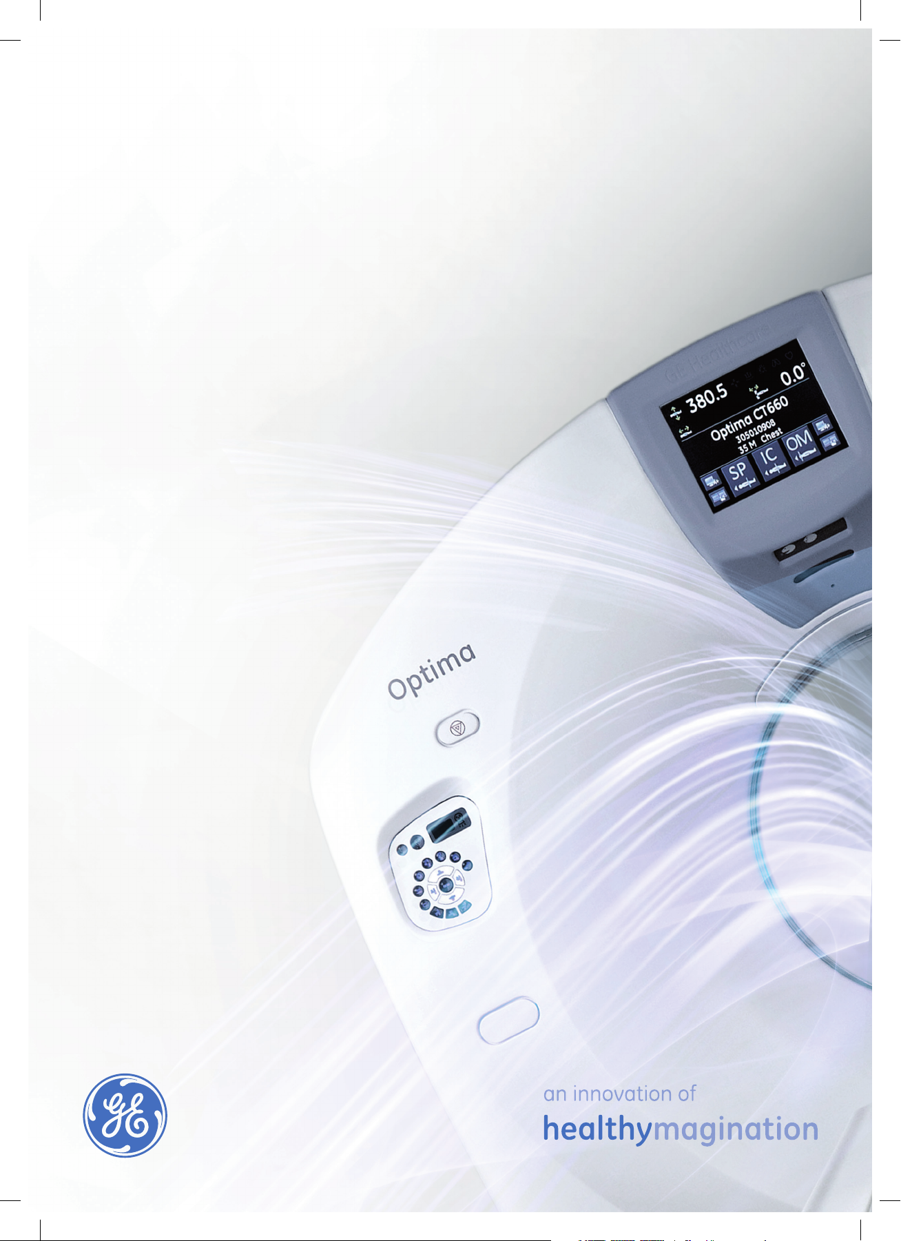
Optima* CT660
Spatial Enhanced

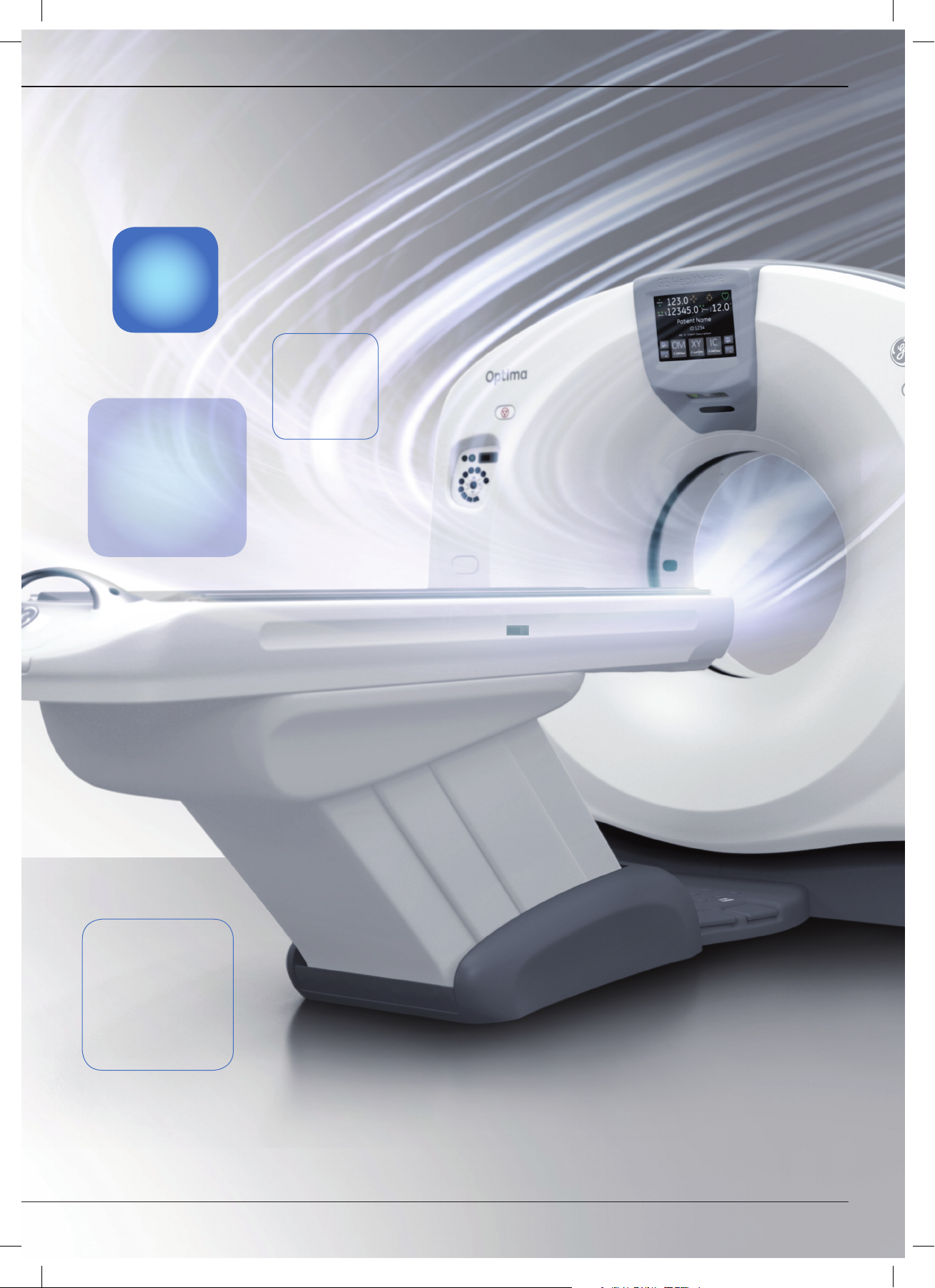
Because you simply want
the best for your patients…
It’s no secret that hospitals and clinicians today are faced with having to do more
with less. Healthcare reform, market uncertainties and changes in the delivery of
patient care including the emergence of Accountability Care Organizations are now
driving purchasing decisions. Yet the need for quality patient care at low dose is
more important than ever.
Using feedback obtained from customers, we’ve created an intelligent CT scanner
with a simplifi ed workfl ow for quick and streamlined operation. Workfl ow features
like ED mode and auto patient positioning, a user-friendly console and synchronized
injection allow users to spend more time focused on patient care and comfort.
The Optima CT660 Spatial Enhanced is a new generation, Volume CT scanner that
brings together diagnostic power and workfl ow effi ciency, enabling fast, high-quality
acquisitions at optimized dose. It helps institution of addresses a new era of
exceptional patient care and operational excellence. Our system solution features
full capabilities in advanced applications such as cardiac, oncology, angiography
and dynamic imaging.
As providers consider options to manage dose, we created Lower Dose by Design —
combining research, training, technology, and clinical practice to achieve high
diagnostic image quality at optimized dose. We are committed to helping you deliver
the highest quality patient care — with features like ASiR*
Check.*
The Optima CT660 Spatial Enhanced also consumes up to 60% less energy than
previous GE CT systems and boasts a 15% lower siting requirement compared to
other 64-channel detector scanners. Over the life of the product, these
substantial savings can translate into lower operational costs.
Because you want the best for your patients - Optima CT660 Spatial
Enhanced
†
, ODM, Optidose* and Dose
Performance
Imaging
Robust
Cardiac
Imaging
Simple
High
to Use
40mm
detector
Clinical
Advanced
Application
Low
Dose
Speed and
Coverage

1.531
†
Helical
pitch
Just 5
touches for
scanning
55fps
Real-time
recon
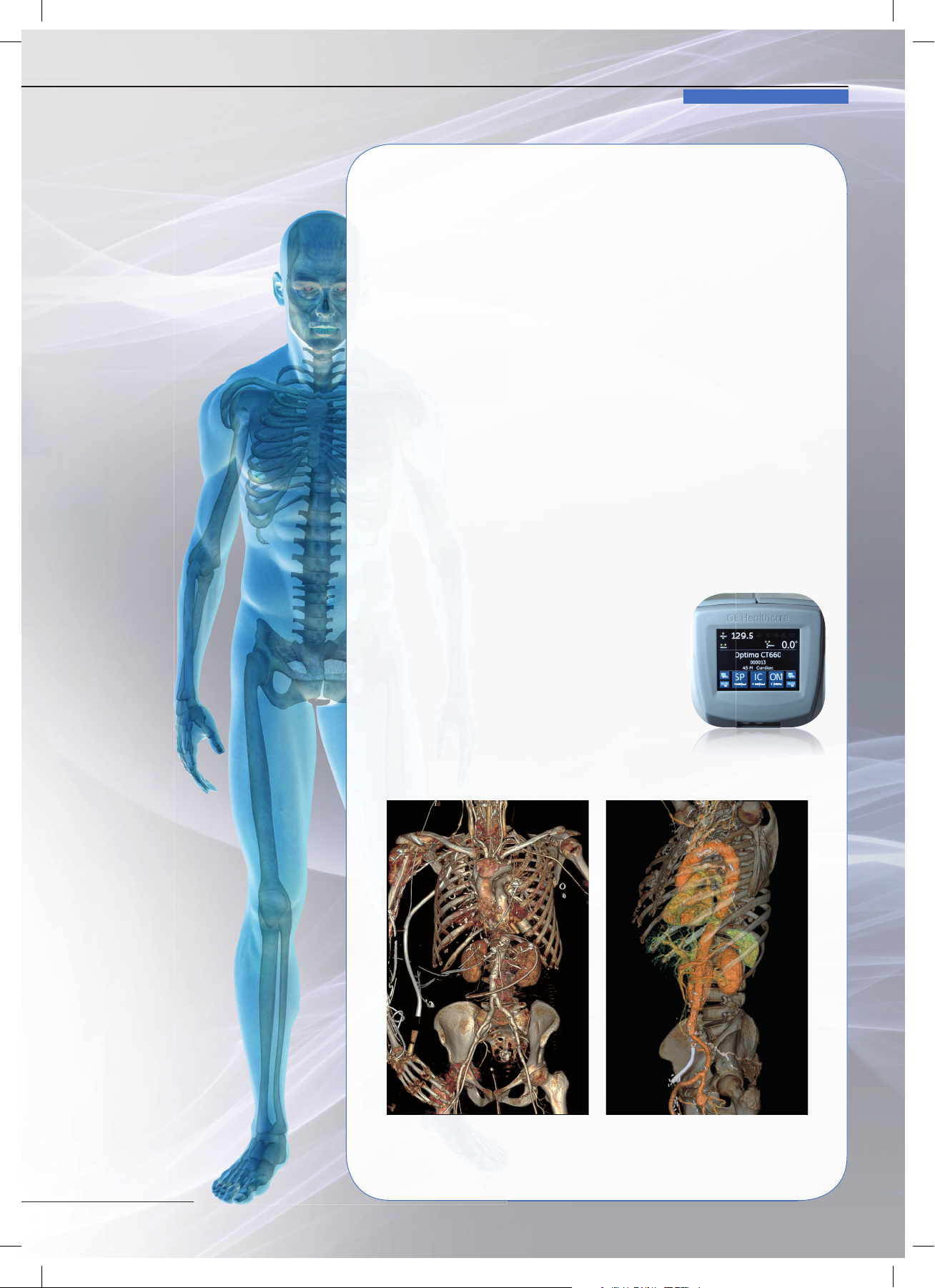
Emergency:
When seconds count
With the Optima* CT660, the user can scan
quickly from the Circle of Willis to the lower limbs,
in one pass with diagnostic image quality and
images reconstructed in real time. The optimized
ergonomics on the scanner improves patient
management and the users can reassure the
patient in the scan room while pre-scanning on
the gantry display. The patient is scanned within
seconds.
Using real time reconstruction (55 fps)**, the
acquired images appear in less than 1 second
on the CT console—enabling a quick diagnosis
and improving triage and door-to-door treatment
times in emergency radiology.
ADVANCED TECHNOLOGY.
Designed to be simple
to use with a streamlined
workfl ow
Automated post-processing at your fi ngertips
The Xtream Display shows basic patient information on the gantry
monitor. As such, the user can confi rm patient information in the scan
room, improving workfl ow and potentially reducing the opportunity
for error. Pre-scanning can be accomplished in as few as fi ve touches.
For example, the Optima* CT660’s exceptional one-stop scanning
mode† provides streamlined workfl ow, shown on the Xtream Display
with phrases such as “Patient selection”, “Protocol selection”, and
“Confi rm”.
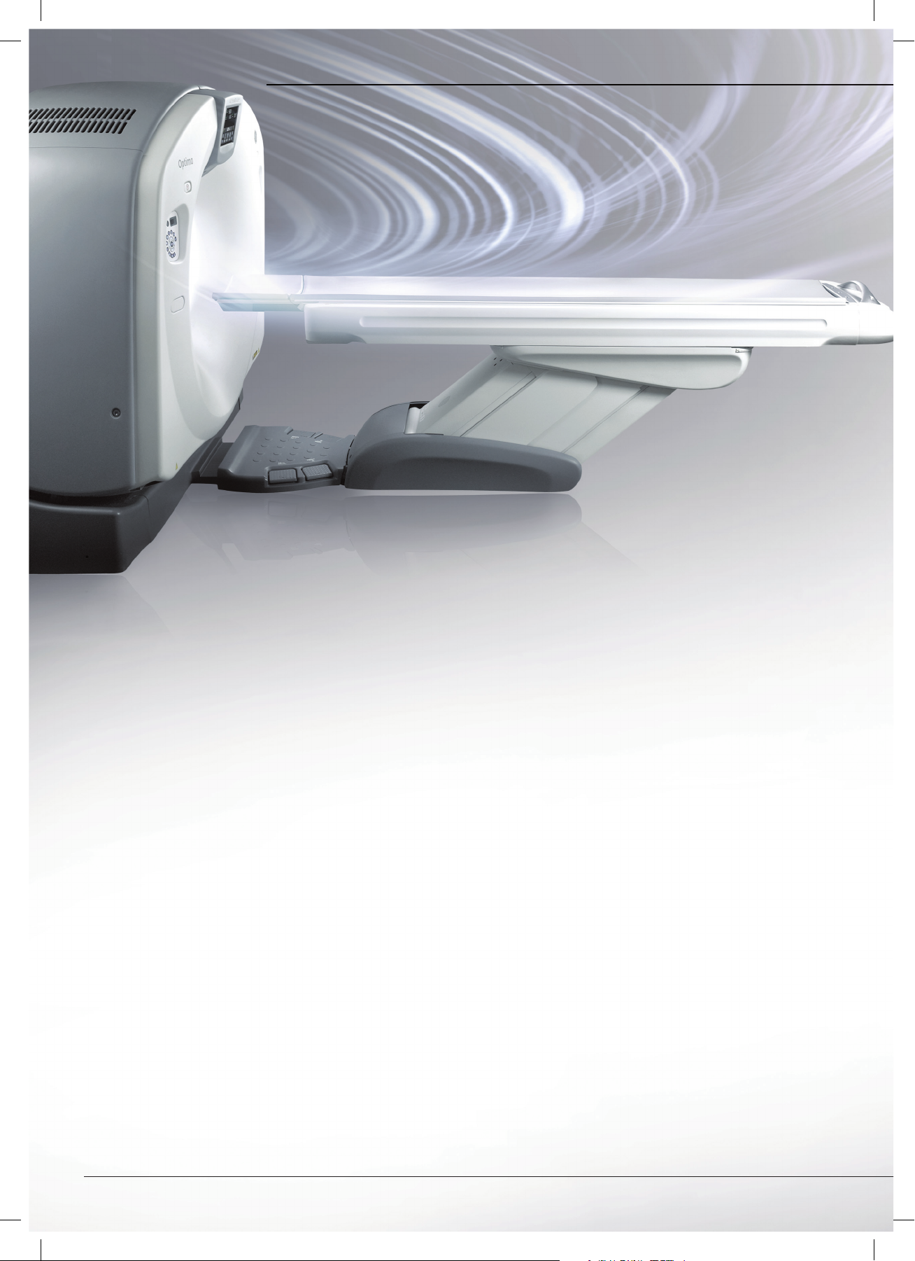
Enhanced Table: Scan a whole-body trauma
without moving the patient
The VT2000** allows patients, weighing up to
227 kg, to be imaged through a long, scannable
range. The Default Patient Positioning provides
semi-automatic positioning according to the
type of exam, reducing manual positioning
and streamlining workfl ow. The Xtream Display
shows pictures that help operators understand
the correct exam position.
12sec at 1500mm scan coverage
Advanced technology
for dynamic studies
Optima* CT660: A partner of choice
for your most critical studies
Cardiovascular: Comprehensive solutions for heart and vessels
assessment
Delivering 40 mm coverage per rotation, featuring
a temporal resolution down to 44 ms, the Optima
CT660 is designed to scan the heart in 5 beats. Its
ample tube power combined with ASiR*† delivers
the image quality demanded, even with large
patients.
Snapshot* Pulse
SnapShot Pulse mode is for low dose imaging of
the coronary arteries. SnapShot Pulse can also be
used to image structures that are near to the heart
and may be affected by heart motion such as
thoracic aorta’s or pulmonary arteries. Prospective
Gating based SnapShot Pulse achieves up to 83%
dose reduction compared to ECG gated helical
acquisition mode.
ASiR may help clinicians achieve dose reductions
of up to 40%. Customers using ASiR have
demonstrated excellent diagnostic image quality
at low dose across exam types and body regions.
 Loading...