

Page 1

Eagle 1000 Patient Monitor
Servicing Instructions
227 468 01 SA(e) Revision G
Page 2

Caution:
During repairs/service interventions, observe the protective measures against damage due
to ESD.
· GE Medical Systems Information Technologies (GEMS IT) considers itself
responsible for the effects on safety, reliability, and performance of the equipment, only
if:
- assembly operations, extensions, readjustments, modifications, or
repairs are carried out by
GE Medical Systems Information Technologies (GEMS IT) or by persons
authorized by GE Medical Systems Information Technologies,
- the electrical installation of the relevant room complies with the applicable
national and local requirements, and
- the instrument is used in accordance with the instructions for use.
• This manual contains service information; operating instructions are provided in the
operators manual of the instrument.
• This manual is in conformity with the instrument at printing date.
• All rights are reserved for instruments, circuits, techniques, and names appearing in the
manual.
© Copyright 1997 - 2003, GE Medical Systems Information Technologies GmbH.
All rights reserved.
Printed in Germany
Page 3

GE Medical Systems EAGLE 1000 Page 3
Information Technologies Servicing Instructions 227 468 01 Rev. G
Contents
1. General overview of EAGLE 1000 ........................................................................................................ 7
1.1 Documentation and nomenclature of Marquette Hellige instrument part numbers .......................... 7
1.1.1 Configuration of instrument part number ............................................................................... 7
1.1.2 Configuration of the PCB par numbers .................................................................................. 7
1.2 Eagle 2000 system overview ............................................................................................................ 9
1.3 System overview............................................................................................................................. 10
1.3.1 Hooking up EAGLE 1000 monitors to PC Central Station SynOpsis 1000 ......................... 10
1.3.2 Patient data transfer from EAGLE 1000 via HELAN to SMU using QuickLink .................... 12
1.3.2.1 Introduction .......................................................................................................... 12
1.3.2.2 Data transfer between Eagle and Gateway ........................................................ 13
1.3.2.3 Connection to the LAN ........................................................................................ 13
1.3.2.4 EAGLE 1000 - QuickLink - HELAN - Examples of the link to networks .............. 15
1.3.2.4.1 Functional structure overview .......................................................... 15
1.3.2.4.2 Main components for the installation of QuickLink: .......................... 15
1.3.2.4.3 EAGLE 1000 - QuickLink ................................................................. 16
1.3.2.4.4 Network link - HELAN Concentrator ................................................ 17
1.3.2.4.5 Network link - HELAN Twisted Pair.................................................. 18
2. System overview .................................................................................................................................. 19
2.1 Technical specifications .................................................................................................................. 19
2.1.1 System interfaces ................................................................................................................ 19
2.1.2 Display ................................................................................................................................. 19
2.1.3 Monitor functions ................................................................................................................. 19
2.1.4 Power supply ....................................................................................................................... 20
2.1.5 Operation and storage ......................................................................................................... 21
2.1.6 ECG and heart rate ............................................................................................................. 21
2.1.7 Oxygen saturation Sp02 ...................................................................................................... 23
2.1.8 Non-invasive blood pressure ............................................................................................... 24
2.1.9 Invasive blood pressure ....................................................................................................... 25
2.1.10 Temperature......................................................................................................................... 27
2.1.11 Trend.................................................................................................................................... 28
2.2 Functional description of PCBs and subassemblies ...................................................................... 28
2.2.1 Eagle 1000 configuration and functional groups ................................................................. 28
2.2.2 PCB ECG Monitor................................................................................................................ 29
2.2.3 System power supply .......................................................................................................... 30
2.2.4 Floating system power supply and signal flow .................................................................... 31
2.2.5 Signal exchange with the processor.................................................................................... 31
2.2.6 Display ................................................................................................................................. 32
2.2.7 Block diagrams .................................................................................................................... 33
2.2.7.1 Functional groups ................................................................................................ 33
2.2.7.2 Floating system supply and signal flow ............................................................... 34
2.2.7.3 System power supply .......................................................................................... 35
2.2.7.4 PROCESSOR block diagram .............................................................................. 36
2.2.7.5 DISPLAY block diagram ...................................................................................... 37
2.2.8 ECG signal preprocessing ...................................................................................................38
2.2.8.1 Functional descriptiony ........................................................................................ 38
2.2.8.2 ECG/Pace Preprocessing ................................................................................... 40
2.2.9 PCB MP 204 Oximeter 930 117 26 ...................................................................................... 41
2.2.9.1 Function ............................................................................................................... 41
2.2.9.2 Figure of pcb MP204 Oximeter ........................................................................... 41
2.2.9.3 Pin assignment .................................................................................................... 42
2.2.10 Non-invasive blood pressure module .................................................................................. 43
2.2.10.1 Function ............................................................................................................... 43
2.2.10.2 Figure of PCB NBP Module NB11: 930 117 15 ................................................... 44
2.2.10.3 Figure of PCB NBP 2000 Module 930 117 93 or 388 032 60 ............................. 45
2.2.10.4 Pin assignment .................................................................................................... 46
2.2.11 PCB Press-Temp 388 031 75/PCB Temp 388 032 04......................................................... 47
Page 4

GE Medical Systems EAGLE 1000 Page 4
Information Technologies Servicing Instructions 227 468 01 Rev.G
2.2.11.1 PRESS module .................................................................................................... 47
2.2.11.2 Temp module ....................................................................................................... 48
2.2.11.3 Internal interfaces ................................................................................................ 49
2.2.11.4 Interfaces to peripherals ...................................................................................... 50
2.2.11.5 TEMPERATURE BLOCK DIAGRAM ................................................................... 51
2.2.11.6 PRESS BLOCK DIAGRAM ................................................................................. 52
2.2.12 Recorder 50 Part No. 218 111 12 ........................................................................................ 53
2.2.12.1 Function ............................................................................................................... 53
2.2.12.2 Recorder block diagram ...................................................................................... 54
2.2.12.3 Internal interfaces ................................................................................................ 55
2.2.12.4 Interfaces to peripherals ...................................................................................... 56
2.2.13 PCB Power Supply 930 117 41 ........................................................................................... 57
2.2.13.1 Function and pin assignment .............................................................................. 57
2.2.13.2 Figure of PCB Power Supply 930 117 41 ............................................................ 58
2.2.14 Membrane keypad ............................................................................................................... 59
2.2.14.1 Pin assignment ........................................................................................................................ 59
2.2.14.2 Membrane keypad 390 001 30 and 390 001 64, circuit diagram ........................ 60
2.3 Pin configuration of panel connectors ............................................................................................ 61
2.3.1 Socket ECG 303 444 28 ......................................................................................................61
2.3.2 Socket SpO2 303 444 29.....................................................................................................62
2.3.3 Socket PRESS 303 444 30 ................................................................................................. 63
2.3.4 Socket Temperature (TEMP) 303 444 31 ............................................................................ 64
2.3.5 Socket on the rear panel of the Eagle 1000 ........................................................................ 65
2.3.6 2-Port Press Adapter 220 102 01 ........................................................................................ 66
2.4 Complete circuit diagram and mechanical diagrams ...................................................................... 67
2.4.1 Systems without a recorder 101 123 .. ................................................................................ 67
2.4.2 Systems with a recorder 101 124 .. ..................................................................................... 67
2.4.3 Complete wiring circuit (S-Plan) .................................................... Please refer to the appendix
2.5 Master Record Index (MRI) ................................................................... Please refer to the appendix
3. Maintenance and technical inspection .............................................................................................. 69
3.1 Visual check .................................................................................................................................... 69
3.2 Checkout Procedures ..................................................................................................................... 70
3.2.1 Recommended testing/calibration equipment and accessories: ......................................... 70
3.2.2 Test preliminaries ................................................................................................................. 71
3.2.3 Operating control and indicator performance checks.......................................................... 71
3.2.4 ECG performance check ..................................................................................................... 72
3.2.4.1 Patient cable identification ................................................................................... 72
3.2.4.2 ECG Simulator settings ....................................................................................... 72
3.2.4.3 Analysis of the ECG signal, HR value and QRS beep ........................................ 72
3.2.4.4 Testing alarm limits, recorder start, audible alarm activation .............................. 73
3.2.4.5 Testing the input amplifier offset .......................................................................... 73
3.2.4.6 Testing pacing detection ...................................................................................... 73
3.2.4.7 Testing the lead fail detection .............................................................................. 74
3.2.5 NI blood pressure performance check ................................................................................ 74
3.2.5.1 Leakage test ........................................................................................................ 74
3.2.5.2 Testing display errors .......................................................................................... 74
3.2.5.3 Testing the deflation pressure threshold ............................................................. 76
3.2.5.4 Dynamic blood pressure measurement .............................................................. 76
3.2.6 SpO2 performance check .................................................................................................... 76
3.2.6.1 Simulator setting .................................................................................................. 76
3.2.6.2 Monitor settings ................................................................................................... 76
3.2.6.3 Displaying waveforms and parameter readings .................................................. 76
3.2.6.4 Testing display accuracy of SpO2 and pulse rate ............................................... 77
3.2.7 Invasive blood pressure performance check (Vers. 2.0 and higher) ................................... 77
3.2.8 Temperature performance check (Vers. 2.0 or higher) ........................................................ 78
3.2.9 RS232 performance check (Vers. 2.0 or higher) ................................................................. 78
3.2.10 Nurse call output performance check .................................................................................. 79
3.2.11 Testing condition of battery .................................................................................................. 79
Page 5

GE Medical Systems EAGLE 1000 Page 5
Information Technologies Servicing Instructions 227 468 01 Rev. G
3.2.12 Backup battery for the clock ................................................................................................ 79
3.3 Calibration check of the measuring system....................................................................................80
3.3.1 Technical Inspections of the Measuring System ................................................................. 80
3.4 Safety Analysis Test ........................................................................................................................ 81
3.4.1 General introduction ............................................................................................................ 81
3.4.2 Recommended Test Equipment .......................................................................................... 81
3.4.3 Protective Earth Resistance Test......................................................................................... 83
3.4.4 Leakage Current Measurement........................................................................................... 83
3.4.4.1 Enclosure Leakage Current Test ......................................................................... 83
3.4.4.2 Patient Leakage Current Test .............................................................................. 84
3.4.4.3 Enclosure Leakage Current Test (System) ......................................................... 85
4. Instrument service ............................................................................................................................... 86
4.1 Service mode tests ......................................................................................................................... 86
4.2 Instrument performance .................................................................................................................. 87
4.2.1 Start-up performance ........................................................................................................... 87
4.2.2 Operating performance ........................................................................................................ 87
4.2.3 Performance in the event of a malfunction .......................................................................... 87
4.3 Battery management ...................................................................................................................... 88
4.4 Safety information ........................................................................................................................... 89
4.4.1 Insulating foil ........................................................................................................................ 89
4.4.2 High voltage ......................................................................................................................... 89
4.4.3 ESD protection..................................................................................................................... 89
4.5 Eagle 1000 servicing information ................................................................................................... 90
4.5.1 Service repair kit .................................................................................................................. 90
4.5.2 Opening the instrument ....................................................................................................... 90
4.5.3 Removing and reinstalling assemblies ................................................................................ 91
4.5.4 Adjustment procedures ........................................................................................................ 95
4.5.4.1 Location of the adjustment devices on PCB ECG Monitor Index G to I .............. 97
4.5.5 Recorder service ................................................................................................................. 99
4.5.6 PCB Press-Temp 388 031 75, PCB Temp (partly equipped PCB Press-Temp) ................ 101
4.6 Troubleshooting tips...................................................................................................................... 101
4.7 QuickLink test ............................................................................................................................... 105
4.7.1 Technical inspections ......................................................................................................... 105
4.7.2 Tools required .................................................................................................................... 105
4.7.3 Testing QuickLink adapter leading to QuickLink Station and HELAN ............................... 105
4.7.4 Testing QuickLink adapter leading to Eagle 1000 ............................................................. 107
4.7.5 QuickLink test: RS-232 connections ................................................................................. 108
4.7.6 Maintenance and checkout procedures ............................................................................ 109
5 PCB R-Plan, P-Plan .............................................................................................................................111
5.1 PCB ECG Monitor 388 031 19 (V2.0) ........................................................................................... 111
5.2 PCB ECG Monitor 388 031 19 I (V2.01) ........................................................................................111
5.3 PCB Recorder 50: 388 031 70 ...................................................................................................... 111
5.4 PCB Press/Temp 388 031 75 ........................................................................................................ 111
5.5 PCB Temp 388 032 04................................................................................................................... 111
5.6 PCB Press Distributer 388 032 12 ................................................................................................. 111
5.7 Isolation relay Eagle 1000: 388 032 14 ......................................................................................... 111
6. Sparparts List ..................................................................................................................................... 113
7. Miscellaneous, Notes ........................................................................................................................ 117
Appendix............................................................................................................................................. 119
Circuit diagrams, mechanical diagrams ................................................................. Apprendix
Master Record Index (MRI) .................................................................................... Apprendix
Page 6

GE Medical Systems EAGLE 1000 Page 6
Information Technologies Servicing Instructions 227 468 01 Rev.G
Revision History
This manual is subject to the GE Medical Systems Information Technologies change order service. The
revision code, a letter that follows the document part number, changes with every update of the manual.
The initial version of the manual has the letter A.
Part No. Revision Code Date Comment
227 468 01 A 1997-05 Initial Release
227 468 01 B 1997-06 Updates
227 468 01 C 1997-08 Updates
227 468 01 D 1997-12 Updates
227 468 01 E 1998-02 ECO 063757
227 468 01 F 2001-02 ECO 065760
227 468 01 G 2003-02 ECO 068791
Page 7
GE Medical Systems EAGLE 1000 Page 7
Information Technologies Servicing Instructions 227 468 01 Rev. G
1.1 Documentation and nomenclature of Marquette Hellige
instrument part numbers
1.1.1 Configuration of instrument part number
The instrument part number comprises 8 digits, the first 6 digits determining the instrument
type, the last 2 digits the instrument version. The language is determined by configuration,
thus having no influence on the part No.
E.g. Instrument Type Version
EAGLE 1000
without recorder 101 123..
with recorder 101 124..
07 complete system
02 ECG only
1.1.2 Configuration of the PCB par numbers
388 xxx yy Spare part numbers for the operative PCBs.
The instrument documentation, e.g., reference diagrams, circuit diagrams and parts lists
are listed under these part numbers.
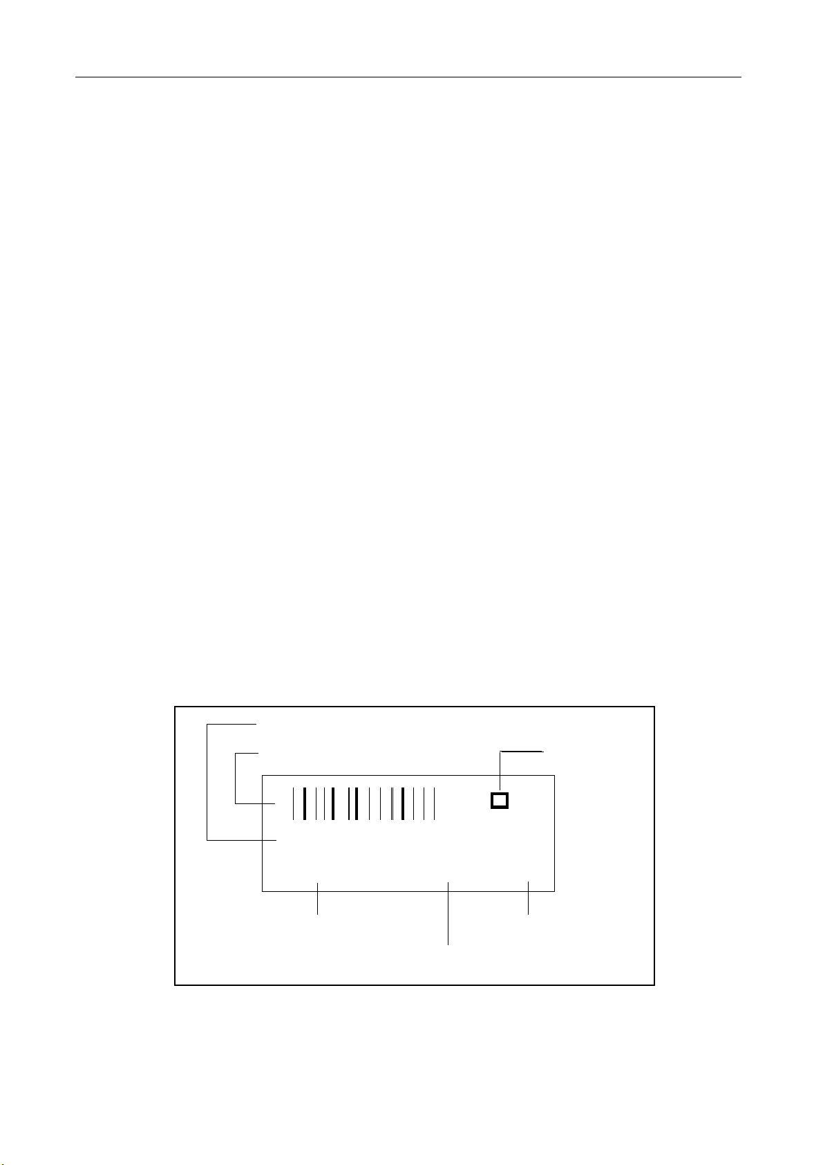
The 388 number is located on the barcode label.
Configuration of the barcode labels:
Consecutive number
Consecutive number barcode Test mark
0000000219
388 011 51 3131 C3
Part No. Parts list index
Test number (batch)
Page 8

GE Medical Systems EAGLE 1000 Page 8
Information Technologies Servicing Instructions 227 468 01 Rev.G
303 xxx yy Spare part numbers for PCBs tested especially thoroughly
303 numbers are only given to PCBs where the level of testing applied to 388 PCBs is
inadequate for implementation when servicing in the field, or where only a complete set of
PCBs can be replaced in the field.
In addition to a barcode label (388 number) 303 part numbers also have an additional
label with a 303 number and are to be found in the spare parts list under this number.
Exchange for defective PCBs
Where servicing is required exchange PCBs are available for the replacement of some
PCBs. When using a exchange PCB, the defective PCB is to be returned to the Freiburg
factory. Replacement PCB part numbers are included in the spare parts list.
1.1.3 Instrument status documentation (nominal status)
Due to the hardware and software combination unambiguous documentation of the instrument assembly status is necessary, also in the event of repairs.
This documentation comprises the following documents and measures:
Master Record Index (MRI)
This document is a component of this instrument documentation.
This document states the combination of permissible hardware and software for a particular instrument version. The permissible PCB Index is given in the Index column with each
update delivered. Further permissible PCB Indexes are given in the compatible column.
The PCB Index can be found in the PCB barcode label.
Product Status Index
This document is created during manufacture. The Product Status Index documents the
hardware/software product status.
Page 9

GE Medical Systems EAGLE 1000 Page 9
Information Technologies Servicing Instructions 227 468 01 Rev. G
1.2 Eagle 2000 system overview
These servicing instructions describe the instrument versions with Hellige circular connectors.
Compact, multi-parameter patient monitor, 12 different models
101 123 02 EAGLE 1002 ECG
101 124 02 EAGLE 1003 ECG Alarm recorder
101 123 03 EAGLE 1004 ECG SPO
101 124 03 EAGLE 1005 ECG SPO
101 123 04 EAGLE 1006 ECG NBP
101 124 04 EAGLE 1007 ECG NBP Alarm recorder
101 123 05 EAGLE 1008 ECG SPO
101 124 05 EAGLE 1009 ECG SPO
101 123 06 EAGLE 1010 ECG SPO
101 124 06 EAGLE 1011 ECG SPO
101 123 07 EAGLE 1014 ECG SPO
101 124 07 EAGLE 1015 ECG SPO
2
2
2
2
2
2
2
2
Alarm recorder
NBP
NBP Alarm recorder
NBP TEMP
NBP TEMP Alarm recorder
NBP TEMP 2 x IBP
NBP TEMP 2 x IBP Alarm recorder
Table of EAGLE 1000 models (NBP = non-invasive blood pressure, IBP = invasive blood
pressure.
Page 10
GE Medical Systems EAGLE 1000 Page 10
Information Technologies Servicing Instructions 227 468 01 Rev.G
1.3 System overview
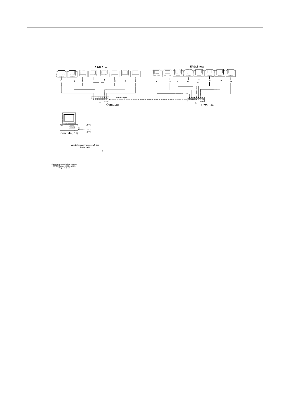
1.3.1 Hooking up EAGLE 1000 monitors to PC Central Station SynOpsis 1000
Eagle 1000 monitors version V2.1 and higher can be connected up to the PC Central
Station SynOpsis 1000. This involves the hooking up of up to eight monitors via a serial
cable with an OctaBus 1000. Up to two OctaBus 1000s can be connected up to the
Central Station. Each OctaBus is connected to the Central Station PC using a printer port.
Alarm release ensues from one of the two OctaBus devices, these being connected appropriately. The OctaBus devices and the Central Station PC monitor controll each others
performance and give an alarm when a malfunction is detected. For configuration see
figure below.
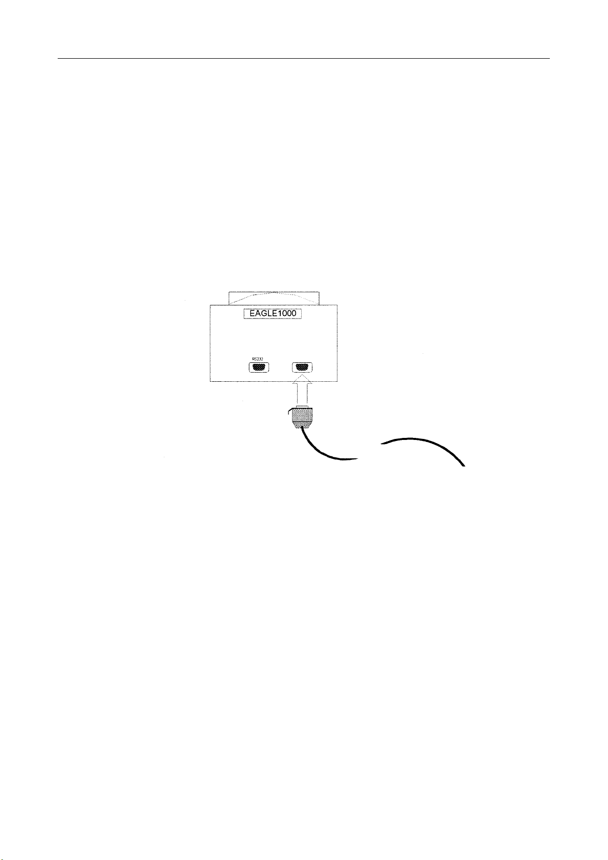
Nurse Call
EAGLE wall outlet
Figure 1. Connection of EAGLE 1000 with current loop output to Nurse Call/ Central Station
socket
Connection
The Nurse Call socket of Eagle 1000 V2.1 or higher is used for hooking up to the Central
Station via the OctaBus. A current loop permits connections over distances of up to 200 m.
Connection to the OctaBus is via an 8-lead cable either from a wall outlet or directly. Refer
to the Installation Instructions for more details.
Page 11
GE Medical Systems EAGLE 1000 Page 11
Information Technologies Servicing Instructions 227 468 01 Rev. G
Central Station PC
Figure 2. Connection of EAGLE 1000 monitors to a PC Central Station SynOpsis 1000 using
2 OctaBus hardware interfaces.
Page 12
GE Medical Systems EAGLE 1000 Page 12
Information Technologies Servicing Instructions 227 468 01 Rev.G
1.3.2 Patient data transfer from EAGLE 1000 via HELAN to SMU using
QuickLink
1.3.2.1 Introduction
To ensure continuous monitoring it is also necessary to be able to record patients vital
signals during relocation and transportation. However, since during patient transfer there is
no connection to the LAN, the trend data are collected and then transferred later as a
block.
After taking over patient monitoring on the transport monitor recording of the data begins.
No connection is being established with the Gateway. The patient remains anonymous,
i.e., his name is not entered into the transport monitor.
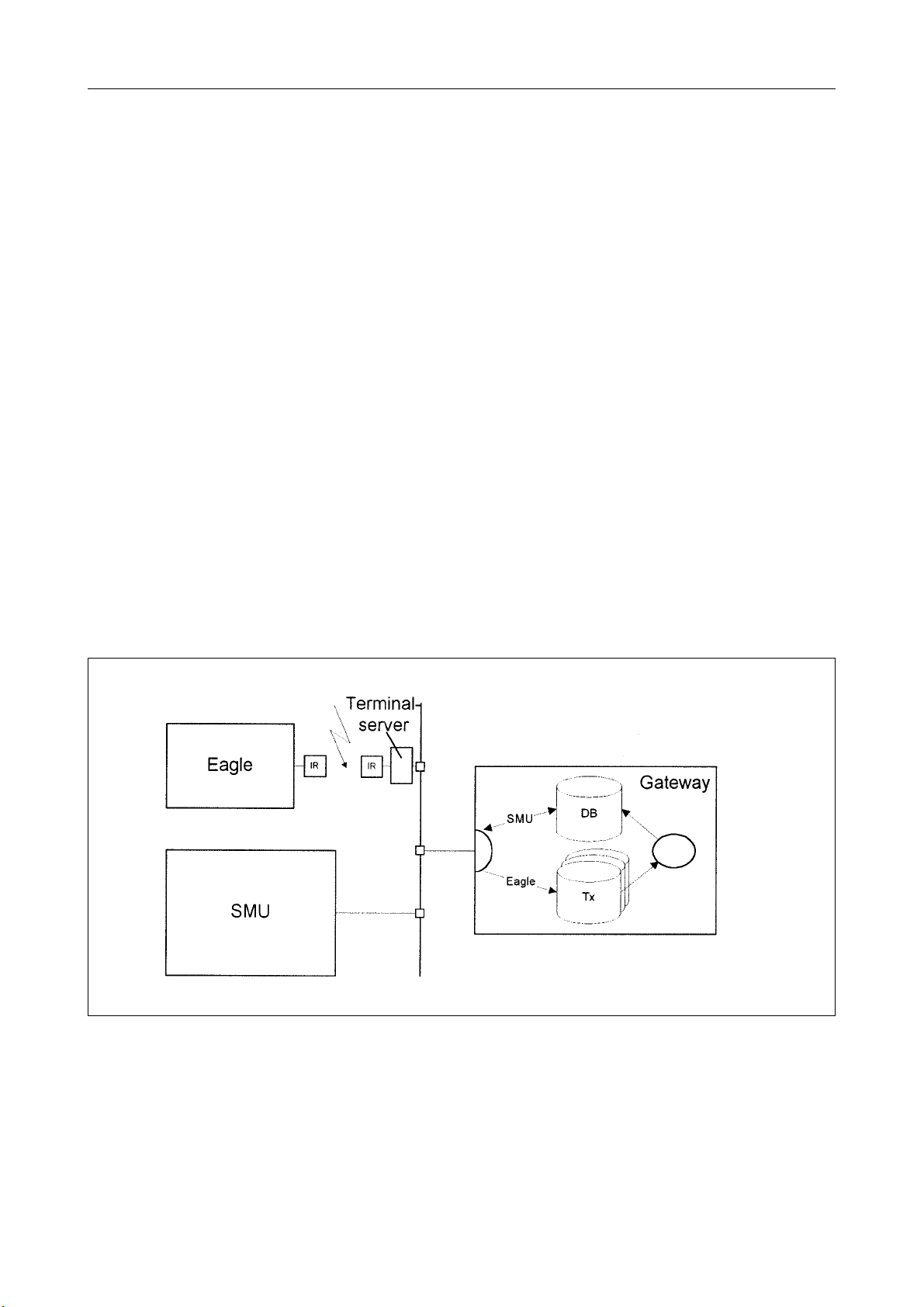
Upon arrival at his new destination the patient is hooked up to the destination monitor and
the transfer of the trend data initiated by the transport monitor operator. The transport
monitor establishes a connection over an IR beam and a terminal server (QuickLink) with
the LAN and transfers the data to the Gateway.
In the Gateway the data are stored in a file bearing a file name indicating the ID of the
transport monitor used. Assignment of patient data is carried out using the operating routines of the destination monitor. The operator selects the transport monitor ID, to which the
transferred patient belonged to. It is now the task of the Gateway to enter the trend data
into the database correctly.
Figure 3 shows the principle of data transfer. The terminal server comprises QuickLink, the
Gateway comprises VICOM TR server or VICOMport + VICOMserver.
Page 13

GE Medical Systems EAGLE 1000 Page 13
Information Technologies Servicing Instructions 227 468 01 Rev. G
1.3.2.2 Data transfer between Eagle and Gateway
Eagle and Gateway will communicate while transferring trend data. Transfer is initiated by
the user request function.
The data are first stored in a file and, when data transfer is complete, arranged in proper
order. The transport monitor waits for the results of this processing procedure.
If an error occurs in the database during this procedure, all the data in the Gateway are
deleted and the entire data must be transferred once more.
When the data transfer is initiated, the time will be sent by the transport monitor real-time
clock. From this the Gateway can identify any possible time misalignment of the internal
clock of the transport monitor and account for this when the trend data are entered.
Sporadic trend data, e.g., non-invasive blood pressure values, are only transferred for the
period of time in which continuous trend data, e.g., ECG, are also available. Earlier or later
parameter readings will be ignored.
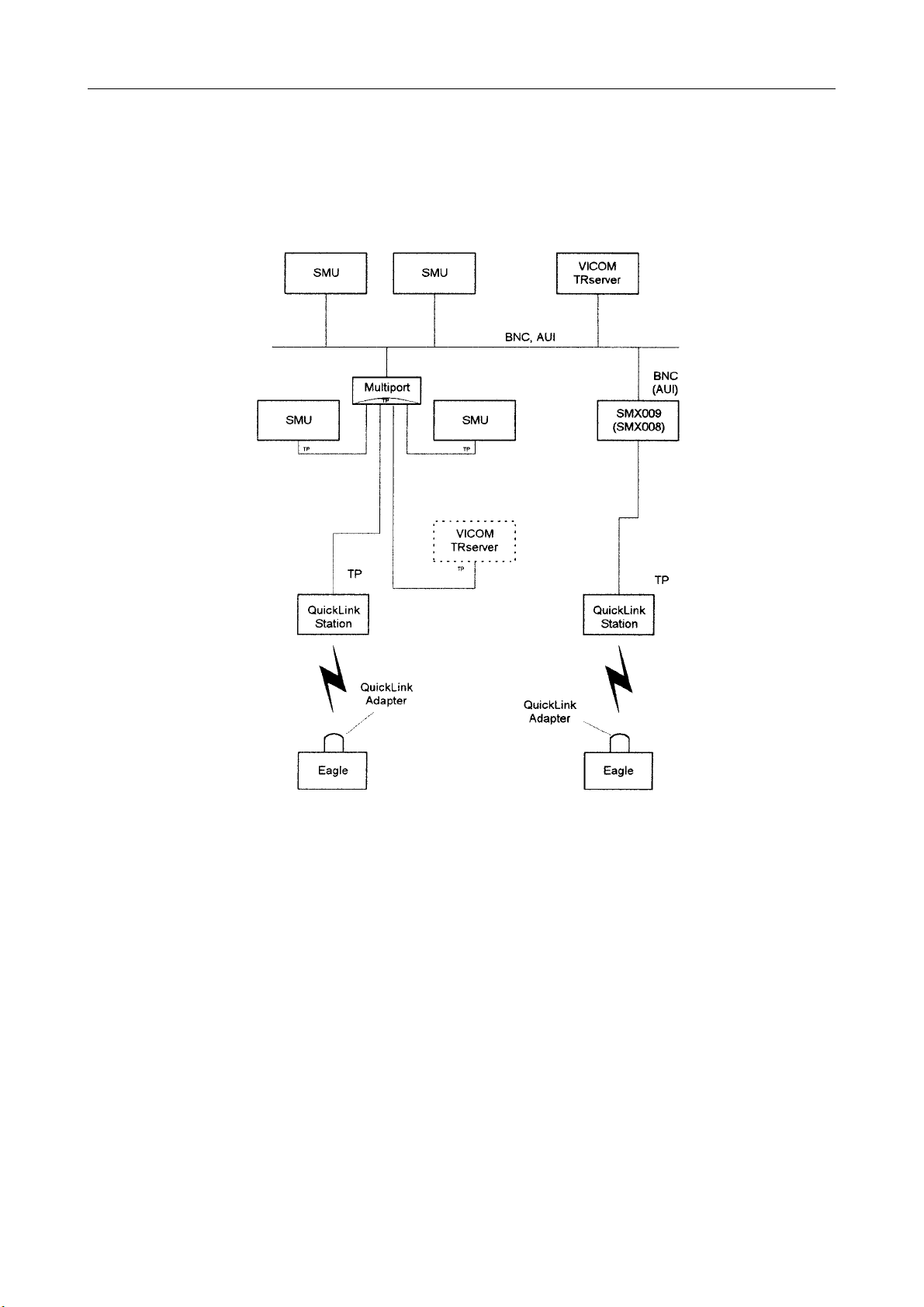
1.3.2.3 Connection to the LAN
The QuickLink Station is connected to a Multiport or an SMX using twisted-pair wiring.
The Multiport can lead to the LAN using BNC or AUI wiring.
The SMX 008 leads to the LAN using AUI wiring, the SMX 009 to the LAN using BNC
wiring.
Page 14
GE Medical Systems EAGLE 1000 Page 14
Information Technologies Servicing Instructions 227 468 01 Rev.G
Configuration with BNC, AUI or twisted pair (TP)
Figure 4. QuickLink - LAN connection
twisted pair
installation option
Page 15

GE Medical Systems EAGLE 1000 Page 15
Information Technologies Servicing Instructions 227 468 01 Rev. G
1.3.2.4 EAGLE 1000 - QuickLink - HELAN - Examples of the link to networks
1.3.2.4.1 Functional structure overview
The link of Eagle 1000 to the networks via QuickLink enables the transfer of patient data
acquired during patient relocation and transportation for continuous and uninterrupted
analysis.
A QuickLink adapter connected to the RS 232 interface of the Eagle 1000 sends the transportation data using IR to a QuickLink Station. The QuickLink Station acts as a terminal
server and is the communication partner to the HELAN network.
The data transferred onto the HELAN are stored on a server which acts as a Gateway
(VICOM TRserver or VICOMport in connection with VICOMserver).
This is where the patient data and the measured values recorded during transportation are
made available for complete documentation on any patient monitor or on a clinical information system.
1.3.2.4.2 Main components for the installation of QuickLink:
Part Number Device Description
216 135 01 QuickLink Adapter IR-Interface, connectable to Eagle 1000
216 134 01 QuickLink Station Station to receive IR signal from QuickLink
adapter incl. RJ45 connection (Twisted
Pair)
216 107 01 SMX 001 Transceiver for AUI
216 133 01 SMX 005 Transceiver for RJ45 (Twisted Pair)
930 117 40 SMX 006 Multiport Repeater (12 x RJ45)
930 117 39 SMX 007 Multiport Repeater (24 x RJ45)
930 117 76 SMX 008 Repeater RJ45 to AUI
930 117 77 SMX 009 Repeater RJ45 to BNC (CheaperNet)
101 130 01 VICOM TRServer R.2.0 Patient data transfer within HELAN
101 131 01 VICOMport R.2.0 Link of HELAN to VICOM Server
101 132 01 VICOMserver R.2.0 Database for HELAN and interface to other
networks
Page 16

GE Medical Systems EAGLE 1000 Page 16
Information Technologies Servicing Instructions 227 468 01 Rev.G
1.3.2.4.3 EAGLE 1000 - QuickLink
Network link - HELAN CheaperNet
SMX 008 + SMX 001 can be used in place of SMX 009 + BNC T-Plug.
Page 17
GE Medical Systems EAGLE 1000 Page 17
Information Technologies Servicing Instructions 227 468 01 Rev. G
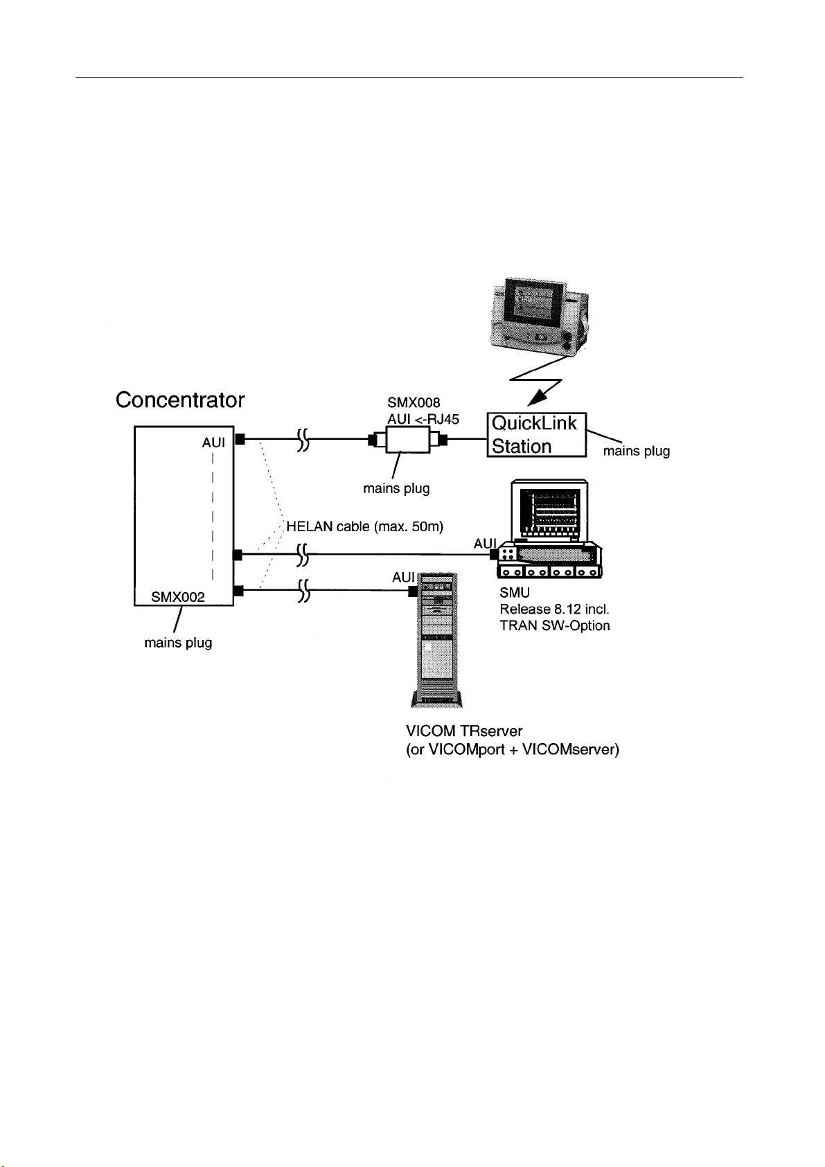
1.3.2.4.4 Network link - HELAN Concentrator
Page 18

GE Medical Systems EAGLE 1000 Page 18
Information Technologies Servicing Instructions 227 468 01 Rev.G
1.3.2.4.5 Network link - HELAN Twisted Pair
Page 19

GE Medical Systems EAGLE 1000 Page 19
Information Technologies Servicing Instructions 227 468 01 Rev. G
2.1 Technical specifications
2.1.1 System interfaces
Interface for Nurse Call and Central Station
* output for control signal in the case of alarm or warnings for connection of an Isolation
Relay 303 444 77, short-circuit-proof
* trigger output (digital impulse, width 1 ms, 0 V to 9.5 to 14.5 V)
* 20 mA current loop output to connect OctaBus to PC Central Station SynOpsis with
V2.1 or higher version
RS 232 interface
* RS 232/V24 interface for connection of QuickLink to HELAN, available from V 2.0 or
higher
2.1.2 Display
* size: 6-inch diagonal (14 cm)
* type: monochrome liquid-crystal display
* resolution: 320 by 240 pixels
Waveform display
* 3 channels
* sweep speed of 25 mm/s
* erase-bar mode
2.1.3 Monitor functions
Operating modes
* test, monitoring enabled/disabled, service mode, data transfer (download)
Alarm system
* text on display, alarm LED, Alarms OFF LED, audible alarm signal (can be disabled
via softkey or from the defaults menu)
Data storage in case of line failure or empty battery
* at least 5 minutes
Page 20

GE Medical Systems EAGLE 1000 Page 20
Information Technologies Servicing Instructions 227 468 01 Rev.G
2.1.4 Power supply
* from the power line, monitor design in IEC protection class I
Potential equalization
* potential equalization connection according to DIN 42 801(measurement in or at the
heart) or connection for function ground (interference elimination during general cardio-
logical examinations)
Rated voltage range from 95 to 240 V AC
* operating voltage range from 85 to 264 V, 49 to 65 Hz
* rated current from 0.15 to 0.3 A
Internal power supply (rechargeable battery)
* automatic toggling from line to battery operation and vice versa
* integrated charge circuit with automatic adaption of charge characteristics
* fast battery charging to 80% of its capacity within 3.5 hours, afterwards
* normal charging to 100% of its capacity within 8 hours, afterwards
* continuous trickle charging by means of charge pulses to prevent the memory effect
Charge balance
measurement of the charge and load current
compensation for battery self-discharge
graphic indication of battery capacity
Alarms
warning when battery provides power for only 5 minutes of operation (without
recorder)
automatic switch-off at 10.0 V to prevent overdischarging
Capacity of the rechargeable battery
2000 mAh, 12 volts
Charging indicator
LED
Operating time
approx. 3 hours (typical operating time with fully charged battery at 23 °C, at a
battery temperature of 60 °C the capacity may be reduced to 70%)
(min. 2 hours at 25 °C ambient temperature and new, fully charged battery)
Page 21

GE Medical Systems EAGLE 1000 Page 21
Information Technologies Servicing Instructions 227 468 01 Rev. G
2.1.5 Operation and storage
Operational readiness
after approximately 5 seconds
Operating position
vertical (free circulation of air under the unit and ventilation openings must not be ob-
structed)
Environmental conditions
Operation
* ambient temperature between +10 °C and +40 °C
* battery charging temperature between +10 °C and +40 °C
* relative humidity between 30% and 75%, no condensation
* atmospheric pressure between 700 and 1060 hPa
Storage
* ambient temperature between -30 °C and +60 °C
* relative humidity between 10 and 100%, no condensation
* atmospheric pressure between 500 and 1060 hPa
Dimensions
width 262 mm
height 215 mm
depth 160 mm
weight 2 kg to 4.4 kg (depending on monitor model)
2.1.6 ECG and heart rate
ECG signal inputs
Electrode connection for simultaneous transfer of 2 ECG signals: ECG 1 and ECG 2 via
lead selector (7 standard lead configurations each).
Signal inputs for 3-lead and 5-lead patient cables with connections for electrode leads;
buffer amplifier with protective circuit and HF filter; floating input circuit (isolated patient
connection of the CF type, IEC); overvoltage protection for all inputs, detection of pacing
pulses, monitoring of electrode-to-skin contact impedance (specifications without leads):
differential input impedance for DC > 65 MOhms, for f<=100 Hz > 6 MOhms
common-mode input impedance referred to RL
for DC > 10 GOhms, for f<=100 Hz > 50 MOhms
Page 22

GE Medical Systems EAGLE 1000 Page 22
Information Technologies Servicing Instructions 227 468 01 Rev.G
overrange limit (e.g. for polarization voltages) for differential signals >0.6 V, for signals
referred to RL > 1.0 V
common-mode rejection referred to RL for f<= 50 Hz approx. 80 dB RL referred to
chassis >110 dB
patient leakage current (rms values): under normal conditions <0.005 mA, in fault condi
tion (e.g. patient in contact with mains voltage) <0.020 mA
quiescent input current <40 nA
non-destructive load for electrode connections referred to RL ± 50 V, referred to chas-
sis ± 1000 V
pulse-voltage resistance of all electrode connections and of the RL-connection referred
to chassis (either polarity, e.g. defibrillation, electrocautery) 5000 V
Signal processing
The signal in channel 1 is processed. All specifications refer to a signal free of inter
ference.
Amplification, QRS detection, digitization, display
HR measuring range 15 to 300 bpm, error ± 1 BPM (EC 13)
ECG cut-off frequency >=100 Hz
differential signal range ± 4.5 mV
ECG resolution 0.005 mV
sampling rate 1000/1200 Hz
noise below 0.04 mV
time constant for ECG transfer 0.3 s (0.5 Hz)
HR averaging for displayed value: weighted average from the most recent RR intervals
display update interval: 2 seconds
trigger sensitivity 0.25 mV to 0.30 mV for pulse width of 40 ms
max. rate stabilization of 6 seconds for RR interval change of 95%
alarm delay 4 seconds
asystole alarm 4 seconds
3 signal sizes for monitor display in freeze mode or else on the recorder trace: 5 10
20 mm/mV
1-mV calibration signal on waveform and recorder trace
detection of pacing pulses (these specifications refer to an ECG signal free of inter-
ference)
pulse duration dp>approx. 0.2 ms, <40 ms
pace marker independent of polarity
pulse amplitude ap±10 to ±700 mV
over/undershoot ao±1 mV
time constant to 25 to 100 ms
Page 23

GE Medical Systems EAGLE 1000 Page 23
Information Technologies Servicing Instructions 227 468 01 Rev. G
alarm limits adjustment range: 20 to 250 BPM
trigger output on Nurse Call connection nominally 12 V 1 ms, delay max. 25 ms, tole-
rance 9.5 V to 15.5 V
rate measurement
derivation of trigger pulses from the ECG signal or from the pressure waveform or from
the Sp02 signal with automatic adaption of the trigger threshold or blanking of pacing
pulses; calculation of the mean rate; 3-digit display on the monitor screen
2.1.7 Oxygen saturation Sp0
2
Signal input
Connection for one Sp02 probe, isolated from ground, floating input circuit (isolated patient
connection of the CF type, IEC).
Used in conjunction with the approved probes, the module is protected from voltages
generated by defibrillation discharges and electrocautery.
Signal processing
dual-wavelength pulse oximeter
Sp0
measuring range 1 to 100%
2
PR measuring range 20 to 250 BPM
resolution 1% Sp0
2
deviations from actual value (% Sp02 = 1st standard deviation) in combination with:
standard finger probe (DS 100A)
between 70 and 100%: ±3 digits
disposable probe for adults (D25)
between 70 and 100%: ±2 digits
disposable probe for children (OXI D20)
between 70 and 100%: ±2 digits
disposable probe for babies (J-20)
between 70 and 95%: ±2 digits
disposable probe for neonates (N-25)
between 70 and 95%: ±2 digits
unspecified between 0 and 69% Sp0
Plethysmogram
accuracy of the pulse rate measurement
±3 pulses/min between 20 and 150 pulses/min
±2% between 150 and 250 pulses/min
2
Page 24

GE Medical Systems EAGLE 1000 Page 24
Information Technologies Servicing Instructions 227 468 01 Rev.G
2.1.8 Non-invasive blood pressure
Signal input
Single-lumen tubing system, type CF signal input (IEC 601-1), patient is electrically not
connected to the system, thus the system is protected from defibrillation discharges and
electrosurgery voltages).
Signal processing
Oscillometric method:
stepwise pressure decrease, zero pressure reference prior to each measurement
Two measuring modes: adult
neonate
mmHg
adult systolic 25 to 255
diastolic 10 to 220
mean 18 to 255
neonate systolic 20 to 155
diastolic 5 to 110
mean 10 to 130
The specifications are met for pulse rates between 40 and 240 BPM (adult/neonate).
Safety circuits
Redundant monitoring of the cuff pressure by means of a second independent pressure
sensor; cuff deflation immediately on exceeding max. cuff pressure, on detection of faults
during a measurement and on power loss.
Max. cuff pressure
adult 300 mmHg
neonate 160 mmHg
Special features
When the cuff is properly applied the module recognizes in the adult mode whether the
cuff matches the selected range. If the wrong cuff is applied, the measurement is interrupted.
Page 25

GE Medical Systems EAGLE 1000 Page 25
Information Technologies Servicing Instructions 227 468 01 Rev. G
Cuff inflation pressure
adult 160 mmHg
neonate 100 mmHg
Adaptive cuff inflation pressure
After a measurement the inflation pressure of the subsequent measurement depends on
the previous measuring result: it is either 1.8 times the previous mean pressure or 15%
above the previous inflation pressure (whichever is less).
Measurement cycles
Selectable intervals for automatic measurement cycles:
2, 5, 10, 15, 30, 60 minutes
Measuring accuracy
P
sys/Pdia
<= 8 mmHg, 1st standard deviation
error limits for cuff pressure via the pressure transducer ±3 mmHg
Measurement duration
adult: 90 seconds max.
neonate: 70 seconds max.
typical duration for undisturbed measurement: 30 seconds
Technical inspection, calibration
Technical inspection must be carried out once a year, calibrations every two years (refer to
section 3 Maintenance and technical inspection).
2.1.9 Invasive blood pressure
Signal inputs
2 connections which are both isolated from ground (type CF according to IEC 601-1) for
pressure transducers with ohmic strain-gauge bridge, diagonal resistance of 200 Ohms to
10 kOhms with a sensitivity of 0.005 mV/mmHg per V of supply voltage:
supply voltage for transducer:
5 volts DC
At 50 and 60 Hz:
differential input impedance > 100 kOhms, real component 500 kOhms
common-mode input impedance > 100 kOhms
common-mode rejection > 60 dB up to 60 Hz
Page 26

GE Medical Systems EAGLE 1000 Page 26
Information Technologies Servicing Instructions 227 468 01 Rev.G
Connections
direct connection for measurement of one pressure
connection via adapter for measurement of two pressures
Display
pressure waveform with 25 mm/s sweep for both channels
pressure waveform for the following pressure sites: arterial (ART1, ART2), pulmonary
artery (PA), venous (VEN), left-atrial (LAP), intracranial (ICP) for ART, PA and LAP the
systolic, diastolic and mean values can be displayed
display update interval: 2 seconds
Signal processing
Pressure
measuring range -30 to +300 mmHg
resolution 0.25 mmHg, internal
cut-off frequency 12 Hz
accuracy ±2 mmHg ±1% of actual value
zero drift < ±0.1 mmHg/°C
5 minutes to warm up for full accuracy
Zero balancing
automatic compensation of the transducer value for a zero pressure in both channels,
every channel to be zeroed is checked for absence of pulse signals
balancing initiated after key press > 2 seconds
balancing range ±100 mmHg
storage of the balancing value for 5 minutes after power failure or when monitor is
switched off
deviation < ±1 mmHg
initiation only when signal modulation < ±1 mmHg
Page 27

GE Medical Systems EAGLE 1000 Page 27
Information Technologies Servicing Instructions 227 468 01 Rev. G
2.1.10 Temperature
Signal input
Input isolated from ground (type CF according to IEC 601-1) for connection of a temperature probe from the Marquette HELLIGE accessory range (these probes conform to the
YSI 400 or Exacon 4000 series; as these probes have jack plug contacts which are accessible when not connected, they were additionally tested with 1500 V
cal saline according to IEC 601-1)
Signal processing
Together with two test readings and a correction value the temperature reading is processed for regular self-tests and for fine-balancing by multiplexing in a common amplification
circuit:
measuring range 0 to 44 °C
error limits for module without probe: between 0 and 25 °C ±0.2 °C, between 25 and
44 °C ±0.1 °C
display update interval: 2 seconds
5 minutes to warm up for full accuracy
50 Hz in physiologi-
rms
Functional safety
Continuous self-tests prevent display of values outside the calibration error limits; when
service screen is called up the test values of 38.8 °C ±0.1 °C and 4.0 °C ±0.2 °C are displayed.
Technical inspection, calibration
Calibration tests must be carried out on the monitor every two years (refer to section 3
Maintenance and technical inspection), technical inspection once a year.
Page 28

GE Medical Systems EAGLE 1000 Page 28
Information Technologies Servicing Instructions 227 468 01 Rev.G
2.1.11 Trend
* storage of parameter values
* tabular trend, NBP table depending on NBP intervals
* graphic trend for each parameter
* storage of trend values every 20 seconds or after NBP measurement
* display of trends via softkeys
* automatic display of a time scale and of reference lines (labelled)
* zoom function for selection of other trend periods (graphic: 45 minutes, 3 hours,
24 hours; tabular: 1 minute, 5 minutes, 15 minutes)
* trend memory can be cleared with a softkey, or the memory is cleared automatically
when the monitor is switched off for more than 5 minutes
2.2 Functional description of PCBs and subassemblies
2.2.1 Eagle 1000 configuration and functional groups
see diagram Functional groups
The basic Eagle 1000 patient monitor version comprises the large PCB ECG Monitor
installed in the system on the front side of an L-shaped metal plate. In addition to the ECG
input circuit, the instrument control system is also located on this PCB.
The keypad, display screen, battery and the PCB Power Supply are connected to it. Also
connected are the ECG input socket and the two rear panel outputs RS-232 and Nurse
Call.
The keypad and CRT have conductor foil connections and care must be taken when connecting them up and sharply bending them avoided.
The battery is installed in the bottom side of the L-shaped metal plate.
The power supply is located inside the rear panel of the system next to the power supply
socket.
The recorder, if there is one, is mounted on the base plate and connected to the PCB
ECG Monitor.
Page 29

GE Medical Systems EAGLE 1000 Page 29
Information Technologies Servicing Instructions 227 468 01 Rev. G
In the models with additional features the PCB ECG Monitor also acts as a motherboard. The PCB Oximeter, the PCB for non-invasive blood pressure and the PCB for
temperature or for temperature and 2 invasive blood pressures can be added to this
board.
In conformity with Cardiac floating there is a high-impedance isolating transformer referred separately to the ECG input module, the PCB Oximeter, the PCB module for temperature and the PCB module for 2 invasive blood pressures. Both pressure measuring components are electrically connected. These PCBs are insulated from the non-floating components of the system and each other by spacing and insulating foil.
Warning: It is thus important that when disconnecting or replacing PCBs in the
system to ensure PCBs and circuits are correctly installed and the insulating foil positioned accordingly.
2.2.2 PCB ECG Monitor
The PCB ECG monitor appears as a block diagram in the PCB circuit diagrams. The
circuits are arranged so that (viewing Eagle 1000 from the rear) the floating ECG Processing is located in the left-hand third, in the remainder all the other functions:
Floating Non-Floating
x Display Controller
x ECG Controller
x Interface Output
x ECG Filter x Controller
x Power Supply (instrument, battery, floating)
x ECG Input x Interface Input
This means that ECG Processing and the Central System signal processing and system
controller are implemented on this PCB. The description of each is given below, indicating
the connected modules and their functions.
The PCB plugs and sockets are shown in the S-Plan of the system plans and are allocated as listed below. This allocation is useful in finding the pin configuration in the P circuit
diagrams of the PCB ECG Monitor.
Page 30

GE Medical Systems EAGLE 1000 Page 30
Information Technologies Servicing Instructions 227 468 01 Rev.G
Name Connection Denotation Page P-Plan Subtitle
AA Conductor Foil Display 9 Display
AB For Optrex LCD only 9 Display
AC Cable Battery 6 Power
AD Cable PCB Power Supply 6 Power
AE Cable LCD Backlight 9 Display
AF Flat cable Recorder 8 Interface_Out
AG Flat cable PCB Non-Invasive Blood 5 Interface_In
Pressure
AH Flat cable PCB Oximeter (Sp02) 5 Interface_In
AI Debugging Connector only 7 Control
AJ Stackable Connector PCB Press/Temp 5 Interface_In
AK Flat cable ECG Input 2 ECG_Input
AM Conductor Foil Membrane Keypad 5 Interface_In
AN Flat cable RS 232 External Port 8 Interface_Out
AO Flat cable Nurse Call Adapter 8 Interface_Out
2.2.3 System power supply
see diagram System Power Supply
The power supply is in protection class I with a protective ground terminal. A primaryregulated wide-range power supply unit, delivering 15.5 V DC, precludes the necessity of a
power toggle switch. This unit also acts as a charger for the 12-V system battery. All control functions are regulated on the PCB ECG Monitor. A charge controller measures the
battery current across a resistance referred to zero, activates the front panel charge indicator and communicates with the processor. Should the output voltage of the power unit
fail to reach 15.5 V, the battery will not be charged to full capacity.
The battery meets the temporary high current demand of the recorder and the PCB NonInvasive Blood Pressure as well as current peaks and is also essential for retaining data.
As the battery continuously is discharged, this is indicated by an 11.5V comparator, which
triggers the warning display Battery blinking and the audible alarm signal sounding.
When the charge drops below 10 V after storing the data the 10V comparator switches off
the system to protect the battery.
Page 31

GE Medical Systems EAGLE 1000 Page 31
Information Technologies Servicing Instructions 227 468 01 Rev. G
Otherwise, the system is switched on and off with the appropriate button on the front panel, which, just like the 10V comparator, exerts an effect via the processor on the ON/OFF
interface with its FET switch.
A 5V analog voltage +5VREF is generated with a linear controller from the battery voltage
supplied when it is switched on. A switching controller generates the 5V digital voltage
from the battery voltage supplied when it is switched on. Apart from this the battery voltage
supply is used directly and unregulated for oscillators and other components requiring a
power supply.
2.2.4 Floating system power supply and signal flow
Power supply
see diagram Floating system supply and signal flow
The floating system is supplied from a central 100-kHz oscillator on the PCB ECG Moni
tor. This delivers the supply voltage stabilized on a HF transformer, i.e., individual power
fluctuations on some PCBs are not fully compensated. The individual floating components
receive power via the primary HF transformers connected in parallel. Isolation is effected
in these floating transformers. These each generate ±7.5V ±5%. In turn, in each case a
linear +5V ±2% analog/digital voltage is obtained from these voltages.
Signal flow
see diagram Floating system supply and signal flow.
Apart from the non-invasive blood pressure all patient input circuits are high-impedancefloating circuits. In terms of signals, these are isolated in both directions via optoelectronic couplers. All other circuit components are not isolated and referred to circuit zero.
Furthermore, there is a separate ground contact to enable potential equalization.
The non-invasive blood pressure input is isolated by means of a non-electronic patient
input.
2.2.5 Signal exchange with the processor
see Processor block diagram
The processor on the PCB ECG Monitor comprises a controller with a separate 32kHz
quartz crystal, watch dog, RAM and EPROM. The controller is in communication with all of
the following units.
Page 32

GE Medical Systems EAGLE 1000 Page 32
Information Technologies Servicing Instructions 227 468 01 Rev.G
The keypad is used to enter data, the beeper to give alarms and acknowledgment sig
nals. The recorder interface leads to the integrated recorder and the CPU interface to the
liquid-crystal screen.
The RS 232 interface acts as a system output, it is mainly used for sending signals to the
QuickLink to output data to the HELAN after patient relocation and transportation.
The Nurse Call interface leads to the nurse call adapter. A nurse call system can be
connected by means of the nurse call relay for the Eagle 1000. From Version 2.1 and
higher, a 20 mA current loop is available to connect the Eagle to an OctaBus of a SynOpsis Central Station.
The patient data inputs are entered via three serial interfaces:
The QSPI interface allows the transfer of ECG, temperature and the two invasive blood
pressures via the 16 Qued Processor Interface signals. One chip select signal each is
generated for each of the three signal groups. A clock signal synchronizes the transfer of
the two Master In - Slave Out and Master Out - Slave In signals.
The PCBs Oximeter and Non-Invasive Blood Pressure are each connected separately via
an asynchronous interface. This has both an Rx and a Tx cable as well as a reset cable.
Another EC.Syn signal is provided for the oximeter (Sp02), another RESETB signal for the
non-invasive blood pressure (NBP).
2.2.6 Display
see Display block diagram
All of the elements shown apart from the block display are located on the PCB ECG
Monitor.
The LCD display is controlled by an LCD controller. This works together with an external
3.6MHz oscillator and external display RAM. The control of the LCD controller is effected
via the CPU interface.
The LCD display is controlled by a -23V power supply. This voltage is acquired from the
battery voltage supply. The contrast on the PCB ECG Monitor must be adjusted.
The display has a reverse video feature which is supplied with 300 V AC at 30 to 40 Hz
from a CCFT supply. In open-circuit operation this is approx 565 V.
Warning: Avoid contact with this voltage where it is exposed on the PCB ECG
Monitor.
Page 33

GE Medical Systems EAGLE 1000 Page 33
Information Technologies Servicing Instructions 227 468 01 Rev. G
2.2.7 Block diagrams
2.2.7.1 Functional groups
Keypad
Tastenfeld
NBP
Press-Temp
Display
Bildschirm
AC AD
Schw.Ruf
Nuse Call
Power Supply
Stromversorgung
RS 232
AD
230 V~
115 V AC
AC
Recorder
Registriergerät
EKG / ECG
Oxymeter ECG-Monitor
Batterie/Battery
Page 34

GE Medical Systems EAGLE 1000 Page 34
Information Technologies Servicing Instructions 227 468 01 Rev.G
2.2.7.2 Floating system supply and signal flow
2
CALL
BEEPER RS 232 NURSE DISPLAY
KEYPAD
PROCESSOR
QSPI
RECORDER
NIBP
Asyn
Asyn
SPO
Float.
15 V
Supply
100 kHz
ISOLATION
±7.5 V
FLOATING NON-FLOATING
SENSORS
+5 V
each
PRESS1
PRESS2
TEMP
ECG
Page 35

GE Medical Systems EAGLE 1000 Page 35
Information Technologies Servicing Instructions 227 468 01 Rev. G
2.2.7.3 System power supply
Processor
SW
<11.5 V Lo. Bat. Warn.
OFF
<10 V
9.5...15.5 V
+12 V
ON-
+5 V REF
+5 V
+5 VSWGenerator
ON/OFF
OFF
+12 V
Front
Charge Control
Charge Battery
+15.5 V
Mains
99 V AC
264 V AC
Page 36

GE Medical Systems EAGLE 1000 Page 36
Information Technologies Servicing Instructions 227 468 01 Rev.G
2.2.7.4 PROCESSOR block diagram
Display
TIME OF
RECORDER IF
CPU-IF
RS 232-IF
Nurse-Call-IF
DAY CLOCK
BEEPER / KEYPAD
Watch
Dog
CONTROLLER
MEMORY
32 kHz
QSPI-IF
-Asyn-IF
2
SPO
NIBP-Asyn-IF
ECG
Temp.
BPress.
Page 37

GE Medical Systems EAGLE 1000 Page 37
Information Technologies Servicing Instructions 227 468 01 Rev. G
2.2.7.5 DISPLAY block diagram
LCD
DISPLAY
Hinterleuchtung
back lighting
EE
U
565 V/300 V
30...40 kHz
CCFT
SUPPLY
-23 V
+5V
+12 V
LCD-CONTROL
CPU IF
RAM
CONTRAST
~
3,6 MHz
Page 38

GE Medical Systems EAGLE 1000 Page 38
Information Technologies Servicing Instructions 227 468 01 Rev.G
2.2.8 ECG signal preprocessing
2.2.8.1 Functional description
The block diagram ECG/Pace Preprocessing shows the functional setup.
ECG input
In the Eagle 1000 ECG signal acquirement and processing takes place in one of the
remaining functional groups electrically separated from the others on the motherboard.
The functions of this component comprise analog processing of the ECG signals and a
QRS detector to monitor the cardiac rhythm.
All control functions are monitored by a processor, which exchanges data and commands
with the ECG circuit via the optoelectronic coupler.
Before it becomes operative the ECG circuit must be initialized. This is effected by the
processor in accordance with the selected accessories. The processor receives ID signals
called LET1, LET2 and LET3, identifying the nature of the connected accessories and,
hence, determines which ECG leads are available and which connections need to made
to the patient.
For this reason, the lead selector is preset to identify the patient cables used and to test
whether all the electrode leads are connected correctly. Every disconnected (electrode)
lead produces an error signal which is evaluated by the processor so that such electrodes
are not used for ECG monitoring.
ECG signal transfer
If fully operational electrodes are correctly connected to the EAGLE 1000, the ECG signal
is first amplified to 1.1 V per 1 mV as an analog signal, then transferred as a digital signal
via the optoelectronic coupler.
During analog signal amplification signal transfer is interrupted in the event of pacemaker
pulses and the arithmetic mean value is transferred as a baseline in place of the ECG
signal. The digital data are generated 1000 times a second from each of the transferred
amplitude values.
ECGs up to max. ±4.5 mV are digitized completely.
Three-lead patient cables provide only one ECG lead (lead I, II or III) on channel 1, connecting a five-lead cable enables EAGLE 100X to provide one lead each on two channels.
The options are leads I, II, III, AVR, AVL, AVF, V.
Page 39

GE Medical Systems EAGLE 1000 Page 39
Information Technologies Servicing Instructions 227 468 01 Rev. G
Heart rate monitoring
A QRS detector in the ECG circuit detects the beats. This detector filters and triggers the
signal components typical of QRS from the channel 1 ECG signals when their amplitude is
greater than a threshold value arising in the prior amplitudes.
The processor receives a signal for every beat detected by the ECG circuit, calculates the
heart rate from these and triggers an alarm when the heart rate is outside the programmed
limits.
Pacing detection
Signal segments with a steep rising or falling edge are suppressed as long as their amplitude exceeds a value of 6 mV. This suppression is limited to a time period of about 50 ms.
During this period the baseline is recorded and a pacing-pulse message is passed on to
the processor, which puts a marker for this event on the display.
Page 40

GE Medical Systems EAGLE 1000 Page 40
Information Technologies Servicing Instructions 227 468 01 Rev.G
2.2.8.2 ECG/Pace Preprocessing
SR
+12 V
Processor
SR_ON
SW. Regulator
Status
Reg.
HR
TRIG.
Dir.
Pace
Pace
ECG1
PACE
DETECT
+BLOCK
QSPI
Gain
Offset
ECG 2
ADC
OPTO ISOL.
3.6 MHz
~
Gain
Offset
Reg
Control
-7.5 V
+5 V
+7.5 V
Lead Fault
Detection
Pat. Cable
coding
Codes
ECG 1
Electrode Ampl.
ECG 2
Lead Selector
Protective circuit
3/5 Electrodes
Page 41

GE Medical Systems EAGLE 1000 Page 41
Information Technologies Servicing Instructions 227 468 01 Rev. G
2.2.9 PCB MP 204 Oximeter 930 117 26
2.2.9.1 Function
The PCB Oximeter acquires and automatically processes the Sp02 signal. Via an inter
nal asynchronous connection it passes the data on oxygen saturation and pulse rate on
to the PCB ECG Monitor. The entire PCB Oximeter is floating (CF). The floating separation for power supply and communication is located on the PCB ECG Monitor.
2.2.9.2 Figure of pcb MP204 Oximeter
V
JP1
JP5
AH
388 031 19
Lpl MP204-Oxymeter Board
930 117 26
Page 42

GE Medical Systems EAGLE 1000 Page 42
Information Technologies Servicing Instructions 227 468 01 Rev.G
2.2.9.3 Pin assignment
Sensor interface connector pin assignment (JP1)
Pin 1: Anodes (detector)
Pin 2: no connection
Pin 3: no connection
Pin 4: Cathode (detector shield) shield
Pin 5: ground (RCAL return)
Pin 6: RCAL
Pin 7: -LED
Pin 8: no connection
Pin 9: +LED
Pin 10: ground
Host interface pin assignment (JP5)
Pin 1: VCC (+5V)
Pin 2: ground
Pin 3: ground
Pin 4: +7.5V
Pin 5: ground
Pin 6: -7.5V
Pin 7: unused
Pin 8: TX Transmit data (output from module)
Pin 9: RX Receive data (input to module)
Pin 10: CTS_ Clear to send (input to module, active low)
Pin 11: RESET_ (Reset input, active low)
Pin 12: ground
Pin 13: ECG sync input, active high
Pin 14: ground
Page 43

GE Medical Systems EAGLE 1000 Page 43
Information Technologies Servicing Instructions 227 468 01 Rev. G
2.2.10 Non-invasive blood pressure module
2.2.10.1 Function
The PCB NBP acquires and automatically processes the non-invasive blood pressure
signal. Via an internal asynchronous connection it passes the non-invasive blood pressure data on to the PCB ECG Monitor. From here it receives a reset signal as well as a
separately controlled pump power supply for safety reasons. The PCB is not electrically
isolated as isolation is effected by the pneumatic signal input.
Warning: For safety requirements, when replacing the PCB it is important to use
the flat strip cable of correct length to match the version used.
Page 44

GE Medical Systems EAGLE 1000 Page 44
Information Technologies Servicing Instructions 227 468 01 Rev.G
2.2.10.2 Figure of PCB NBP Module NB11: 930 117 15
Use wire set 383 273 30
PCP NBP Module NB11
Page 45

GE Medical Systems EAGLE 1000 Page 45
Information Technologies Servicing Instructions 227 468 01 Rev. G
2.2.10.3 Figure of PCB NBP 2000 Module 930 117 93 or 388 032 60
Use wire set 383 273 65
Page 46

GE Medical Systems EAGLE 1000 Page 46
Information Technologies Servicing Instructions 227 468 01 Rev.G
2.2.10.4 Pin assignment
NBP pin connections to PCB ECG Monitor
Plug-in connector 2 rows of 5 pins
1 +7 V
2 +7 V
p
p
Pump power supply, host-controlled
Pump power supply, host-controlled
3 + 7V Power supply for PCB NBP
4 GND
5 GND
6 GND
7 KEY Plug coding
8 RESET Reset, host-controlled
9 Rx Receive
10 Tx Transmit
Page 47

GE Medical Systems EAGLE 1000 Page 47
Information Technologies Servicing Instructions 227 468 01 Rev. G
2.2.11 PCB Press-Temp 388 031 75/PCB Temp 388 032 04
General information
The PCB PRESS-TEMP enables measurement of the two electrically isolated parameters
temperature (one temperature) and invasive blood pressure (two pressures,both isolated
together). These parameters are connected along with the ECG to the processor by means of a QSPI interface. The signals are isolated by optoelectronic couplers.
The power supply is fed in via one HF transformer each for PRESS and TEMP from a
common oscillator, which also supplies power to the ECG circuit and the SpO2 module.
See block diagram Floating system supply and signal flow.
The PCB is equipped with precision components (and an adjustment program in the case
of TEMP) so that a potentiometer adjustment is not required.
The individual floating measurement circuits are shielded on both sides of the PCB by a
metal cover. For PCBs which do not have an insulating cover over the two floating parts
the PCB is underlayed with a sheet of insulating foil. These insulation precautions are
important for safety reasons!
2.2.11.1 PRESS module
The circuit is described in the block diagram Press block diagram.
Power supply
The floating power supply for PRESS uses an HF transformer connected in parallel with
the other floating transformers. The corresponding oscillator delivers a closed-loop controlled transformer output voltage, which may vary somewhat depending on the load, from the
unregulated battery voltage. This supplies the amplifier and the connection detector with
±7.5 V.
The ADC and digital circuit are supplied by a voltage controller with a stabilized 5 V, in the
form of an analog and a decoupled digital power supply. When the 5V power supply drops
below 4.75 V an error signal interrupts the chip select to ADC so that no incorrect parameter readings can be transferred.
Probe detection
Each PRESS channel is assigned an ID by means of a resistor coding system which, even
in operation via an adapter, informs the system which channel the probe is connected to or
whether two probes are connected up. This is transmitted to the pro
cessor by a probe ID amplifier as an analog signal via the ADC.
If no probe is connected, this is detected by the probe connection comparator and dis
ables the 5V power supply to save power. The processor is informed of this by the MISO
signal being reset to zero (optoelectronic coupler under continuous current).
Page 48

GE Medical Systems EAGLE 1000 Page 48
Information Technologies Servicing Instructions 227 468 01 Rev.G
ADC operation
The ADC receives all the information and transfers the parameter readings via a
bidirectional serial interface. Although this provides a clock to operate the interface
functions, it does not provide the clock for conversion. This is obtained as a local asyn
chronous signal from a 4MHz oscillator, whose output is divided into two times 2 MHz.
PRESS amplifier
An integrated precision instrument amplifier acts as an amplifier for the pressure signaling
each channel, which amplifies the signal by a factor of approx. 200 and has an earthed
neutral over ¼ of the ADC range. A signal feedback to the input capacitors maintains the
common mode rejection.
12 Hz filter
Downline from the amplifier there is one 12V low-pass filter each. This filter is a Bessel
low-pass Grade II filter so that jumps in pressure are transferred exactly as registered.
2.2.11.2 Temp module
The circuit is shown in the block circuit diagram Temperature block diagram.
Power supply
The floating power supply for TEMP uses an HF transformer connected in parallel with
the other floating transformers, from the unregulated battery voltage. The corresponding
oscillator delivers a closed-loop controlled transformer output voltage, which may vary
somewhat depending on the load. This supplies the amplifier and the connection detector
with ±7.5 V.
The ADC and digital circuit are supplied by a voltage controller with a stabilized 5 V, in the
form of an analog and a decoupled digital power supply. When the 5V power supply drops
below 4.75 V an error signal interrupts the chip select to ADC so that no incorrect parameter readings can be transferred.
Probe detection
If no probe is connected, this is detected by the probe connection comparator with 3
threshold values, which differ between Hellige and Marquette front panel connectors,and
disables the 5V power supply to save power. The processor is informed of this by the
MISO signal being reset to zero (continuous current to optoelectronic coupler).
Page 49

GE Medical Systems EAGLE 1000 Page 49
Information Technologies Servicing Instructions 227 468 01 Rev. G
ADC operation
The ADC receives all the information and transfers the parameter readings via a
bidirectional serial interface. Although this provides a clock to operate the interface functions, it does not provide the clock for conversion. This is obtained as a local asynchronous signal from a 4MHz oscillator, whose output is divided into two times 2 MHz.
TEMP amplifier
An integrated precision instrument amplifier acts as an amplifier for the temperature signal, which alternately receives the measurement signal from the temperature halfbridge, two control values and a reference value for offset correction from the precision
resistor network via a multiplexer. The multiplexer is controlled along with the ADC of the
serial interface by a PAL.
2.2.11.3 Internal interfaces
Mechanical interfaces
The PCB PRESS-TEMP is plugged into the PCB ECG Monitor mechanically. The floating
power supply, i.e., the oscillator connections and the QSPI signal paths to the processor
are via this plug-through connector. This is located at the middle of the floating section of
the PCB.
Electronic interfaces
Internal communication is mediated by a QSPI interface.
JJ/1 SCLK Serial clock
JJ/2 MOSI Master Out Slave In signal
JJ/3 PCS2_ Chip select PRESS
JJ/4 PCS3_ Chip select TEMP
JJ/5 MISO Master In Slave Out signal
JJ/6 JJ/7 +5 V
JJ/8 GND
JJ/9 +12 VH Transformer power supply
JJ/10 SR_VSW Transformer power switch
Page 50

GE Medical Systems EAGLE 1000 Page 50
Information Technologies Servicing Instructions 227 468 01 Rev.G
2.2.11.4 Interfaces to peripherals
Mechanical interfaces
There is an interface for the patient connections of the three connectable probes (2 x
PRESS, 1 x TEMP).
PRESS:
The measurement input to the two pressure amplifiers is connected floating to the front
panel via the elbow plug-in connector P1 top right along the shortest path via ferrite toroidal cores.
The single pressure input of the system with HELLIGE connectors only permits one pressure transducer to be connected directly to PRESS1. Two invasive pressures can be
measured by inserting an adapter bearing two transducer interfaces to this system input.
This can be affixed to the system using a Velcro strip.
TEMP:
The measurement input to the temperature amplifier is connected floating to the front
panel on the other opposite side via the coded left-hand plug-in connector and ferrite
toroidal cores.
Electronic interfaces
PRESS:
The PRESS channels are identified by resistor coding, even when an adapter is interposed in systems with a Marquette Hellige connector. Data transfer is on channel 1 of the
ADC.
TEMP:
In model versions with Marquette Hellige round connectors the jack plug socket is scanned for the position of the normally closed contact to detect the presence of the probe
(voltage at T/8 3/4Uz = 3.53 V (from D7) connected and 1/4Uz = 1.18 V disconnected
against approx.1/2Uz = 2.46 V as the threshold at the comparator.
Page 51

GE Medical Systems EAGLE 1000 Page 51
12345678901234
1
4
1
4
1
4
1
4
12345678901234
4
4
4
4
Information Technologies Servicing Instructions 227 468 01 Rev. G
2.2.11.5 TEMPERATURE BLOCK DIAGRAM
QSPI
INTERFACE
15 V
100 kHz
CS
MOSI
SCLK
OPTO
MISO
234567890123
234567890123
234567890123
234567890123
234567890123
234567890123
234567890123
234567890123
ISOLATORS
CON-
TROL
4/2
MHz
ADC
0
A
U
5 V ±7.5 V
STAB.
FLOATING NON FLOATING
D
U
V
to CS
OFF ERR
PAL
MUX
SENSOR NOT PLUG:
RN
NTC
CODES
SENSOR
Page 52

GE Medical Systems EAGLE 1000 Page 52
4
4
4
4
4
4
4
4
Information Technologies Servicing Instructions 227 468 01 Rev.G
2.2.11.6 PRESS BLOCK DIAGRAM
QSPI
INTER-
FACE
15 V
100 kHz
SCLK
MOSI
4/2
OPTO
MHz
ISOLATORS
MISO
ADC
0
2
CS
234567890123
234567890123
234567890123
234567890123
234567890123
234567890123
234567890123
234567890123
FLOATING NON FLOATING
±7.5 V
1
ERR
+5 V
STAB.
OFF
<12 Hz
PRESS 1
<12 Hz
SENSOR
NOT PLUG.
PRESS 2
SENSOR
CODE
U
D
A
U
Page 53

GE Medical Systems EAGLE 1000 Page 53
Information Technologies Servicing Instructions 227 468 01 Rev. G
2.2.12 Recorder 50 Part No. 218 111 12
Construction
The Recorder 50 is a compact module comprising a drive and drive electronics. The drive
essentially comprises 2 shells in which the DC motor-transmission-speedo unit, rubber
cylinder, thermal-array printhead, paper container with paper feed flap are assembled.
The complete drive electronics is located on the PCB Recorder 50 in the drive.
2.2.12.1 Function
The circuit setup is shown in the circuit block diagram Recorder block diagram The two
main recorder components are the printhead and the motor for paper advance.
The printhead power supply comes from the through-connected battery voltage,
increased to +22 V by a transformer.
The printhead controller acts on the printhead and the motor indirectly. It can be reset by
the host processor or by the printhead electronics.
The printhead controller acquires its data from the host processor and returns relevant
information. It acts on the printhead via a protective circuit which measures the print
head temperature and processes this. This protective circuit also receives information on
the requested heating pulse duration, which can be adjusted, and which can be modified
in accordance with the printhead temperature.
The Eagle 1000 uses only one motor speed, 25 mm/s. This is preset by the motor controller and can be adjusted.
The motor current signal MISEN, which is passed on to the host processor, is used to
protect the motor.
Page 54

GE Medical Systems EAGLE 1000 Page 54
Information Technologies Servicing Instructions 227 468 01 Rev.G
2.2.12.2 Recorder block diagram
HEAD
PRINT
Protective Circuit
+22 V
HEAT
PULSE
Power
Supply
+12 V
HEAT
PULSE
DURA.
Motor
Motor
Control
VELOCITY
RESET
REC_RES
HEAD
PRINT
CONTROL
Processor
Motor Current Signal
MISEN
Page 55

GE Medical Systems EAGLE 1000 Page 55
Information Technologies Servicing Instructions 227 468 01 Rev. G
2.2.12.3 Internal interfaces
Mechanical interfaces
- 30-lead flat strip cable with cased socket housing to printhead
- 4-lead male multipoint connector with coding to the motor
Electronic interfaces
Printhead interface
The thermal-array printhead is connected with a 30-lead flat strip cable to the PCB DIGITAL. Serial loading of data ensues via the KDATA cables, using the clock signal KCLOCK
into the printhead shift register. Parallel data is transferred to the printhead latch register
with the KLATCH signal. The heating period is controlled by the KSTRB cable.
The printer controller receives an analog signal, which varies according to the temperature
of the printhead, via cables TERM1 and TERM2.
Via signals delivered by the VDD1 cable when not printing the thermal-array printhead is
put into power save mode.
The remaining leads are used to supply voltage to the printhead. Refer to P-Plan PCB
Recorder for pin assignment.
Motor interface
The motor is connected to the male multipoint connector on the printer controller via a 4lead plug-in socket. The motor is connected between +22V and Motor+, the speedo generator between MOTGEN- and MOTGEN+.
Refer to P-Plan PCB Recorder for pin assignment.
Page 56

GE Medical Systems EAGLE 1000 Page 56
Information Technologies Servicing Instructions 227 468 01 Rev.G
2.2.12.4 Interfaces to peripherals
Mechanical interfaces
The interface for peripherals comprises a 38-cm flat strip cable soldered to the PCB with a
34-pin Berg female connector. The connector is coded at Pin 25
AA/1 GND
AA/2 to 9 D_REC_0 - D_REC_7 8-bit databus Input
AA/10 GND
AA/11 DATSTRB_ Data strobe Input
AA/12 GA Sampling value/alphanumeric change-over Input
AA/13 SELCHAN Waveform channel selection
Input
AA/14 MARKER_ Marker triggering Input
AA/15 RASTER Raster activation Input
AA/16 SD One/two-channel toggling Input
AA/17 REC_RES Reset Input
AA/18 GND
AA/19 GRAPHR Waveform sample acknowledgment Output
AA/20 ALPHAR Text and control character acknowledgment Output
AA/21 PVALARM_ Motor overcurrent and checksum alarm Output
AA/22 MISEN_ Motor current alarm direct Output
AA/23 OVLOAD_ Overtemperature alarm Output
AA/24 GND Logic ground Input
AA/25 COD Connector coding
AA/26 +5V 5V power supply Input
AA/27 to 30 GNDR Ground Input
AA/31 to 34 +12V 12V power supply Input
Page 57

GE Medical Systems EAGLE 1000 Page 57
Information Technologies Servicing Instructions 227 468 01 Rev. G
2.2.13 PCB Power Supply 930 117 41
2.2.13.1 Function and pin assignment
The PCB Power Supply comprises a primary clock-rated wide-range power supply unit
with an output voltage separated by a transformer. Thus a voltage selector is not
necessary. The power supply unit fullfills leakage current requirements according to IEC
601 and UL 544. It is tested according to IEC, UL and CSA.
PIN CHART
J1
Pin 1 AC Live
Pin 2 AC Neutral
J 2
Pin 1 +Vout
Pin 2 +Vout
Pin 3 +Vout
Pin 4 Return
Pin 5 Return
Pin 6 Return
P1
Pin 1 Safety Earth Ground
AC = 95 to 240 V AC rated voltage (85 to 264 operating voltage)
Frequency 49 to 65 Hz
Dielectric strength 1500 V AC
+Vout = +15.5 V under load
(+15.6 V in idling mode)
Page 58

GE Medical Systems EAGLE 1000 Page 58
Information Technologies Servicing Instructions 227 468 01 Rev.G
2.2.13.2 Figure of PCB Power Supply 930 117 41
0.22 (5.59)
J2
OUTPUT ADJUST
MAXIMUM
HEIGHT 1.2
COMPONENT
IO
5.00
(127)
0.158 (3.98) DIA
(4 PL)
CS
STANDOFF
NON-METALLIC
(NOTE 6)
J1
T1
2A, 250 V AC
P1
4.55
(115.8)
3.00
(76.2)
Page 59

GE Medical Systems EAGLE 1000 Page 59
Information Technologies Servicing Instructions 227 468 01 Rev. G
2.2.14 Membrane keypad
The proper function of the membrane keypad can partly be tested by operating routines
listed below under Test.
2.2.14.1 Pin assignment
Pin assignment of keypad connector F at socket AM of the PCB ECG Monitor.
Pin Denotation Signal Test
1 Screen
2 Alarm On/Off key TA_AL_ONOFF_ Actuate in alarm situation
3 System On/Off button TA_ONOFF_ Switch on, switch off
4 LED Alarm Off LE_AL_OFF Without having an alarm, press alarm key
5 LED Alarm LE_ALARM Initiate alarm situation
6 LED Sound Off LE_TON_OFF In alarm situation menu 2 x Next Menu, press
audible alarm signal ON/OFF
7 LED Charge Battery LE_BATLAD Disconnect / reconnect mains plug
8 Ground for LED GND_LED Test a LED
9 Ground for keys GND_TASTEN Test a key
10 F1 key TA_F1_ Test directly in menu and in level 2
11 F2 key TA_F2_ dto.
12 F3 key TA_F3_ dto
13 F4 key TA_F4_ dto
14 F5 key TA_F5_ dto
Page 60

GE Medical Systems EAGLE 1000 Page 60
Information Technologies Servicing Instructions 227 468 01 Rev.G
2.2.14.2 Membrane keypad 390 001 30 and 390 001 64, circuit diagram
Page 61

GE Medical Systems EAGLE 1000 Page 61
Information Technologies Servicing Instructions 227 468 01 Rev. G
2.3 Pin configuration of panel connectors
2.3.1 Socket ECG 303 444 28
Marquette Hellige input socket, circular version
Green mark on plug
5-Lead Cable 3-Lead Cable
1 Input R R
2 Input C L
3 Input F F
4 Input N --5 Input L --6 Ident Cable Connected Ident Cable Connected
Front view
7 GND floating GND floating
8 --- --9 Ident 5-Lead Cable --10 Screen Screen
Page 62

GE Medical Systems EAGLE 1000 Page 62
Information Technologies Servicing Instructions 227 468 01 Rev.G
2.3.2 Socket SpO2 303 444 29
Marquette Hellige input socket, circular version
Blue mark on plug
On device
1 Anode Photodetector
2nc
3nc
4 Cathode Photodetector
5 GND
6 R(cal)
7 LED - (Red LED Cathode, IR_LED Anode)
8nc
9 LED + (Red LED Anode, IR_LED Cathode)
10 GND Screen
Front view
Page 63

GE Medical Systems EAGLE 1000 Page 63
Information Technologies Servicing Instructions 227 468 01 Rev. G
2.3.3 Socket PRESS 303 444 30
Marquette Hellige input socket, circular version
Red mark on plug
On device On Press Distributer
1 pos. Signal PRESS1 pos. Signal PRESS 1
2 neg. Signal PRESS 1 neg. Signal PRESS 1
3 pos. Signal PRESS 2 --4 Ident to PRESS Distributor Ident Wrong Probe
5 neg. Signal PRESS 2 --6 pos. excitation voltage pos. excitation voltage
Front view
7 neg. excitation voltage neg. excitation voltage
8 Ident Probe Press 1 Connected Ident Probe Connected
9 GND BP --10 Screen Screen
Page 64

GE Medical Systems EAGLE 1000 Page 64
Information Technologies Servicing Instructions 227 468 01 Rev.G
2.3.4 Socket Temperature (TEMP) 303 444 31
10-Pin Connector Socket Signal
T to PCB TE to System
1 ----\ GND_AT
T/1 to T/2
2 ----/ NTC Pulldown
3 ---------------- 2 NTC Input
4 ---------------- 2 NTC Input
5 ---------------- 1 GND_AT
6 Coding
7 ---------------- 1 GND_AT
8 ---------------- 3 Plug-in contact detection by break contact jack
9 ----\ Plug-in contact detection threshold value
T/9 to T/10
10 ----/ Reference plug-in contact detection threshold value
T
Code
Coding slot
blue
T/4
T/5
T/3
T/7
Rear view
Rear view
TE
Page 65

GE Medical Systems EAGLE 1000 Page 65
Information Technologies Servicing Instructions 227 468 01 Rev. G
2.3.5 Socket on the rear panel of the Eagle 1000
5
9
RS 232 Connector
1 +12.5V ±3V <100mA short circuit-proof
2 RxD
3 TxD
4 (-12V V24)
5 GND
6-
7 Connected to Pin 1
1
6
8 Nurse Call*
to 10V ±3.5V <50mA short circuit-proof
9-
Nurse Call Connector
1 to 12.5V ±3V <100mA short circuit-proof
2 ECG Trigger (Delay max. 25ms,
Pulse +12.5V ±3V <15mA 1 ms)
3 Alarm (ALARM_open Collector
max. 20V, max. 20mA load)
4-
5-
6 TxD_20mA Current Loop*
7 GND
8 Nurse Call
to +10V ±3.5V <50mA short circuit-proof
9 Pause Alarm (KUE_open Collector)
* for V2.1 or higher
Page 66

GE Medical Systems EAGLE 1000 Page 66
Information Technologies Servicing Instructions 227 468 01 Rev.G
2.3.6 2-Port Press Adapter 220 102 01
For pin assignment refer to PRESS as well as PCB Press Distributer 388 032 12 P
Page 67

GE Medical Systems EAGLE 1000 Page 67
Information Technologies Servicing Instructions 227 468 01 Rev. G
2.4 Complete circuit diagram and mechanical diagrams
2.4.1 Systems without a recorder 101 123 ..
2.4.2 Systems with a recorder 101 124 ..
2.4.3 Complete wiring circuit (S-Plan)
Please refer to the appendix
Page 68

GE Medical Systems EAGLE 1000 Page 68
Information Technologies Servicing Instructions 227 468 01 Rev.G
2.5 Master Record Index (MRI)
Please refer to the appendix
Page 69

GE Medical Systems EAGLE 1000 Page 69
Information Technologies Servicing Instructions 227 468 01 Rev. G
- Maintenance intervals
The manufacturer recommends monitor is inspected once annually.
The inspection involves a Visual Inspection, Checkout Procedures and Safety Ana-
lysis Tests.
- Intervals for Calibration Check of the measuring systems
This tests are required in Germany and have to be performed according domestic laws.
Accuracy tests of the measuring systems for temperature and non-invasive blood pressure
have to be performed every 2 years.
Important! The following inspections should only be performed by persons with adequate training and experience.
Notes! According the following check out procedures, the current function and safety of
the equipment will be checked.
They serve the experienced technician when inspecting the monitor.
A knowledge of how to operate the monitor in compliance with the operators
manual is assumed.
The tests are based on the test equipment described below. Where possible,
the tests should be carried out using the customers accessories to detect any
defective accessories automatically.
Using test equipment other than those named below may require changes in
the tests and tolerance specifications.
The suggested tubing fittings to check the NI blood pressure parameter must be
adapted accordingly for use with other manometers and cuffs.
These checklists were compiled with an Eagle 1015 Vers. 2.0, which has the
maximum number of possible options of the Eagle 10.. series.
When testing an Eagle 10.. with less options, the corresponding tests should
not be carried out.
3.1 Visual check
Check that
- fuse links have the rating proclaimed by the manufacturer;
- safety labels and inscriptions on the device are clearly legible;
- the mechanical condition will allow the device to be put to further use;
- any soiling has no effect on safety of operation.
Page 70

GE Medical Systems EAGLE 1000 Page 70
Information Technologies Servicing Instructions 227 468 01 Rev.G
3.2 Checkout Procedures
Note: The F keys F1-F5 change their function depending on the selected menu.
When the following testing instructions refer to such a text this is displayed in square
brackets [....].
The numbers given in parentheses (..) of operating controls and socket connections refer
to the diagram on the front and rear panels of the monitor in the users manual.
3.2.1 Recommended testing/calibration equipment and accessories:
For monitor models 10112301 - 10112307
10112401 - 10112407
1X Multiparameter simulator Lionheart
ECG
1X 3-lead patient cable 223 287 01
1X 3-lead patient trunk cable 223 288 01
1X Set of electrode leads 384 011 34
Invasive blood pressure
1X Blood pressure simulation cable Lionheart with
Hellige connector
1X Dual transducer cable 220 102 01
Non-invasive blood pressure
1X Digital pressure manometer Diptron 3 plus
Measuring range 500 mmHg
Accuracy 0.04% FS
Parts for the pneumatic circuit comprising:
1m Tubing 822 046 30
1X Fastening coupling 929 166 35
1X Coupling 929 166 34
2X T-piece 929 167 02
1X Pumpball with deflation valve, connecting nipple 3 mm - 5mm
1X Connecting tubing 216 118 01
1X Blood pressure cuff 217 319 04
SpO2
1X SpO2 simulator 408 610 - 001
1X Simulator cable SPO2 223 417 01
1X Finger probe 701 240 21
1X Connection cable for SpO2 probe 303 443 58
Temperature
1X Temperature simulator 220 104 01
Page 71

GE Medical Systems EAGLE 1000 Page 71
Information Technologies Servicing Instructions 227 468 01 Rev. G
Miscellaneous
1X Isolation relay 303 444 77
1X RS232 cable as depicted
3.2.2 Test preliminaries
Connect the monitor to the poweroutlet and switch it on.
The F keys are the 5 keys below the LCD display with F1 to the left and F5 to the right.
1. Actuate buttons F1 and F5 simultaneously to display the monitor defaults menu.
Note down the customers settings in the monitor defaults menu or make a hard copy
with the printer.
Caution! After completing the test, reactivate the customers settings.
2. Enable the factory settings by activating the menu option Factory Settings and pressing
F5 [Enter].
To save and execute this setting press the following keys:
F3 ------------------------> [EXIT]
F1 ------------------------> [Apply and Save]
Now retrieve the monitor defaults menu and adjust the following settings:
- enabling the required language
- enabling the function alarm printout
- enabling the function Alarms OFF perman.
To save and execute the settings proceed as before and press the F3 key [EXIT] followed by F1 [Apply and Save].
3.2.3 Operating control and indicator performance checks
Check that the keys F1 to F5 are functioning correctly.
The yellow battery LED should be permanently illuminated, when the power plug is disconnected the LED gets dark.
Press the key (8) (Silence Alarm), the yellow LED (9) should be on.
Page 72

GE Medical Systems EAGLE 1000 Page 72
Information Technologies Servicing Instructions 227 468 01 Rev.G
3.2.4 ECG performance check
3.2.4.1 Patient cable identification
Connect the 3-lead patient cable to the monitor.
Press the F1 key [ECG] and then press the [Channel 1] key. It should only be possible to
select leads I, II and III.
Connect up the 5-lead patient cable and hold down the [Channel 1] key.
It should now be possible to select from:
I, II, III, AvR, AvL, AvF and V.
3.2.4.2 ECG Simulator settings
Switch on the simulator.
Connect up 5-lead patient cable with the following correct electrode configuration and
program the appropriate settings:
IEC cable
red ----------> RA
yellow ----------> LA
green ----------> LL
black ----------> RL
white ----------> C/V1
Amplitude 1mV
Heart rate (RATE) 60
3.2.4.3 Analysis of the ECG signal, HR value and QRS beep
Select the various leads for [Channel 1] and [Channel 2], all possible leads should be
available.
The ECG waveforms must be noise-free.
The heart rate of 60 bpm +/-2 bpm appears on the display.
In the main menu actuate the F5 [Next Menu] button, until you reach the menu op
tion [QRS/Pulse Beep]. Enable function by pressing the F3 key [QRS/Pulse Beep].
When [HR Source ECG] is enabled an acoustic impulse will be triggered after every QRS
complex.
Now disable the acoustic impulse.
Enable the square wave impulse function on the ECG pulse generator. Select Lead II for
[Channel 1] and [Cheannel 2]. The square wave impulse waveform should correspond to
the amplitude of the displayed 1 mV reference pulse (only valid for the above-mentioned
pulse generator).
Page 73

GE Medical Systems EAGLE 1000 Page 73
Information Technologies Servicing Instructions 227 468 01 Rev. G
3.2.4.4 Testing alarm limits, recorder start, audible alarm activation
Select lead I for channel 1 and lead II for channel 2.
Enable the device monitoring system, the yellow indicator (9) must be disabled.
Switch the ECG pulse generator to a heart rate of 180.
The HR alarm should appear on the screen, the alarm sounds, the red LED (7) illuminates
and the recorder starts an alarm printout of approx. 20 s duration.
Starting from Main Menu, press the F5 [Next Menu] button 2 times and actuate F2 [Alarm
Tone ON] for [Alarm Tone OFF] to appear.
The audible alarm should now be silenced, the LED (6) illuminated.
Press the Silence Alarm button (8) once, the red LED (7) goes off. Press the button (8)
again, the yellow LED illuminates, and the message Alarms OFF appears on the screen.
The alarm recording must contain the following information.
2 ECG waveforms, 1mV reference pulse, leads selected, date, time of day, ECG sensitivity, filter setting and vital value block.
Set the HR on the multifunction pulse generator to 60/s.
Enable the audible alarm with 2 times button F5 [Next Menu] and pressing F2
[ Alarm Tone OFF ] for [ Alarm Tone ON ] to appear.
3.2.4.5 Testing the input amplifier offset
Power off the ECG pulse generator.
Select sensitivity 5mm/mV, 10mm/mV and 20mm/mV in sequence. The baseline offset
should not exceed 4mm.
3.2.4.6 Testing pacing detection
Multifunction simulator settings:
Enable simulator.
Connect an external resistance of 10kohm (1%) to the simulator between the BNC connector (HI LEVEL OUT) and V4 port and select simulator setting Pace and 2mV.
Switch channel 1 of the monitor to lead II. Select display sensitivity 5mm/mV.
Black bars should now be superimposed over the pacemaker pulses.
Page 74

GE Medical Systems EAGLE 1000 Page 74
Information Technologies Servicing Instructions 227 468 01 Rev.G
3.2.4.7 Testing the lead fail detection
Reset HR on the simulator to 60.
Reactivate monitoring with the silence audible alarm button (8), yellow LED goes off.
Select lead V for channel 1.
Detach the electrodes LA, LL, RA and C one after the other but only one. After detaching
each separate electrode the message Lead Fail appears and an intermittent alarm
signal can be heard.
3.2.5 NI blood pressure performance check
3.2.5.1 Leakage test
Cuff, pressure manometer, tubing, T-piece and pumpball are to be connected up as
shown in diagrams Pneumatic Circuit for NBP Performance Check.
The cuff should be rolled up tightly, preferably around a piece of PVC tubing of external
diameter 12cm.
Press F1 and F5 simultaneously.
Press F3 and F4 simultaneously. Ensure the bulb control is open.
Keep F5 depressed until mano mode is displayed on the screen.
Using the pumpball inflate to 220 mmHg and wait for about 30 s. From now on, the drop
down of pressure must be less than 4 mmHg/min.
3.2.5.2 Testing display errors
Increase pressure and then deflate. Test the tolerance of the displayed values with the
following pressures:
50 mmHg, 100 mmHg, 150 mmHg, 200 mmHg, 250 mmHg
The displayed value for the Eagle 1000 has to be within the tolerance limits of +/- 3 mmHg.
When using a pressure manometer other than the one recommended its error tolerance
limits are to be taken into account.
Page 75

GE Medical Systems EAGLE 1000 Page 75
Information Technologies Servicing Instructions 227 468 01 Rev. G
Pneumatic Circuit for NBP Performance Check
NI Blood pressure for instrument version:
101 123 05 - 101 123 07
101 124 05 - 101 124 07
Connecting
tubing
216 118 01
-
+
Piece of 12-cm
diameter PVC
pipe or compara-
ble object
Pressure cuff p/n
217 319 04 or
different size
SpO
2
ECG
SNr. 929 166 34
Pumpball with
deflation valve
Diptron 3 plus
pressure mano-
meter or compa-
rable manometer
Coupling
Fastening
coupling
p/n. 929 166 35
T-piece p/n
929 167 02
Tubing, approx.
1 m
p/n 822 046 30
Page 76

GE Medical Systems EAGLE 1000 Page 76
Information Technologies Servicing Instructions 227 468 01 Rev.G
3.2.5.3 Testing the deflation pressure threshold
At the adult setting (Adult appears on the manometer mode screen):
Increasing the pressure to approximately 300 mmHg the cuff will be deflated automatically.
At the baby setting (Baby appears on the manometer mode screen):
To activate the baby mode terminate the manometer mode by pressing F3 and F4 simultaneously.
Select parameter NIP in the main menu and in the submenu toggle from [Adult] to [Baby]
with the F2 button.
Now reactivate the manometer mode.
Increase the pressure with the pumpball, at approximately 160 mmHg the cuff will be
deflated automatically.
Quit the manometer mode by pressing F3 and F4 simultaneously.
3.2.5.4 Dynamic blood pressure measurement
Apply cuff on yourself and measure blood pressure. Sys, Map and Dia parameter readings
are measured and assessed for their plausibility.
3.2.6 SpO2 performance check
3.2.6.1 Simulator setting
Test the SpO2 parameter with the marquette SpO2 simulator as follows:
- Switch simulator power switch to off.
- Connect connector cable up between simulator and bedside monitor.
- Set SpO2 to 95.5% using the white Nellcor scale.
- Set pulse rate to 100 bpm.
- Select mode on Nellcor.
- Activate power switch.
3.2.6.2 Monitor settings
- Activate pulse frequency display by pressing F2 [SpO2], followed by F3 until
[HR Source Pulse] appears on the screen.
3.2.6.3 Displaying waveforms and parameter readings
- A sinus wave SpO2 waveform should appear on the screen.
- An SpO2 parameter reading of 92%-95% should be displayed.
- The pulse rate displayed should lie between 96 and 104 bpm.
Page 77

GE Medical Systems EAGLE 1000 Page 77
Information Technologies Servicing Instructions 227 468 01 Rev. G
3.2.6.4 Testing display accuracy of SpO2 and pulse rate
Check displayed values on the LCD screen using the following simulator settings:
Simulator value SpO2 Value displayed
95.5% 92% - 99%
90.6% 87% - 94%
85.5% 82% - 89%
Simulator value pulse rate Value displayed
70 bpm 66 bpm - 74 bpm
100 bpm 96 bpm - 104 bpm
160 bpm 155 bpm - 165 bpm
Activate interference test on the simulator by pressing key and keep it pressed, the SpO2
value must remain displayed on the screen.
Disconnect the SpO2 plug from the monitor. The message No SpO2 passive appears,
accompanied by an intermittent audible alarm signal.
Press (8) button, alarm is silenced.
Connect up finger probe and check it is working correctly.
3.2.7 Invasive blood pressure performance check (Vers. 2.0 and higher)
Comment: This test is to be applied to both blood pressure channels. For devices with
rounded connectors use the dual transducer cable.
Activate Alarms OFF, LED (9) should be illuminated.
Preparing the multifunction simulator:
Plug pressure simulator cable into simulator input Blood Pressure 1 (120/80).
Settings:
Polarity -----------> pos.
Sensitivity -----------> 5uV/V
mmHg -----------> 0
Connect up simulator to monitor and press F3 [NIP/PRESS] in the main menu, followed
by selecting [Mode INVASIVE] with F4 and perform zero adjustment. Select the following
pressure values and check display is within tolerance limits.
200 mmHg +/- 6 mmHg
100 mmHg +/- 4 mmHg
30 mmHg +/- 3 mmHg
Set simulator to wave and activate pressure waveform on the monitor. A pulsating pressure waveform displaying the following digital values should appear on the screen:
Page 78

GE Medical Systems EAGLE 1000 Page 78
Information Technologies Servicing Instructions 227 468 01 Rev.G
Systolic value approx. 120 mmHg
Diastolic value approx. 80 mmHg
Mean pressure approx. 93 mmHg
3.2.8 Temperature performance check (Vers. 2.0 or higher)
Plug the temperature simulator into the Eagle 1000, select the different positions and
check them according the following list:
Positions on simulator Displayed value
43.8°C 43.7°C - 43.9°C
38.8°C 38.7°C - 38.9°C
30.0°C 29.9°C - 30.1°C
4.0°C 3.8°C - 4.2°C
open Probe
short Probe
3.2.9 RS232 performance check (Vers. 2.0 or higher)
With infrared transmitter plugged in check transfer of patient data to QuickLink.
When no infrared transmitter is plugged in the RS232 interface can be tested separately.
Enter Service Menu on Monitor:
Press F1 and F5 simultaneously.
Press F3 and F4 simultaneously.
Prerequisites:
PC with RS232 port and Windows 3.11
RS232 interface cable configured as follows:
9-pin Sub D male 9-pin Sub D female
2 --------------> 3
3 --------------> 2
5 --------------> 5
Activate terminal emulation on PC.
Program following settings on PC:
Initiate the Terminal program from the Accessories window.
Select the menu option Settings and from this the submenu Data Transfer and adjust the
parameters as follows:
Baud rate 9600
Data bits 8
Stop bits 1
Parity none
Protocol none
Interface interface used COM1 or COM2
Page 79

GE Medical Systems EAGLE 1000 Page 79
Information Technologies Servicing Instructions 227 468 01 Rev. G
Exit menu with OK.Call up the Settings menu and then the submenu Terminal Settings,
the Local Echo option must be disabled.
Exit menu with OK, the window with the Terminal bar is enabled and the cursor blinks in
the left-hand corner of the window.
Enter a series of numbers with the PC keyboard, no characters should appear on the
screen.
Program following settings on the Eagle Monitor:
Keep F3 depressed until RS232 Test is displayed on the screen.
Enter another series of numbers with the PC keyboard, these should now appear on the
screen.
To exit the service menu press F3 and F4 buttons on the monitor simultaneously.
3.2.10 Nurse call output performance check
Initiate monitoring, LED (9) is off.
Connect nurse call relay, exceed adjusted HR alarm limit, monitor gives an alarm signal.
The nurse call relay should switch in.
Check trigger output on nurse call connector pin 7 (GND) Pin 2 (Signal), for active ECG
trigger signal a positive pulse of pulse width 1ms and amplitude 9.5-14.5V should be measured.
For devices of version 2.1 and higher, the following additional test needs to be performed:
Devices connected to a PC Central Station SynOpsis 1000 must be checked for correct
transfer of ECG waveforms, vital signs and audio alarm signals.
3.2.11 Testing condition of battery
The condition of the battery is determined by discharging, fully recharging (12 h), and
subsequent discharging. If the operating life is less than 1.5 h, the battery should be replaced.
The battery generally needs replacing every 3 years. More information is given at 4.3
Battery management.
3.2.12 Backup battery for the clock
The lithium battery used to run the clock has a service life of approx. 5 years. The cutoff voltage is 2.5 V at 0.005 mA. If, after the instrument has been switched off for about a
minute, the time and the date do not appear on the configuration screen or on the recording, the lithium battery is completely depleted and must be replaced immediately.
In the case of a cyclic startup failure refer to 4.6 Troubleshooting tips.
Page 80

GE Medical Systems EAGLE 1000 Page 80
Information Technologies Servicing Instructions 227 468 01 Rev.G
3.3 Calibration check of the measuring system
This tests are required in Germany and have to be performed according domestic laws.
The measurement systems for non invasive blood pressure and temperature have to be
checked every 2 years for their accuracy according the following procedure:
3.3.1 Technical Inspections of the Measuring System
The measuring system for noninvasive blood pressure and temperature must be inspected
every two years.
For these inspections, the following devices should be used:
For NIBP digital manometer Diptron 3 plus (measuring range: 500 mmHg)
For TEMP temperature simulator 220 104 01
Conditions: Please note that the measurement standards used for these inspections
must provide an adequate degree of accuracy. These measurement
standards must be traceable to a national or European standard.
Proceed as follows to perform the technical inspections of the measuring system:
- NIBP
The conditions stated above must be fulfilled and the functional test for NIBP as per 3.2.5
must be completed.
Step 1: Leakage Text
Step 2: Testing display errors
- TEMP
The conditions stated above must be fulfilled and the functional test for TEMP as per 3.2.8
must be completed.
Page 81

GE Medical Systems EAGLE 1000 Page 81
Information Technologies Servicing Instructions 227 468 01 Rev. G
3.4 Safety Analysis Test
3.4.1 General introduction
The suggested Safety Analysis Tests refer to the international standard IEC 601-1.
The test are generallay performed with Safety Testers, on most of them, the measuring
circuits according IEC 601 are already implemented.
The following is a general description of the tests. For the handling of your Safety Tester
follow the user manual.
The tests may be performed under normal ambient conditions of temperature, humidity
and pressure and with line voltage. The leakage currents correspond to 110 % of rated
voltage for the tested unit. Most Safety Testers take this into account, otherwise the measured values have to be calculated.
In countries like Germany adequate tests according DIN VDE 0751 may be performed.
3.4.2 Recommended Test Equipment
- Safety Tester for measurements according to IEC 601.
- Testing connector according to the following drawings.
Page 82

GE Medical Systems EAGLE 1000 Page 82
Information Technologies Servicing Instructions 227 468 01 Rev.G
Testing connector for measuring patient leakage current
The following wiring of testing connectors is recommended for the technical inspection of
module input type BF (body floating) and type CF (cardiac floating).
The catalog numbers of the Marquette Hellige components are given below.
When making the connectors observe the following configurations:
The diagrams of the plug inserts in this document are always as viewed from the inside of
the connector shell.
- Testing connector for the temperature input
1 x 2-pin jack plug 91530000
1 x Telephone socket or 91534800
matching isolated plug connection for testing case
The two jack plug poles are short-circuited with one another and connected to the
telephone socket.
9
10
8
6
3
12
8
6
3
1
7
4
5
9
10
7
4
5
2
- Testing connector for ECG input
1* Connector 91541378
10* Contact pins 91541394
1* Telephone socket or 91534800
matching isolated plug connection for testing case
Short-circuit all 10 contact pins with one another and connect to
telephone socket.
- Testing connector for SaO2 input
1* Connector 91541871
7* Contact pins 91541394
1 * Telephone socket or 91534800
matching isolated plug connection for testing case
6* 10 kohm resistor 92184443
Connect pin 10 directly to the telephone socket.
Connect pins 1,4,5,6,7,9 via 10 kohm resistor to telephone socket.
- Testing connector for PRESS input
1* Testing connector parts 91541376
10
8
6
3
1
9
7
4
5
2
7* 1 kohm resistor 92184430
9* Contact pins 91541394
1* Telephone socket or 91534800
matching isolated plug connection for testing case
Connect pins 7 and 10 directly to telephone socket.
Connect pins 1,2,3,4,6,8,9 separately via 1 kohm resistor to telephone socket.
Page 83

GE Medical Systems EAGLE 1000 Page 83
Information Technologies Servicing Instructions 227 468 01 Rev. G
3.4.3 Protective Earth Resistance Test
This test determines whether the monitor has a power ground fault.
- The protective earth resistance from power connector to any grounded exposed metal
surface is measured.
- Specs. of test circuit: AC current source 50 Hz/60 Hz of at least 10 A up to 25 A with
limited output voltage of 6 V.
- If resistance is greater than 100 mOhm, the unit fails this test.
3.4.4 Leakage Current Measurement
To perform the suggested measurements, the unit under test has to be separated from
any interconnection to a system. If the unit is part of a system, extended tests according to
IEC 601-1-1 have to be performed. If the unit is connected to a SynOpsis also proceed
3.4.4.3. The following diagram shows the Measuring Circuit [M] required for leakage cur-
rent. The reading in mV corresponds to µA (leakage current). The Safety Testers generally
work with this Measuring Circuit [M] and the displayed values are already converted to
leakage current.
10 k
1 k
0,015 µF
mV
Measuring
instrument/
Calibrator
(R
>1 M Ω)
i
3.4.4.1 Enclosure Leakage Current Test
This test is performed to measure leakage current from chassis to ground during normal
conditions (N.C.) and single fault conditions (S.F.C.).
In all cases, the leakage current is measured from any exposed conductive parts to
ground, the unit under test has to be switched on.
Connect the unit under test to your Safety Tester.
- During normal conditions (N.C.), referrfing to the electrical diagram, measurements
have to be done under the following conditions:
* Polarity switch NORM and RVS
* GND switch GND closed
* S1 closed
- During single fault conditions (S.F.C.), referring to the electrical diagram, the measurements have to be done under following conditions:
* Polarity switch NORM and RVS
* GND switch GND open
* S1 closed
Page 84

GE Medical Systems EAGLE 1000 Page 84
Information Technologies Servicing Instructions 227 468 01 Rev.G
Test has failed if the measured values are greater than:
N.C. S.F.C.
100 µA 500 µA
300 µA (U.S. requirements)
Electrical diagram for Enclosure Leakage Current Test
Safety Tester
S1
[M]
NORM/RVS
GND
POWER CORD
UNIT
UNDER
TEST
Probe to exposed chassis
HIGH
LOW
GND
POWER CORD
3.4.4.2 Patient Leakage Current Test
This test performs a leakage current test under single fault conditions (S.F.C.) depending
on domestic power outlet with 115 or 230 V ac as source into the floating inputs.
- The following signals have a separate floating input and have to be tested separately.
ECG
SpO
2
Temp
IBP (Blood Pressure 1 and 2 together)
In all cases, the leakage current is measured from input jack of unit under test to ground.
For testing the ECG input, a patient cable or test plug, with all leads or pins connected
together, is used. For testing SpO2-, Temp- and IBP-input, test plugs are used.
Connect the unit under test to your Safety Tester.
- Referring to the electrical diagram, measurements have to be done under the following
conditions:
* Polarity switch NORM and RVS
* GND switch GND closed
Test has failed if the measured values are greater than 50 mA
Page 85

GE Medical Systems EAGLE 1000 Page 85
Information Technologies Servicing Instructions 227 468 01 Rev. G
Electrical Diagram for Patient Leakage Current Test
Safety Tester
S1
NORM
[M]
RVS
GND
PATIN JACK
POWER CORD
UNIT
UNDER
TEST
patient cable
or test plug
HIGH
LOW
GND
POWER CORD
R
For protection of the test person, the following values of resistor R may be used:
Typ BF 22 kOhm (120 to 130 V)
47 kOhm (220 to 240 V)
Typ CF 100 kOhm (220 to 240 V)
3.4.4.3 Enclosure Leakage Current Test (System)
If the Eagle is connected to SynOpsis the enclosure leakage current has to be measured
additional according chapter 3.4.4.1.
Doing this measurement, the Eagle needs to be connected (hard wired) to SynOpsis.
Page 86

GE Medical Systems EAGLE 1000 Page 86
Information Technologies Servicing Instructions 227 468 01 Rev.G
4.1 Service mode tests
Using F3 // F4 call up the service mode from the configuration mode, the latter with F1 //
F5.
Key test with short press, actuate key function by pressing key for about 2 seconds. Quit
service mode with F3 // F4.
Instrument configuration is identified as display is generated on the service screen, do not
quit prematurely.
Key F1 / Print
Printhead test
The entire screen should show
diagonal lines
Key F2 /Dump
Screen printout
In the case of TEMP the control values
are shown.
Key F3 / RS 232 Enables interface to be tested
4. 0 °C
Name
Key F4 / -
Key F5 / Mano Measurement of a steady pressure in NBP
Page 87

GE Medical Systems EAGLE 1000 Page 87
Information Technologies Servicing Instructions 227 468 01 Rev. G
4.2 Instrument performance
4.2.1 Start-up performance
After switching on, the software performs a start-up self-test. There is an internal and an
external test. Only in the event of a malfunction, in the case of the internal test, whenever
still possible, a corresponding error screen appears and an audible technical malfunction
alarm signal is activated.
Internal test:
- CPU test, internal watch dog test, LCD RAM test, RAM test, ROM CRC test and
EPROM test
External test:
- output beep (audible alarm signal test)
- output chessboard pattern on the screen
- output software version on the screen
- external watch dog test with time base test
- afterwards the power up screen appears on the display
4.2.2 Operating performance
During normal operation important system data are checked every 5 seconds. In the event
of an error this activates a warm start and the audible technical malfunction alarm signal is
activated.
4.2.3 Performance in the event of a malfunction
during start-up
After switching on, the software performs a start-up self-test. In the event of an error,
whenever still possible, one of the following messages appears on the error screen and
the audible technical malfunction alarm signal is activated:
Error in RAM, Error in ROM Error in permanent memory (EEPROM) Error in time
base Exception error during start-up Permanent start-up/shutdown error and QSPI
error.
There is now no alternative but to switch the instrument off.
In the case of a cyclic startup failure refer to 4.6 Troubleshooting tips.
during normal operation
During normal operation the working memory and backing storage are checked every 5
seconds by means of the checksum. If a malfunction is detected, this triggers a warm
start.
In the event of exceptions (deviations from normal processor operation, e.g., bus error,
address error, illegal command, division by zero, etc.) this also triggers a warm start.
Page 88

GE Medical Systems EAGLE 1000 Page 88
Information Technologies Servicing Instructions 227 468 01 Rev.G
Warm start
During a warm start the system is reinitialized and the configuration data are loaded. They
are stored in the RAM (in normal memory and backing memory) and in the EEPROM and
are protected in both cases by a checksum. Provided the checksum is correct, the data
will be downloaded from the RAM. If the test fails, testing continues in the sequence indicated above. If the data on the EEPROM are also defective, the fac
tory default data are loaded. In the event of repeated start-up/shutdown (more than twice
within 20 seconds) the error screen will appear, see above.
4.3 Battery management
Battery connection
After disconnecting and reconnecting the battery the instrument has to be started up without a power input until the battery is depleted. Otherwise, the battery is considered depleted and, depending on the actual charge, strongly overcharged.
Battery replacement
When the operating time of the instrument with a full battery is only less than 1.5 hours it
has to be replaced. The charging time to 80% capacity is approx. 3.2 hours, to 100%
approx. 12 hours.
Battery test for version V2.1 and higher
Discharge battery (switch on instrument without power line) until the audible alarm signal
sounds and the flashing message Battery appears. When this event occurs the battery
display reading is saved and after the plugging in of the power line can be seen in the
service screen. If the value is greater than 40 %, for instruments which have not been in
use for a long period, the battery can be formed by charging and depleting the battery
again. Otherwise, the battery has exceeded its service life and must be replaced.
When starting up the instrument after disconnecting the battery from the PCB ECG Monitor it must be operated without power input until the battery is depleted. Otherwise, depending on the actual battery charge, battery overcharging may occur.
* A new battery should be depleted and charged 3 times to achieve full capacity.
* To maintain the battery capacity for a long time the battery should be depleted and
charged at least once a month.
Page 89

GE Medical Systems EAGLE 1000 Page 89
Information Technologies Servicing Instructions 227 468 01 Rev. G
Backup battery for the clock
The lithium battery used to run the clock has a service life of approx. 5 years. The cutoff voltage is 2.5 V at 0.005 mA. If, after the instrument has been switched off for about a
minute, the time and the date do not appear correctly on the configuration screen or on the
recording, the lithium battery is completely depleted and must be replaced immediately.
In the case of a cyclic startup failure refer to 4.6 Troubleshooting tips.
4.4 Safety information
4.4.1 Insulating foil
The insulating foil in the instrument serves to isolate the floating components, i.e., to ensure the required air gaps. This foil is thus of great significance from a viewpoint of safety
and must, under all circumstances be put back into the instrument when it is being reassembled. Pieces of insulating foil are located. (Where the pcb is equipped).
x under the PCB ECG Monitor
x under the PCB Oximeter
x under the PCB Press/Temp in the case of PCBs Press/Temp or Temp without isolating
foil around the PCB
x twice round the PCB PRESS/TEMP or
x around the pcb Temp
x around the power unit
4.4.2 High voltage
On the PCB ECG Monitor upper right when looking at the reverse side, next to the buzzer
is the CCFT supply for the LCD Reverse Video. In stand-by mode this generates 565 V AC
at 30 to 40 kHz. Sparking as a result of contact with this voltage causes un
pleasant burns! It is thus important to exercise caution when touching this PCB.
4.4.3 ESD protection
All instrument PCBs bear semiconductors which must be protected from electrostatic
discharge. When working on open instruments and when handling PCBs it is thus im
portant to observe the ESD safety precautions. It is especially important that service technicians always establish contact between the PCB and ground before touching a component or a cable.
When working on an open device or on PCBs use an ESD protective underlay. This
should be connected to a non-fused earth conductor potential. During battery operation
the instrument non-fused earth conductor connector must be connected to the ESD protective underlay. The service technician must be connected to the ESD protectiveunderlay
via an armband. Use an ESD protective travel bag to transport PCBs.
Page 90

GE Medical Systems EAGLE 1000 Page 90
Information Technologies Servicing Instructions 227 468 01 Rev.G
4.5 Eagle 1000 servicing information
Note: Refer to section 6 for the spare parts list. With regard to spare part and replacement
part numbers please refer to section 1.1.2.
Important: After servicing is complete the instrument must be tested as outlined in sections 3.2 (Performance check) and 3.4 (Safety Analysis Tests). After working on PCB NBP
and PCB Press/Temp or PCB Temp the technical inspections of the measuring values /
calibration are to be performed as described in section 3.3.
4.5.1 Service repair kit
Goosenecked tweezers
Pozidrive screwdriver
Type Pozidrive, sizes 1 and 2 for 2.5 to 3 and 4 mm screws
Slotted head screw screwdriver 6 mm for battery screws
Offset screwdriver 2.5 mm for instrument adjustment
Socket wrench 5.5 mm for hexagonal spacers
Claw spanners 5 mm, 6 mm and 8 mm for hexagonal bolts to Sub-D socket, spacer
Knife to detach keypad
Petrolether for keypad installation (remove adhesive)
Alcohol for display and keypad installation (degreases)
Loctite 242 to secure the battery screws
ESD packaging for PCBs
ESD underlay with ESD armband
4.5.2 Opening the instrument
Preliminaries
Before doing any servicing switch off the instrument and disconnect the power plug.
Lay the instrument with the front panel face down on a piece of strong paper on an even
surface.
Remove the QuickLink adapter if there is one
Undo the four screws which hold the cover and remove it.
Disconnect the plug on the instrument RS 232 port.
Undo the two countersunk head screws which hold the QuickLink adapter together with
the instrument RS 232 connector socket.
Page 91

GE Medical Systems EAGLE 1000 Page 91
Information Technologies Servicing Instructions 227 468 01 Rev. G
Remove instrument cover
Undo the 2 fastening screws on the rear panel cover plate and remove it.
To remove the instrument cover undo the 4 screws on the instrument baseplate, 3 on the
rear panel and one under the handle. Remove cover. In instruments with a recorder pull
the metal plating on the recorder side slightly outward.
In instruments version V2.0 or higher with opening above the sockets on the rear panel
unscrew the two sockets for RS 232 and Nurse Call on the rear panel and push them into
the inside of the instrument.
Undo the 3 screws on the rear panel and fold this out to the back side. Disconnect the
plug coming from the power unit to the PCB ECG Monitor. For instruments V2.0 or higher
the rear panel can now be removed.
4.5.3 Removing and reinstalling assemblies
Power unit
The power unit bears an additional primary fast-acting fuse, whose rating is printed on the
PCB. To remove the board disconnect the contacts to the protective earth, the line filter
and to the PCB ECG Monitor. Undo the spring catches on the spacers and remove the
PCB Power Supply.
When reinstalling the new PCB the insulating envelope must once again be positioned
under the PCB on the spacers, with the opening facing toward the RS 232 and Nurse Call
sockets. Make sure that the connection to the PCB ECG Monitor is to the righthand connector 15VPS.
Recorder
To remove the recorder undo the 3 countersunk head screws on the instrument base and
disconnect the flat strip plug-in connector on the PCB ECG Monitor.
There are spare parts available to repair the recorder. For quick repair we recommend to
exchange subassemblies.
When reinstalling the recorder care must be taken to lay the flat strip cable correctly over
the battery.
Page 92

GE Medical Systems EAGLE 1000 Page 92
Information Technologies Servicing Instructions 227 468 01 Rev.G
Battery
To remove the battery disconnect the plug-in connector on the PCB ECG Monitor coming
from the battery.
In instruments V2.0 or higher undo the two screws on the aluminum board over the battery, exposing the battery.
When reinstalling the battery place the two washers under the battery so that the flat strip
cables to the RS 232 and Nurse Call are not pinched (ensure that this is avoided).
Place the aluminum plate above the battery and insert both screws, so that their tips come
out at the bottom of the battery. Now place the battery upon the screw sockets to meet the
screw threads.
Attention! Fasten screws only after the threads have met. Otherwise battery isolation might be destroyed resulting in a short circuit!
Put loctite on the screw ends to avoid loosening after screwing on.
When removing the battery in instruments prior to V2.0 the two cable grips must be cut
open and the battery freed from the adhesive surfaces on the instrument baseplate.
When reinstalling the battery 2 new cable grips and 2 pieces of 1 mm thick Scotch Mount
double-sided polystyrene adhesive tape are required to ensure that the two flat strip cables do not get pinched under the battery.
When reinstalling the battery ensure that the battery connection to the ECG input side
emerges from the battery block on the left-hand side and is plugged into the left-hand
socket BATT on the PCB ECG Monitor.
When putting the instrument back into operation it should not be power-line operated until
the battery is depleted; otherwise, the battery would be overcharged.
PCB Oximeter for SpO
2
When removing this module disconnect the plug coming from the panel socket after cutting the cable grip. Hold on to the hexagonal supports (8 or 6 mm) with spring catches with
a spanner and undo the 3 screws. Disconnect the flat strip cable to the PCB ECG Monitor.
For safety reasons, when reinstalling the module make sure the insulating foil Isolation
PCB SpO2 is inserted under the PCB. Use a new cable grip to attach the cable leading to
the panel socket.
Caution! The end of the cable grip must point toward the PCB for it not to get into the
recorder gear drive.
Page 93

GE Medical Systems EAGLE 1000 Page 93
Information Technologies Servicing Instructions 227 468 01 Rev. G
PCB NBP Module
To remove this module disconnect the flat strip cable to the PCB ECG Monitor. Disconnect the tubing coming from the front panel plate. Undo the 4 fastening screws. Remove
the PCB.
When reinstalling the module make sure that the tubing to the front panel plate cannot be
pinched by the instrument cover. Ensure that the correct part No. of the flat strip cable
matches the PCB part No. as the length of the cable is relevant with regard to safety!
PCB PRESS/TEMP or PCB TEMP
To remove this module disconnect the flat strip connectors from the sockets TEMP and
PRESS. Undo the two nuts with the 5.5 mm socket wrench. Undo the two posidrive
screws. Pull out the PCB vertically to the PCB ECG Monitor from the middle male multi
point pass through connector.
For safety reasons, when reinstalling the PCB ensure that in versions without an insulat
ing wrapping around the floating components the insulating foil is put back under the PCB.
PCB ECG Monitor
To remove this PCB disconnect the remaining plug-in connectors. These are:
- that from the input socket ECG
- the foil strip contact to the display under the grip recess. This requires undoing the
securing mechanism by lifting the plug frame at the right and at the left and carefully
pulling out the foil strip cable
- the 4-pin socket to display back lighting
- the keypad contact to the membrane keypad next to the EPROM on the right-hand
side. Undo securing mechanism in the direction of the keypad cable.
- the flat strip cable to the Nurse Call socket
- the flat strip cable to the RS 232 socket
- the flat strip cable to PCB Oximeter SpO
2
- the flat strip cable to PCB NBP
Undo the posidrive screws and the hexagonal bolts with tapholes, depending on the instrument model. The hexagonal bolts with threaded pipes remain where they are.
For safety reasons, when reinstalling this PCB insert the insulation foil under the ECG
input module. Do not put a kink in the foil strip cable.
Attention: After replacing the PCB ECG Monitor activate the service mode to ini-
tialize the PCB.
Page 94

GE Medical Systems EAGLE 1000 Page 94
Information Technologies Servicing Instructions 227 468 01 Rev.G
Filter screen and display
Caution!
The filter screen at the front is stuck to the LCD display! Both components may be damaged during their removal. For this reason proceed as follows:
To remove the filter screen undo the two plastite posidrive screws and the hexagonal
spacers on the instrument baseplate next to the foil strip cable to the display and pull the
baseplate through the upper slits. When removing the baseplate be careful not to damage
the display foil strip cable and the cable to the backlight circuit. Using a blunt object press
firmly on both sides beyond the PCB Display on the filter screen until this becomes free
after about 30 seconds.
Now you can pull the display away towards the front after removing the 4 Plastite Philips
screws (display 930 117 17) or the 4 M3x6 screws (display 20003727-00x).
When mounting a display with a flex foil cable, fold the cable such that only about 5 cm (2
inches) protrude into the device through the aperture in the enclosure. Do not break the
cable, as this would cause a defect.
Screw the display in firmly first of all, turning the self-cutting screws in the direction they
were turned when being loosened until they click in to avoid cutting a second thread
when being tightened.
Before sticking the metal frame of the display and the filter screen degrease with alcohol
or loctite adhesive remover. Then remove the protective foil from the display. Avoid contamination with dust or fluff. Stick a new adhesive frame onto the metal frame of the display.
Place the filter screen into the recess in the casing and press down firmly.
Membrane keypad
To remove the keypad detach it with a knife applied at the corner adjacent to the filter
screen without damaging the filter screen or the edge of the casing.
Before reinstalling the keypad remove any old adhesive with petrolether. Degrease the
housing recess. Remove the protective foil from the new membrane keypad. Insert the
keypad cable through the slit in the casing. Place the keypad precisely in the casing recess and press down firmly. Avoid putting a kink into the keypad cable as this would lead
to a malfunction!
Page 95

GE Medical Systems EAGLE 1000 Page 95
Information Technologies Servicing Instructions 227 468 01 Rev. G
Sockets for signal inputs
Circular connectors:
To remove the sockets undo the 2 posidrive plastite screws in each case from the in
side.
When reinstalling each socket insert the wide lug upwards. In the case of the temperature
input socket direct the connections inwards and upwards. First of all, turn the selfcutting screws in the direction they were turned when being loosened until they click in to
avoid cutting a second thread when being tightened.
For instruments with sockets not attached by screws, the front panel should be replaced
when they are renewed, in this case field replacement is not recommended.
4.5.4 Adjustment procedures
PCB ECG Monitor
ECG ADJUST
OFFSET_1, OFFSET_2:
1 Connect a 5-lead patient cable to the Eagle 1000 and short circuit the ECG inputs.
2 Switch the Eagle 1000 on.
3 Select DISPLAY in the menu (accessed with the Next Menu key). Select Channel 2
ECG and Channel 3 OFF.
4 Select ECG in the menu (accessed with the Prev Menu. Select leads Channel_1 II
and Channel_2 II.
5 Press mm/mV several times so that 5, 10, 20, 5... appear consecutively. Adjust Chan-
nel 1 with R518 and Channel 2 with R549 so that when pressing the mm/mV key
several times steps disappear in the zero line on the screen.
GAIN_1, GAIN_2
Have the instrument BIO_TEK Multiparameter Simulator LIONHEART at hand.
1 Connect a 5-lead cable to the Eagle 1000 and the ECG inputs to the LIONHEART
simulator. Switch on the simulator and select ECG AMPLITUDE 1.0 mV. Select
square wave.
2 Switch on the Eagle 1000. Select the configuration menu by pressing the F1 and F5
keys simultaneously. Select muscle filter Off and line filter Off. After selecting Exit
select Apply and save.
Page 96

GE Medical Systems EAGLE 1000 Page 96
Information Technologies Servicing Instructions 227 468 01 Rev.G
3-4 As described above under offset.
5a) Eagle 1000 without recorder:
In the ECG menu set softkey mm/mV to 10. Measure the 1-mV marker on the
screen. Adjust this amplitude of the square wave edge on the screen using R517 for
channel 1 and R530 for channel 2. Provided the adjustment has been carried out
correctly, after actuating Freeze the amplitude of the square wave edges is 10 mm.
5b) Eagle 1000 with recorder:
In the ECG menu set softkey mm/mV to 10. Select Print. Adjust 10 mm amplitude of the square wave edge using R517 for channel 1 and R530 for channel 2 in the
recording.
DISPLAY ADJUST
Adjustment procedure for HOSIDEN display 930 117 17 or 2009071-001
CONTRAST ADJUSTMENT
Select DISPLAY in the menu (can be called up with the Next Menu key). Press Contrast- until the darkest setting has been attained. Using R776 adjust the contrast so that
the display image is still just visible.
DISPLAY CONTRAST SETTING
Keep Contrast+ depressed until the background noise viewed on the screen vertically
disappears. When the contrast setting is increased further this leads to bloom on the display.
Adjustment procedure for HITACHI SP14Q002-A1 display 2003727-001 or -004
Press F1 and F5 together and write down the actual monitor defaults. Set status for
DEFAULTS to on, then press ENTER. Now press EXIT and APPLY and SAVE.
Switch unit off and on again, now you have the manufacturer default contrast of the
display. With R776, adjust for best contrast when looking straight to the front to the display.
After this is done, check the monitor defaults with the written down defaults and change
them back if necessary.
Page 97

GE Medical Systems EAGLE 1000 Page 97
Information Technologies Servicing Instructions 227 468 01 Rev. G
4.5.4.1 Location of the adjustment devices on PCB ECG Monitor Index G to I
Page 98

GE Medical Systems EAGLE 1000 Page 98
Information Technologies Servicing Instructions 227 468 01 Rev.G
Location of the adjustment devices on PCB ECG Monitor from Index J
2003727- 001
for DISPLAY-TYPE
930 117 17
2003727- 004
DISPLAY
CONTRAST
Page 99

GE Medical Systems EAGLE 1000 Page 99
Information Technologies Servicing Instructions 227 468 01 Rev. G
4.5.5 Recorder service
Motor adjustment
What to adjust What to mea- How Where to How big and What else to
or to check? sure with? Test mode turn? exact? consider?
Motor speed Check mea- Press Soft- Speed ad- <1 % V = 25 mm/s
surements of key Print juster for
grid printout for a moment R 503 Eagle 1000
with ruler
Heating time adjustment
What to adjust What to mea- How Where to How big and What else to printhead
or to check? sure with? Test mode turn? exact? consider? legend
Heating time Connect oscil- Press Soft- Heating time referred to Adjustment
loscope to key Print adjuster printhead made at
MP 2/1 and for a moment R 504 temperature 25 mm/s.
MP 1/1 (GND) 22 °C: Caution:
printhead
warms up
during
printing
450 µs ±1 % R520
550 µs ±1 % R650
Checking the +22 V supply voltage
What to adjust What to mea- How How big and What else to
or to check? sure with? Test mode exact? consider?
+22 V power Voltmeter Between +22 V +22 V ±5 % Recorder dissupply and GND (C 500) abled
Overtemperature protection
There is a protective circuit which protects the printhead from being overloaded. At a substrate temperature exceeding approx. 58 °C the printhead switches off.
Page 100

GE Medical Systems EAGLE 1000 Page 100
Information Technologies Servicing Instructions 227 468 01 Rev.G
Location of the Recorder 50 adjustment items
 Loading...