


Extraoral Imaging
The Orthophos family
dentsplysirona.com/orthophosfamily

02 I 03
The Orthophos family for extraoral imaging
As versatile as your practice workflow, the Orthophos family ensures that you can image, diagnose, and treat with confidence while increasing case acceptance in your practice. Each of the models offer you the full expertise of Dentsply Sirona, the best image quality and programs to support your needs. From entry-level digital radiography to the highest level of specialization, you’re provided with optimal support in a variety of clinical tasks.
Orthophos SL: The complete solution with the best image quality for practices that want to do more
Orthophos S: The high-performance 2D/3D X-ray unit with a comprehensive range of capabilities for every practice Orthophos E: The preferred entry-level 2D unit for practices new to digital and looking for a great value
Here’s what makes our family so unique:
Outstanding image quality
Thanks to true innovation, the Orthophos family provides impressive and unparalleled sharp images
Our unique autofocus
The autofocus function for sharp, detailed images, even in anatomically difficult cases
Our 3D offer
The appropriate volume, optional upgradability and a program for every indication
(from Ø 5 cm x 5.5 cm to Ø 11 cm x 10 cm)
The Direct Conversion Sensor
Our unique DCS sensor with autofocus function for images with outstanding sharpness
The patented occlusal bite block
Maximum consistency and reproducibility in patient positioning
Fully flexible with Low Dose and HD
From 3D exposures in the dose range of
a 2D X-ray, to high-definition images with a resolution of up to 80 µm
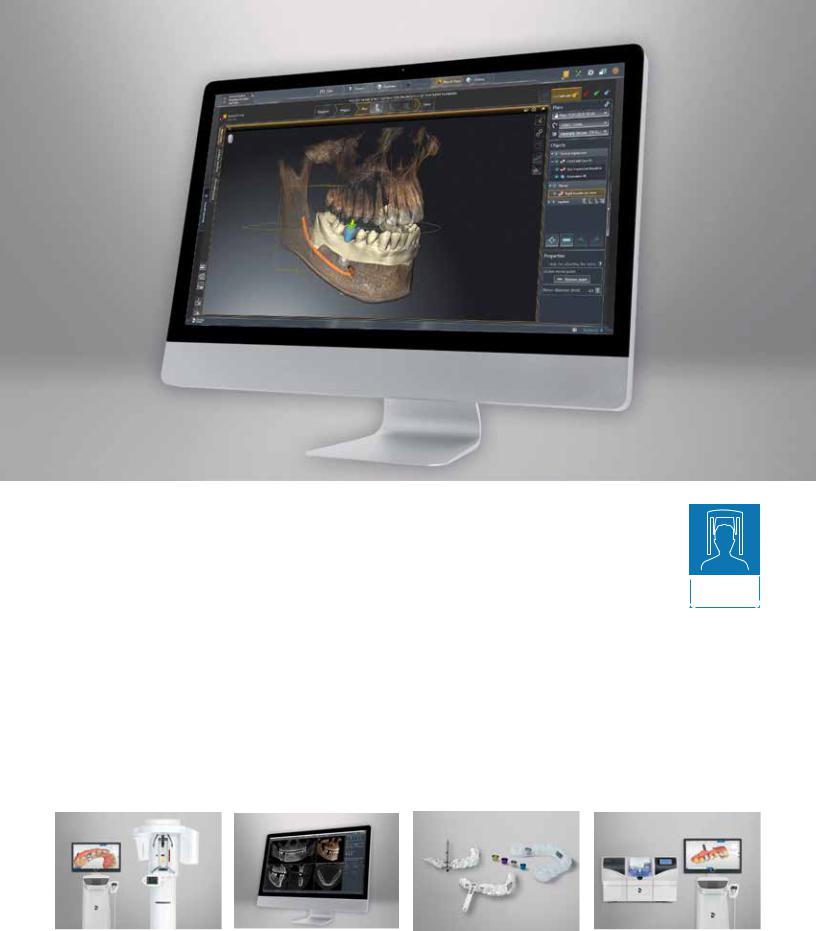
Clinical tasks for the Orthophos family
SICAT Implant
Modern and intuitive implantology. Your way.
Whether you are an advanced implantologist or just getting started, SICAT Implant effectively guides you through the implantology workflow with ease and efficiency.
Featuring seamless integration between the software and hardware, you get a truly unique user experience and an effective partner for your implant workflow. SICAT Implant supports true precision and accuracy during the implant planning process, which helps the clinician to foresee any unfavorable complications prior to treatment, providing peace of mind and confidence during surgery.
In combination with the numerous surgical guide options, which can be ordered directly in the software, sent out to a local lab, or milled directly in-house with your CEREC milling unit, you are supported until the final placement of the implant.
SL 3D S 3D
Scan |
Plan |
Place |
Restore |
SICAT Implant pending FDA 510K approval
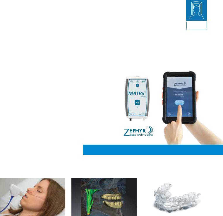
The Simple Sleep Solution
MATRx plus™* with SICAT Air and OPTISLEEP is a complete 3D solution for the analysis and appliance-based treatment of Obstructive Sleep Apnea (OSA).
The Simple Sleep Solution integrates comparative airway images using a Dentsply Sirona 3D Imaging system and expands the SICAT Air and OPTISLEEP workflow by adding MATRx plus, making it the only solution offering a complete workflow from airway analysis, diagnostics of OSA, oral appliance study, to the treatment of OSA with a custom-fitted oral appliance.
MATRx plus™
An innovative, easy-to-use tablet-based and cloud-connected home sleep testing system, MATRx plus simplifies patient selection for oral appliance therapy by identifying responders and their effective target protrusive position in advance of appliance fitting. This position and the respective
jaw relation are exactly transmitted into the final appliance with SICAT Air via a fully digital workflow.
•Cloud-based connectivity streamlines sleep physician study analysis, interpretation, and OSA diagnosis
•Knowing who to treat gives your patient
confidence in therapy
• Accurate prediction of target protrusion eliminates guesswork and saves chairtime
•MATRx plus comes standard with Orthophos SL 3D-Ai
04 I 05
SL 3D
S 3D
1 |
|
2 |
|
|
|
|
|
|
|
|
|
||
|
|
|
|
|
|
|
•Baseline 3D X-ray scan with a Dentsply Sirona 3D X-ray system for upper airway analysis.
•MATRx plus system dispensed for at-home sleep test to assist in OSA diagnostics and to predict therapeutic response and target protrusive position for OPTISLEEP appliance.
•3D X-ray scan with the patient wearing the MATRx plus titration trays at target position and capturing of the optical surface scan data of the patient’s upper and lower jaw. Subsequent fusion with the 3D data within SICAT Air.
•Ordering of OPTISLEEP therapeutic appliance in a completely digital workflow.
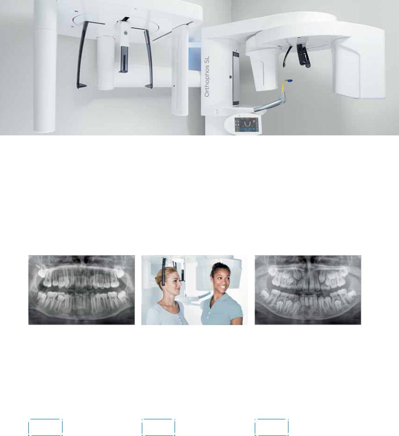
Orthodontics
Versatility, well-chosen programs and outstanding image quality are just a few of the characteristics that make each member of the Orthophos family a perfect partner in your practice. In Orthodontics, they offer safe and efficient treatment using the ALARA principle – and support you in reaching an accurate diagnosis efficiently and with optimal clinical support.
Clear case presentation helps improve overall patient communication and treatment acceptance.
Dedicated programs for young patients
The horizontally and vertically reduced children’s panoramic program delivers high-definition images at the lowest dose.
Ceph arm
Carpus image, PA and AP, as well as lateral ceph with the additional possibility of upper and occipital collimator for additional dose reduction.
Quick shot function
It reduces the capture time and dose. This facilitates, for example, working with children in panoramic and ceph images.
|
|
|
|
|
All |
|
SL 2D /3D |
SL 2D /3D |
|
models |
|
S 2D /3D |
S 2D /3D |
|

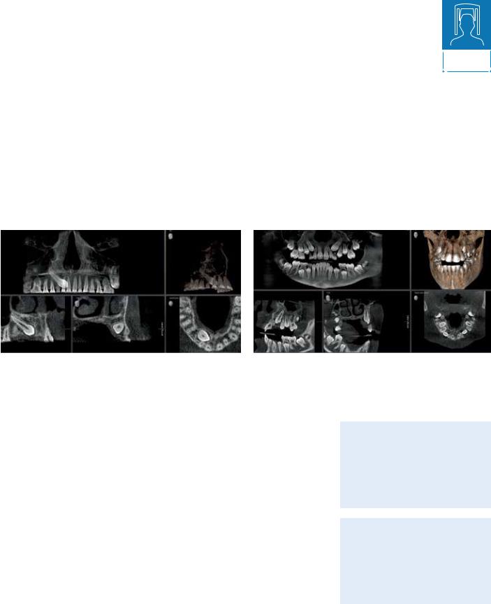
3D endodontics
Does your practice offer endodontic treatments? This can often come with many challenges. Emergency patients that need treatment for elusive canals and unpredictable pathology are just a few of the issues that you may be faced with. 3D imaging visualizes hidden structures, reveals clinical issues and makes it possible to address each situation individually.
No more surprises
SICAT Endo is a CBCT-based software providing you the ability to create a clear map detailing the route you will take into the canal, preparing you for any difficult anatomical structures that exist through realistic and detailed information.
06 I 07
SL 3D
S 3D

Custom 3D image
Digital imaging offers unbeatable benefits for every dental practice, creating a new standard for quality dental care. Whether overlaid teeth, unexpected canals, hidden roots or in the case of orthodontic surgery, a lower dose option for children, 3D images are invaluable in a variety of clinical tasks. In addition, they simplify patient communication for greater acceptance of your treatment proposal.

08 I 09
Your advantages at a glance:
•3D allows you to see structures that are often hidden in 2D X-rays
•Increased diagnostic confidence
•Clearer treatment presentation to patients
•Improved practice offering and as a result, increased growth and profitability
•Eliminates the need to refer your patient out for a CBCT scan
Custom 3D image
Precisely your volume –
More possibilities for your practice
SL 3D
When it comes to volume size, dose and image quality, every clinical case S 3D has its own individual requirements. The Orthophos family combines
image quality and versatility. Choose the appropriate volume for your needs: From the focused Ø 5 cm x 5.5 cm volume to the Ø 11 cm x 10 cm volume, which can display the wisdom teeth and upper respiratory tract.
The available volumes of our 3D models at a glance:
Ø 5 cm x 5.5 cm |
Ø 8 cm x 8 cm |
Ø 11 cm x 10 cm |
||
|
|
|
|
|
|
|
|
|
|
|
|
|
|
|
|
|
|
|
|
|
|
|
|
|
|
|
|
|
|
Additional available volumes (varies by model): Ø 8cm x 5.5cm, Ø 11 cm x 8 cm, Ø 11 cm x 7.5 cm
Each volume can be adjusted accordingly in three different modes to adapt to each patient’s unique situation:
High Definition (HD)
Standard Definition (SD)
Low Dose (Low)
10 I 11
MARS –
Metal Artifact
Metal artifacts are a challenge in 3D imaging. Radiopaque objects create shadowing and streaking effects during the threedimensional reconstruction and as a result can often interfere with diagnostics . MARS automatically reduces metal artifacts and facilitates the diagnosis.
Reduction Software
SL 3D
S 3D
without |
with |
MARS |
MARS |
MARS keeps anatomically relevant structures as free of artifacts as possible.
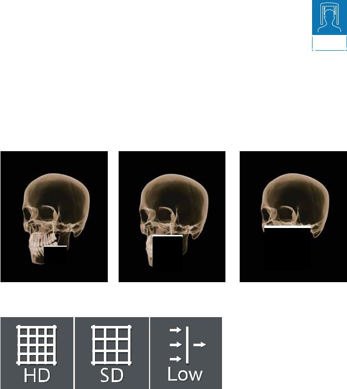
High Definition Mode (HD) – Fine details for safe diagnostics
Standard Definition mode (SD) provides all of your basic clinical information needed for a diagnosis, however in some cases it’s better to further increase the quality of the X-ray image. In endodontics, for example, you might need better visibility of fine structures for treatment planning and implementation. For this purpose, the Orthophos offers you High Definition mode (HD), in which up to 800 individual
images are recorded during one rotation and merged into a lownoise 3D volume with high resolution of up to 80 µm. This mode guarantees a faster and safer diagnosis within the recorded volume.
SL 3D
S 3D
Custom 3D image
Low Dose –
CBCT in the dose range of a 2D image
SL 3D
The optimized Low Dose mode with a dedicated filter allows for the S 3D imaging of dense structures, like bone, at a greatly reduced dose. This
makes Low Dose an efficient option for many clinical tasks – especially for those in orthodontics or implantology. With the two 3D models in the Orthophos family, you’re choosing on a case-by-case basis whether you use high-resolution volumes for fine structures (HD) or a low-dose image for minimal radiation exposure.
Localization of displaced incisor Ø 5 cm x 5.5 cm at 3 μSv |
Tooth position determination Ø 8 cm x 8 cm at 8 μSv |
Low Dose
for a variety of clinical tasks
Tooth position |
|
Implant control in 3D |
determination in 3D |
|
in the dose range of an |
at low dose, especially for |
|
intraoral X-ray |
young, radiation-sensitive |
|
|
patients |
|
|
|
|
|
Program selection for the case-based application using the ALARA (As Low As Reasonable Achievable) principle
Sleep apnea therapy with SICAT Air and OPTISLEEP
12 I 13
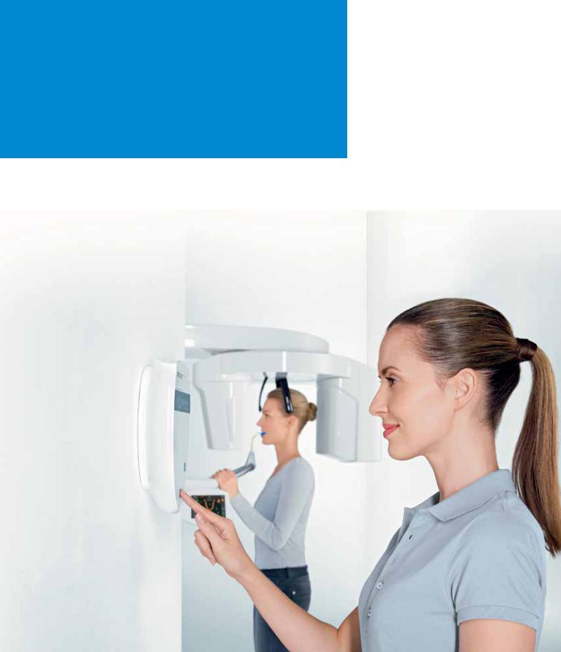
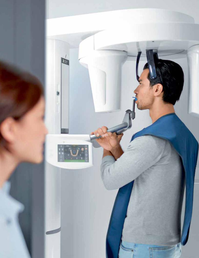
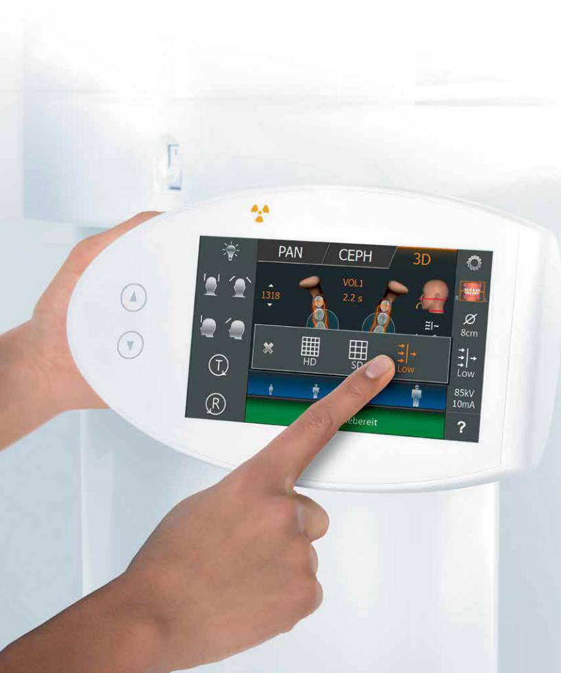
Easy operation, accurate positioning
With the Orthophos line, you capture the best possible image to support your diagnosis and maximize your patient’s experience. Our models offer unique, patented solutions designed to support all of your needs. Optimize your practice’s workflow with intuitive user interfaces and automatic positioning aids to avoid unnecessary secondary exposures.
 Loading...