

Wallach Surgical Devices Summit Doppler Vista AVS L500VAC, Summit Doppler Vista AVS L500VA User Manual

®
REF
User Manual for Vista AVS
L500VA (with Stand and Basket)
L500VAC (Stand and Basket not included)
MAN0014-DFU • Rev. A • 5/13 Wallach Surgical Devices

TABLE OF CONTENTS
(Vista AVS)
Section 1: Introduction........................................................................................ 1
Section 2: Safety Information.............................................................................. 2
Section 3: Description of Product and Controls ............................................... 5
Section 4: Preparation for Use............................................................................. 9
Section 5: Menu Configuration and Set Up ..................................................... 11
Section 6: The Ankle Brachial Index (ABI) Examination .............................. 16
Section 7: The Toe Brachial Index (TBI) Examination .................................. 23
Section 8: The Seated ABI Examination .......................................................... 25
Section 9: Segmental Studies ............................................................................. 27
Section 10: Individual Site Mode......................................................................... 29
Section 11: File Management............................................................................... 30
Section 12: Peripheral Arterial Waveform Interpretation............................... 33
Section 13: Maintenance and Cleaning............................................................... 36
Section 14: Battery Recharging and Replacement............................................ 38
Section 15: Specifications..................................................................................... 40
Section 16: Accessories......................................................................................... 45
Section 17: Troubleshooting Guide..................................................................... 46
Section 18: Clinical References............................................................................ 47
Section 19: AVS ReportTM Software................................................................... 48
Section 20: Warranty ........................................................................................... 64
Section 21: Explanation of Symbols.................................................................... 65
MAN0014-DFU • Rev. A • 5/13 i Wallach Surgical Devices
SECTION 1: Introduction
Thank you for choosing the Vista AVS® from Wallach Surgical Devices. We believe you
have purchased the finest arterial examination system available.
Your total satisfaction is our highest priority. We strive to continually improve our products
and services. Please contact us with any suggestions. We look forward to enjoying a longterm relationship with you!
Wallach Surgical Devices
95 Corporate Drive
Trumbull, CT 06611 USA
Here’s how you can reach us...
Phone: 1-800-243-2463
(203) 799-2000
Fax: (203) 799-2002
Email us at: customerservice@wallachsurgical.com
Visit our website at www.wallachsurgical.com
Important:
Please read this manual carefully and become familiar with the features,
operation, care, and safety requirements of the Vista AVS prior to use. Please note that
while operating the Vista AVS, step-by-step instructions are shown on the display to assist
you through the examination.
Wallach Surgical Devices provides general reimbursement information related to the diagnosis of
peripheral arterial disease as an overview for our customers. It is important to understand that
reimbursement is a complex process and requirements are subject to change without notice. It is the
responsibility of the healthcare provider to determine and submit appropriate codes, charges, and modifiers
for services that are rendered. Prior to filing any claims, customers are advised to contact their third-party
payers for specific coverage, coding and payment information. Wallach Surgical Devices makes no
promise or guarantee of reimbursement by Medicare or any other third-party payer.
Package Contents
The Vista AVS unit includes the following:
- User Manual - 17 cm Cuffs (2) - Ultrasound Gel
- Quick Guides - Printer Paper - Stand with Basket & Knobs (L500VA only)
- ABI Chart - Tape Measure - 8 MHz Bi-Directional Probe
- Hand Controller - Hose Set - AVS Report Software
- Digit Cuff - ABI Report Forms - USB Cable
- 10 cm Cuffs (2) - PPG Probe - Power Supply (+7 VDC) & cable
- 12 cm Cuffs (4) - Training Video
The following are optional and sold separately from the Vista AVS.
- 5 MHz Bi-Directional Probe
- Carrying case
Year of manufacture
located on the device.
U.S. Patent Pending
MAN0014-DFU • Rev. A • 5/13 1 Wallach Surgical Devices

SECTION 2: Safety Information
Intended Use:
This device is intended for detection of blood flow in veins and arteries and as an aid for the
diagnosis of peripheral vascular disease.
Contraindications:
WARNINGS
• The ultrasound probes are not to be used on or near the eyes.
• This device is for use only on intact skin.
• This device is not intended for use with HF surgical equipment.
• This device is not intended for fetal use.
General Warnings:
WARNINGS
• The Vista AVS is for use by qualified personnel only. Read the User Manual
before use.
• Carefully route all cables and tubing to reduce the possibility of patient entanglement
or strangulation.
• Do not allow the patient to operate any portion of the equipment, including the
hand-held controller.
• Do not place the equipment in any position that would allow it to roll, fall, or collide
with the user or patient.
• Do not use equipment that is damaged or malfunctioning. Seek appropriate service
when needed. Inspect equipment regularly for signs of damage. Use alternate
equipment if needed.
• Do not connect Luer fittings from Summit Doppler equipment into any other
equipment.
• Confirm the setting of the real-time clock prior to saving patient data.
• Any equipment connected to the USB data port must be configured to comply with
IEC 60601-1. By connecting additional equipment to the USB data port, the user is
configuring a medical system and the user is responsible for ensuring the overall
system compliance. Connected equipment must be certified to the applicable IEC
standard (i.e. IEC 950 for data processing equipment, IEC 60601-1 for medical
equipment). Contact the technical service department for more information.
MAN0014-DFU • Rev. A • 5/13 2 Wallach Surgical Devices

General Cautions:
CAUTIONS
• Do not plug the probe cables into a telephone system, computer, or any equipment
other than the Vista AVS.
• Read the Maintenance and Cleaning Section (Section 13) before cleaning.
• U.S. Federal law restricts this device to sale by or on the order of a licensed
practitioner.
• Do not drop or mishandle the Vista AVS main unit, probes, hand controller or other
accessories. Damage may occur.
Limitations of Noninvasive Vascular Tests:
In Current Noninvasive Vascular Diagnosis (Chapter 13, Page 208), Ali F. AbuRahma and Edward B. Diethrich note
the following limitations of arterial leg Doppler examinations.
• Falsely high segmental pressure readings in areas with calcified arteries
• Artificially elevated high-thigh pressure in very large or obese patients
• Difficult interpretation of segmental pressures in patients with multilevel occlusive
disease
• Difficulty in interpretation of high-thigh readings
• False-negative results in patients with mild vascular occlusive disease who have
normal resting ankle pressures
Safety of Ultrasound:
The Vista AVS Doppler probes were designed to be safe and effective. However, the risk
from some hazards cannot be eliminated completely. Instead, they are reduced to a level
that is As Low As Reasonably Achievable (ALARA). Prudent use of this device in
accordance with the principle of ALARA includes minimizing the duration of the patient’s
exposure to ultrasound to the extent practical.
Clinical Safety
Approved by the American Institute of Ultrasound - March 1997, October 1982
Diagnostic ultrasound has been in use since the late 1950s. Given its known benefits
and recognized efficacy for medical diagnosis, including use during human pregnancy, the
American Institute of Ultrasound in Medicine herein addresses the clinical safety of such
use:
There are no confirmed biological effects on patients or instrument operators caused by
exposures from present diagnostic ultrasound instruments. Although the possibility exists
that such biological effects may be identified in the future, current data indicates that the
benefits to patients of the prudent use of diagnostic ultrasound outweigh the risks, if any,
that may be present.
MAN0014-DFU • Rev. A • 5/13 3 Wallach Surgical Devices

Safety in Training and Research
Approved by the American Institute of Ultrasound - March 1997, March 1983
Diagnostic ultrasound has been in use since the late 1950s. There are no confirmed adverse
biological effects on patients resulting from this usage. Although no hazard has been
identified that would preclude the prudent and conservative use of diagnostic ultrasound in
education and research, experience from normal diagnostic practice may or may not be
relevant to extended exposure times and altered exposure conditions. It is therefore
considered appropriate to make the following recommendation:
In those special situations in which examinations are to be carried out for purposes other
than direct medical benefit to the individual being examined, the subject should be informed
of the anticipated exposure conditions, and of how these compare with conditions for
normal diagnostic practice.
MAN0014-DFU • Rev. A • 5/13 4 Wallach Surgical Devices

SECTION 3: Description of Product and Controls
Description of Unit
Vista AVS is a physiologic exam system designed to aid the clinician in the diagnosis
The
of peripheral arterial disease (P.A.D.). The unit includes a sensitive, bi-directional Doppler
system, arterial photoplethysmograph (PPG), and a pressure system that provides inflation,
controlled deflation, and pulse volume recording (PVR) capabilities.
The
Vista AVS is well suited for the ankle brachial index (ABI) examination, the gold
standard for the diagnosis of P.A.D. The ABI compares the systolic blood pressure at the
ankles with the systolic pressure at the brachial arteries. A significantly reduced ankle
pressure results in a low (<0.9) ABI value, which indicates P.A.D. Systolic ankle pressures
are obtained with a pressure cuff and audio Doppler probe. ABI measurements are
discussed in detail in Section 6.
A single level, lower extremity arterial exam (CPT® 93922) includes the ABI pressures,
calculated index, and arterial physiologic waveforms. Two types of waveform modalities
are provided on the Vista AVS: continuous-wave (CW), bi-directional Doppler and PVR.
Both of these waveform modalities meet the requirements of CPT 93922. Although both
modalities have significant clinical utility, it is generally not necessary to include both PVR
and Doppler waveforms in reimbursement documentation for CPT 93922 - either one is
sufficient. Waveform analysis is discussed in Section 12.
The Vista AVS is designed to perform segmental studies to compare three or more lower
limb pressures to the brachial pressures. This procedure is reimbursable under CPT code
93923 as a non-invasive, physiologic study of upper or lower extremity arteries, multiple
levels or with provocative functional maneuvers, complete bilateral study.
The Doppler waveform is a graph with a vertical axis (Doppler frequency shift, or pitch)
proportional to the velocity of arterial blood flow. Flow toward the probe is indicated above
the baseline. Flow away from the probe is indicated below the baseline. The 8 MHz,
bi-directional probe is best for superficial vessels and all-around use. The optional 5 MHz,
bi-directional probe is used for deeper vessels and with some obese patients.
Pulse Volume Recording (PVR) is a form of plethysmography, which is an indirect method
of limb volume measurement. A pressure cuff is applied to the limb and inflated to 65 mmHg
to detect the minute fluctuations in limb volume that occur with each heart beat. The PVR
waveform’s contour is a qualitative indicator of presence or absence of peripheral arterial
disease. This type of PVR does not permit calibration by injection of a known air volume
and is used for arterial waveform analysis.
Photoplethysmography (PPG) is an optoelectronic technique for detecting the small changes
of blood volume that occur in the capillary bed. Infrared (IR) light is emitted by the PPG
probe into the skin. Light reflected from the underlying tissue is received by a detector and
converted to an electrical signal. Since blood attenuates IR light at a higher level than the
surrounding tissue, the signal’s pulse contours are determined by the arterial blood supply.
MAN0014-DFU • Rev. A • 5/13 5 Wallach Surgical Devices

This type of PPG system is primarily for arterial pulse detection. In conjunction with a
digit cuff, arterial PPG is quite useful for toe pressure measurement, which is an additional
diagnostic tool when the arteries at the ankle are noncompressible (ABI > 1.30) due to
calcification. Calcified arteries are prevalent among patients with diabetes or kidney disease,
but the small arteries of the toes seldom become calcified. When the Vista AVS is
configured for PPG, the system automatically calculates the toe brachial index (TBI).
A low TBI (<0.65) indicates an arterial obstruction. The TBI is discussed in Section 7.
The Vista AVS includes everything that is needed to conduct fast, efficient peripheral
arterial examinations including display, printer, Doppler probes, PPG probe, pressure hose,
limb pressure cuffs, and a digit cuff. The instrument operates from either its internal battery
pack or from an external medical grade power supply at 100-240 VAC.
Controls and Indicators:
Main Unit
ON/OFF - Turns the unit on or off with momentary actuation
FEED - Printer feeds paper while pressed
CHARGING
- On: Charging
- Flashing: Charging complete (trickle charging while flashing)
LOW BATT - Flashing: Low battery
PAPER RELEASE
- Blue button under paper dispenser: Opens printer cover
MAN0014-DFU • Rev. A • 5/13 6 Wallach Surgical Devices
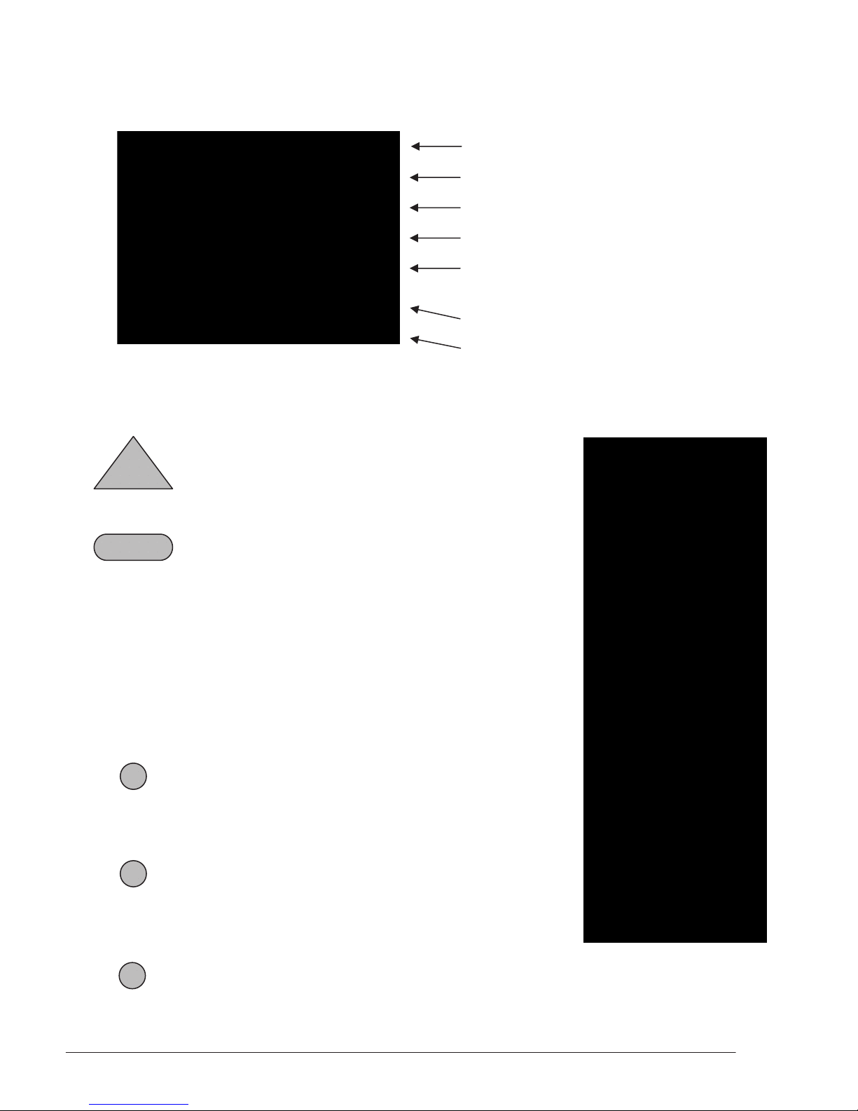
Display Screen - Protocol Location and Markers
Right & Left Brachial Sites
Waveform Sites (Right & Left PT or DP)
Right & Left Ankle Sites (PT or DP)
(Toe Sites when in PPG Mode)
Pressure Gauge (mmHg)
Right & Left ABI
Date and Time
Step-By-Step ABI Instructions
Hand-Held Controller
PUMP - Pump runs while pressed
PUMP
SAVE - During deflation- Stores
SAVE
current cuff pressure and
deflates
- Active waveform- Stores
waveform data
- Frozen waveform- Stores the
frozen waveform
nd
button press- Confirms
- 2
the stored pressure or
waveform
FREEZE - Active waveform- Freezes
the displayed waveform
FREEZE
- Frozen waveform- Starts new
waveform acquisition
SITE - Moves the protocol marker to
SITE
the next location
- Saves data before leaving old
protocol location
SCALE - Changes the waveform scaling
SCALE
MAN0014-DFU • Rev. A • 5/13 7 Wallach Surgical Devices

MUTE - Enables/disables Doppler speaker output
MUTE
PRINT - Prints out the waveform on adhesive-backed label paper
PRINT
ENTER/MENU - Enters the displayed value or makes a menu selection
ENTER/MENU - Also used to open the OPTIONS screen
UP the caliper tool to adjust the pressure values
DOWN the caliper tool to adjust the pressure values
UP - Navigates through the menu in the up direction & moves
DOWN - Navigates through the menu in the down direction & moves
Numeral Mode Character Mode
Numeral 1
1
Numeral 2 A, B, C
2
Numeral 3 D, E, F
3
Numeral 4 G, H, I
4
Numeral 5 J, K, L
5
Numeral 6 M, N, O
6
Numeral 7 P, Q, R
7
Numeral 8 S, T, U
8
Numeral 9 V, W, X
9
Numeral 0 Y, Z
0
Space Space
SPACE
Hyphen Hyphen
-
See Section 5 for information on how to change between Character and Numeral Modes.
MAN0014-DFU • Rev. A • 5/13 8 Wallach Surgical Devices

SECTION 4: Preparation for Use
Tools required: Phillips head screwdriver
1. Unpack the Vista AVS and inspect the unit for external damage.
2. Verify that you have received each of the contents listed on the packing list.
3. Assemble the rolling stand (if applicable) using the instructions provided.
4. Use a Phillips head screwdriver to attach the Vista AVS mounting bracket to the
end of the rolling stand’s pole. Be sure to fit the pole’s alignment pin through the
alignment hole on the bracket.
Use the two stand mounting knobs to attach the mounting bracket to the Vista AVS.
5.
Review the unit and locate each of the controls and connectors (see Section 3 and
6.
information on the next page).
7. Plug the external power supply into the + 7 VDC (Power) connector.
MAN0014-DFU • Rev. A • 5/13 9 Wallach Surgical Devices

VISTA TIPTM:
• For first time use, allow the unit to charge for at least one hour before operating
the Vista AVS from its battery.
Plug the power cord into the external power supply and then into a properly
8.
grounded wall outlet.
9. Plug the Doppler cable into the Doppler probe.
10. Plug the Doppler cable into the Doppler connector. (Blue port)
11. Plug the PPG probe into the PPG connector. (Black port)
VISTA TIP:
• The Doppler and PPG connectors are color coded and physically interchangeable.
If you fail to connect the proper probe, no damage will occur.
12. Attach a cuff to the Luer style hose fitting.
13. Plug the hose quick connect style fitting into the CUFF connector on the connector
panel.
14. Plug the hand-held controller into the HAND CONTROLLER connector.
VISTA TIP:
• To maintain patient safety, it is not possible to conduct an examination while the
Vista AVS is connected to a PC via the USB port.
Connectors
Doppler PPG Hand USB + 7 VDC (Power) Cuff
Controller
MAN0014-DFU • Rev. A • 5/13 10 Wallach Surgical Devices

SECTION 5: Menu Configuration and Set Up
Loading Paper:
Vista AVS is shipped with paper pre-loaded. To load a new roll of paper, press the
The
printer release button. Remove any remaining paper from the old roll and drop in the new
roll as shown below. Close the paper door and push firmly enough to latch the door into
position. Press the FEED button to ensure proper paper alignment.
WARNING
• Printer components become hot during printing. Do not touch the metal pieces
inside the paper holder immediately after printing. After loading paper, press the
ON/OFF switch to begin using the instrument.
Configuring the Examination
Vista AVS can be configured to perform P.A.D. testing in four different automated
The
modes. Pressure can be obtained using either the Doppler at the ankle for typical ABI
testing described in Section 6 or using a PPG at the toes for TBI testing described in Section
7. Additionally, waveforms used to complete the physiologic study can be performed with
either a Doppler or the Pulse Volume Recording (PVR) mode.
1. Press the ENTER/MENU key to open the OPTIONS menu.
2. Press the number 1 key to select the CONFIGURE EXAMINATION.
3. Select the desired configuration for the examination by pressing the appropriate
number:
1 – ABI with PVR Waveform
2 – ABI with Doppler Waveform
3 – TBI with PVR Waveform
4 – TBI with Doppler Waveform
5 – Segmental with PVR Waveform
6 – Segmental with Doppler Waveform
MAN0014-DFU • Rev. A • 5/13 11 Wallach Surgical Devices

VISTA TIP:
• The current configuration mode will be displayed at the bottom of the waveform
upon returning to the main screen.
Setting the Date and Time
Press ENTER/MENU to open the OPTIONS menu.
1.
2. Press the number 2 key to select SYSTEM SETTINGS.
3. Press the number 1 key to set the clock.
4. The clock setting format is:
MM DD YY HH MM SS (Month-Day-Year-Hours-Minutes-Seconds)
5. Use the UP key to move the * cursor over the field that needs to be changed.
6. Use the numerals on the hand-held controller to set the date and time.
7. Press ENTER/MENU or move the * cursor to the end to exit the CLOCK menu.
8. Press the ENTER/MENU key to exit the OPTIONS menu.
Contrast Adjust
To modify the contrast level on the
Vista AVS screen:
Press the ENTER/MENU key to open the menu.
1.
2. Press the number 2 key to select the SYSTEM SETTINGS.
3. Press the number 2 key to increase CONTRAST ADJUST LEVEL one level.
4. Continue to press the number 2 key until desired level is reached.
VISTA TIP:
• If there is flickering or shadowing on the text, the contrast is adjusted too
high. Readjust contrast level by pressing the number 2 key until contrast
level starts over at 1 and slowly increase the contrast level
.
MAN0014-DFU • Rev. A • 5/13 12 Wallach Surgical Devices

Setting the Power Down Status
The Vista AVS can be set to turn off automatically to save battery power. If the automatic
power down is set to ON, the unit will turn off after 20 minutes automatically after the last
key press. If the automatic power down is set to OFF, the unit will not turn off until either
the user presses the ON/OFF button or the battery becomes low.
1. Press ENTER/MENU to open the OPTIONS menu.
2. Press the number 2 key to select SYSTEM SETTINGS.
3. Press the number 3 key to toggle the power down value to ON and OFF.
4. After setting the desired value, press ENTER/MENU to exit the OPTIONS menu.
Setting the Ankle Protocol
The Vista AVS can be set to accommodate protocols that use either one pressure
measurement or two pressure measurements from each ankle. The DUAL ANKLE
PRESSURE mode will be set to OFF for a single ankle pressure. For protocols that use
measurements from both the dorsalis pedis (DP) and the posterior tibia (PT) arteries, select
HIGH or LOW to select the desired protocol with second ankle pressure.
1. Press ENTER/MENU to open the OPTIONS menu.
2. Press the number 2 key to select SYSTEM SETTINGS.
3. Press the number 5 key to toggle the ankle value between OFF, HIGH or LOW.
4. After setting the desired value, press ENTER/MENU to exit the OPTIONS menu.
File Annotation
The File Management SAVE function can use either alpha or numeric characters for saving
a patient filename. To configure the system to the desired function:
Press the ENTER/MENU key to open the OPTIONS menu.
1.
2. Press the number 2 key to select the SYSTEM SETTINGS.
3. Press the number 4 key to toggle between NAME or NUMBER modes.
4. To enter a filename, use the alphanumeric keys on the hand-held controller. Use the
DOWN key to backspace and the UP key to move between different letters on the
same button.
5. Press the SAVE key to save the filename and begin the exam.
MAN0014-DFU • Rev. A • 5/13 13 Wallach Surgical Devices

Selecting a Modality for Obtaining Pressures
Choose between the Doppler probe and the photoplethysmography (PPG) probe to obtain
pressure values for ankle brachial index (ABI) exams, toe brachial index (TBI) exams or
segmental studies:
Press the ENTER/MENU key to open the OPTIONS menu.
1.
2. Press the number 2 key to select the SYSTEM SETTINGS.
3. Press the number 6 key to toggle between DOPPLER and PPG.
The selected pressure modality is indicated in the lower right corner of the display (DOP or
PPG will appear depending on which is selected) as shown below.
Modality –
Doppler probe
VISTA TIP:
• If you’re trying to obtain pressures or waveforms using the Doppler probe but it is not
audible when rubbing the probe tip, the system is probably set in PPG mode. To
change the setting, go to the System Settings menu and select Doppler for obtaining
pressures and waveforms.
MAN0014-DFU • Rev. A • 5/13 14 Wallach Surgical Devices

Entering a Filename Prior to Beginning an Exam
If beginning a new exam by pressing the number 6 key for NEW EXAM – CLEAR ALL
under the OPTIONS screen, the system will display the ENTER FILENAME message.
This message ensures that the user is aware that the patient data will not be automatically
saved. After completing the exam, the user can save the data by following the instructions
provided in the “Saving a File” paragraph in Section 11.
If a current exam is not saved and the user selects either NEW EXAM - CLEAR ALL or
NEW EXAM – KEEP BRACHIALS under the OPTIONS screen, the system will display
the UNSAVED DATA message below to let the user know that the previous data was not
saved.
Press the ENTER/MENU key to ignore this message and start a new exam (the ENTER
FILENAME will appear), or press the
SAVE key to save the data and the SAVE FILE menu
will appear.
MAN0014-DFU • Rev. A • 5/13 15 Wallach Surgical Devices

SECTION 6: The Ankle Brachial Index (ABI) Examination
Preparing the Patient
In a warm room, have the patient take off his/her shoes and socks and rest in a supine
position for approximately 5 minutes prior to taking pressures. The patient should wear
thin, loose fitting clothing. Avoid rolling up sleeves or pant cuffs in such a manner that it
obstructs blood flow. Bulky items such as sweaters should be removed.
Wrap the cuffs snuggly around the arms and ankles as shown below. The edge of the cuff
should be placed approximately 1 to 2 inches above the site of examination. Select the
proper cuff width, equivalent to 40% of the patient’s limb circumference. In general,
average sized adults use 10 cm cuffs, while larger adults use 12 cm cuffs. The most
efficient way to conduct the exam is to wrap all four limbs prior to taking any pressures.
Arm Cuff Leg Cuff
While applying the cuffs, it may be a good time to explain the examination to the patient
and answer any questions.
NOTE: It is very important that the patient remain still and in a supine position for the
duration of the exam.
VISTA TIP:
• The Vista AVS performs the ABI exam in the following order: Right Brachial
Pressure (R-BRA), Right Ankle Waveform (R-WAV), Right Ankle Pressure
(R-ANK), Left Ankle Waveform (L-WAV), Left Ankle Pressure (L-ANK), and Left
Brachial Pressure (L-BRA). You may override this order by using the SITE key to
move between protocol locations.
• Notice that the patient’s right hand side is shown on the left side of the
Vista AVS's screen in order to match what you see when you face the patient.
MAN0014-DFU • Rev. A • 5/13 16 Wallach Surgical Devices

Getting Started
Press the ENTER/MENU key to open the OPTIONS menu. Press the number 6 key to start a
new exam. You may enter a filename at this time by using the keypad. Refer to Section 5
for File Annotation instructions.
VISTA TIP:
• To start a new study on the current patient, press the number 5 key to clear the
data except for brachial pressures. This is useful when it is determined a TBI
will be required following an ABI with noncompressible arteries.
Obtaining the Right Brachial Pressure
Connect the pressure hose to the fitting on the right brachial cuff. Apply a small amount of
gel over the right brachial artery
and place the Doppler probe at approximately 45 degrees,
pointing in the direction toward the shoulder as shown below. Slide the probe laterally
across the arm to find the brachial artery and obtain the best signal possible. Adjust the
volume knob to acquire a proper audio level. Once the best signal is obtained, brace the
hand holding the probe against the patient’s arm to ensure a stable position. Be careful not
to apply too much pressure against the skin. Excessive pressure could occlude the artery.
Next, press and hold the
reaches about 20 mmHg above the occlusion pressure. After the pump stops the
PUMP key to inflate the cuff. Release the key once the pressure
Vista AVS
automatically bleeds the cuff at the correct rate.
Press the
SAVE key at the moment flow returns. This will store the brachial pressure value
in the R-BRA protocol location. This is the systolic pressure. In general, the audible
Doppler signal is a slightly more sensitive indicator of the systolic pressure than the Doppler
waveform display.
MAN0014-DFU • Rev. A • 5/13 17 Wallach Surgical Devices

Doppler Waveform
During Acquisition
of Systolic Pressure
The cuff deflates automatically when
SAVE is pressed. If needed, press PUMP again to
repeat the measurement, or use the arrow keys or numeric keypad to adjust the pressure
value before confirming the value by pressing the
SAVE or SITE key.
Press
SITE or SAVE again to confirm the right brachial pressure and move to the next
protocol location.
VISTA TIP:
• Once a satisfactory pressure has been obtained, pressing
SAVE or SITE will
confirm the stored value and move to the next protocol location.
• During deflation, pressing PUMP again will re-inflate the cuff and allow a new
pressure to be taken.
OBTAINING PRESSURES USING THE PPG PROBE:
In the supine position, ABI pressures can also be obtained using the PPG probe. For
obtaining pressures, apply the PPG probe to an index finger for brachial pressures or the
great toe for ankle pressures. It is important the patient is still and the fingers and toes are
reasonably warm. To obtain brachial pressures, wrap the cuffs around the arms at the
brachial artery site. Place the PPG on the index finger with the blue side of the probe against
the skin. It should be snug to ensure contact, but not too tight to occlude blood flow as
shown below.
It will take a few seconds for the waveform to stabilize on the display after applying
the probe. You may want to consider changing the scale of the display to get a better
view of the waveform by pressing the
PUMP key to inflate the cuff to a pressure approximately 20 mmHg above the
the
SCALE key. Once the waveform is stable, press
pressure until the PPG pulsations disappear. The arm cuff will begin to deflate
automatically once the pump stops.
MAN0014-DFU • Rev. A • 5/13 18 Wallach Surgical Devices

After the first consistent pulsation returns press the FREEZE key to freeze the waveform on
the display. Use the caliper tool by pressing the
UP or DOWN arrow keys to adjust the
pressure reading. Move the caliper to the beginning of the first upward slope at the start of
the pulsation as shown below. This is the systolic pressure. Press the
SAVE key to save the
pressure and move to the next site to be measured.
Refer to Section 7 for instructions for using the PPG probe to obtain toe pressures.
Right Ankle Waveform
Verify that the protocol marker is on R-WAV.
VISTA TIP:
• A single level lower extremity arterial examination normally consists of the ABI
value and either bi-directional Doppler waveforms or PVR waveforms. The
following shows how to set the waveform mode. See Section 12 for information
on waveform interpretation. Note that arterial tests documented with PPG
waveforms are not reimbursable as opposed to Doppler or PVR waveforms.
The Vista AVS can acquire bi-directional Doppler waveforms, pulse volume recording
(PVR) waveforms and photoplethysmography (PPG) waveforms. See Section 5 for
configuring the desired waveform mode.
OBTAINING DOPPLER WAVEFORMS:
Apply a small amount of coupling gel over the site of the artery, either the PT or DP artery.
Place the probe over the artery at an angle of approximately 45 degrees, pointing the probe
tip in the direction toward the calf and knee. Slide the probe slowly across the artery until
the best signal is obtained.
PT DP
MAN0014-DFU • Rev. A • 5/13 19 Wallach Surgical Devices
 Loading...