

Page 1
®
STEINDORFF
POLARIZING Microscope
Model Number
SPMM
User Manual
Page 2
1
®
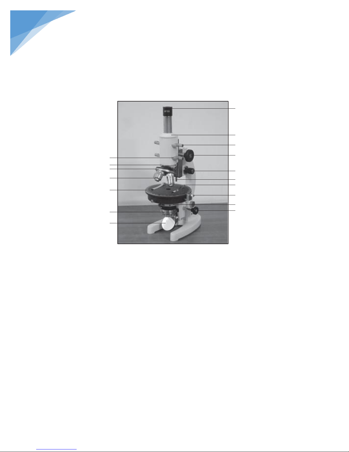
1. Eyepiece
2. Microscope Tube
3. Bertrand Lens
4. Coarse Motion Knobs
5. Fine Motion Knobs
6. Tube Lock
7. Stage Clips
8. Stage Lock
9. Condenser
10. Condenser Knob
11. Reflector
12. Polarizer
13. Rotating Stage
14. Objective
15. Nosepiece
16. Mica & Gypsum Plate
17. Analyzer
1
2
3
4 5 6
7 8 9
10
11
12
13
14
15
16
17
STEINDORFF
PARTS LAY-OUT
LIST OF PARTS
Page 3

2
®
STEINDORFF
The Observation of Orthoscope
Usually to observe a tissue section by Microscope, the condenser should be lower down and the Bertrand lens is taken away from
the light path. Doing like this way, the optical nature of minerals be able to be observed by means of getting in visual field of transmitting
parallel light.
How to Use
Polarizer and Analyzer is to be set on the orthogonal crossed position.
Slide out the analyzer from the light path.
Bertrand lens is also to slide out from the light path.
Without any slide, adjust the Sub Stage Condenser as to become the same brightness in each objectives while observing
through the microscope.
To set with 10X eyepiece and low power Objective.
Insert the Analyzer into the light path, then the field becomes dark.
To rotate the rotating stage having on a slide with specimen, to be observed through the Microscope.
Insert the Gypsum-Plate in order to change a wave length of specimen in 1/4 degree, if required.
Subject to these steps, we may be able to know a figure and color or refractive index of minerals by orthoscope.
Adjustment of Centripetal
The center of the object & rotating stage have to accord with the crossed point of specimen in visual field always. If it is not
correct, it should be adjusted by the centering screws, while rotating the stage. Focus the specimen sharply, find a marked feature in
field view and make it situated at the center of the eyepiece cross line.
The Observation of Conoscope
Set the eyepiece & object, insert an analyzer and polarizer in the light path and adjust the condenser to observe the Conoscope or
interference figure by transmitting light with various magnifications.
How to use
Adjust the Condenser to Upper position.
Insert the Bertrand lens in the light path.
To set with 5X eyepiece & 40X Objective.
To put a specimen on the stage and adjust the focus.
Adjust the intensity of the light/through diaphragm of the condenser.
Page 4

3
®
STEINDORFF
High Power objective 40x is most suitable to get an image of Conoscope.
The condenser might to be put on a higher position for better results.
Maintenance of Lens Surface
Wipe off dust deposited on the lens surface with a soft brush or blow it off with a blower. Lightly and carefully wipe off finger
prints or oily stains with a soft, clean, cotton cloth, gauze, or lens paper moistened with a suitable lens cleaner. Do not
directly saturate the lens surface with cleaners that might creep into the optical system and impair their performance. Never
use strong solvents to clean lens, mirrors and painted surfaces. When wiping the glass surface, do it lightly several times by
sequentially using a new, unused portion of the cloth each time.
When not in use
When the Microscope is not used for a short period, protect it from dust by covering it with a plastic dust cover and store it in a
dry place. When the microscope is not used for a long period, clean it , then detach the objectives and eyepieces, install the
eyepiece tube caps and revolving nosepiece caps and cover it with a vinyl sheet for storage. Put the objectives into the case
and store them together with the eyepieces in a dessicator.
 Loading...