

Page 1

AN2944
Application note
Plethysmograph based on the TS507
Introduction
This application note provides a method to make an analog front-end plethysmograph (from
the ancient greek plethysmos, which means increase), which is an instrument for measuring
changes in volume within an organ or whole body, usually resulting from fluctuations in the
amount of blood or air it contains. In this context, we refer in particular to the fluctuations in
the quantity of blood in blood vessels.
January 2010 Doc ID 15467 Rev 1 1/12
www.st.com
Page 2

Contents AN2944
Contents
1 Description . . . . . . . . . . . . . . . . . . . . . . . . . . . . . . . . . . . . . . . . . . . . . . . . . 4
1.1 Purpose . . . . . . . . . . . . . . . . . . . . . . . . . . . . . . . . . . . . . . . . . . . . . . . . . . . 4
1.2 Theoretical background . . . . . . . . . . . . . . . . . . . . . . . . . . . . . . . . . . . . . . . 4
2 Model for creating a plethysmograph . . . . . . . . . . . . . . . . . . . . . . . . . . . 6
2.1 Getting started . . . . . . . . . . . . . . . . . . . . . . . . . . . . . . . . . . . . . . . . . . . . . . 6
2.2 Sensors . . . . . . . . . . . . . . . . . . . . . . . . . . . . . . . . . . . . . . . . . . . . . . . . . . . 6
2.3 Analog front-end . . . . . . . . . . . . . . . . . . . . . . . . . . . . . . . . . . . . . . . . . . . . . 6
3 Bill of material . . . . . . . . . . . . . . . . . . . . . . . . . . . . . . . . . . . . . . . . . . . . . . 8
4 Results . . . . . . . . . . . . . . . . . . . . . . . . . . . . . . . . . . . . . . . . . . . . . . . . . . . . 9
5 References . . . . . . . . . . . . . . . . . . . . . . . . . . . . . . . . . . . . . . . . . . . . . . . . 10
6 Revision history . . . . . . . . . . . . . . . . . . . . . . . . . . . . . . . . . . . . . . . . . . . 11
2/12 Doc ID 15467 Rev 1
Page 3

AN2944 List of figures
List of figures
Figure 1. Light absorption by hemoglobin at different wavelengths. . . . . . . . . . . . . . . . . . . . . . . . . . . 4
Figure 2. Schematic representation of the pulse wave transit time (PWTT) . . . . . . . . . . . . . . . . . . . . 5
Figure 3. Sensing system . . . . . . . . . . . . . . . . . . . . . . . . . . . . . . . . . . . . . . . . . . . . . . . . . . . . . . . . . . 6
Figure 4. Schematic diagram for the analog front-end plethysmograph . . . . . . . . . . . . . . . . . . . . . . . 7
Figure 5. Analog front-end demonstration board . . . . . . . . . . . . . . . . . . . . . . . . . . . . . . . . . . . . . . . . . 7
Figure 6. Entire system . . . . . . . . . . . . . . . . . . . . . . . . . . . . . . . . . . . . . . . . . . . . . . . . . . . . . . . . . . . . 7
Figure 7. Five seconds recording by plethysmograph of a healthy subject . . . . . . . . . . . . . . . . . . . . . 9
Doc ID 15467 Rev 1 3/12
Page 4
Description AN2944
1 Description
1.1 Purpose
This application note describes a demonstration board which is designed for demonstration
purposes only, and shall not be used as a medical instrument, nor for domestic installation.
The technical data included in this document shall be taken as a guideline.
1.2 Theoretical background
The contraction of the heart causes a pressure wave which moves along the arteries
producing, as a consequence, their expansion during the positive peak. The wave is faster
than the blood flow and its speed reaches a few meters per second. The pulse wave can be
sensed at a limb as well as the wrist or a finger.
The two possibilities for sensing the pulse wave are via a pressure sensor or through an
optoelectronic plethysmograph which uses the physical mechanism of light absorption.
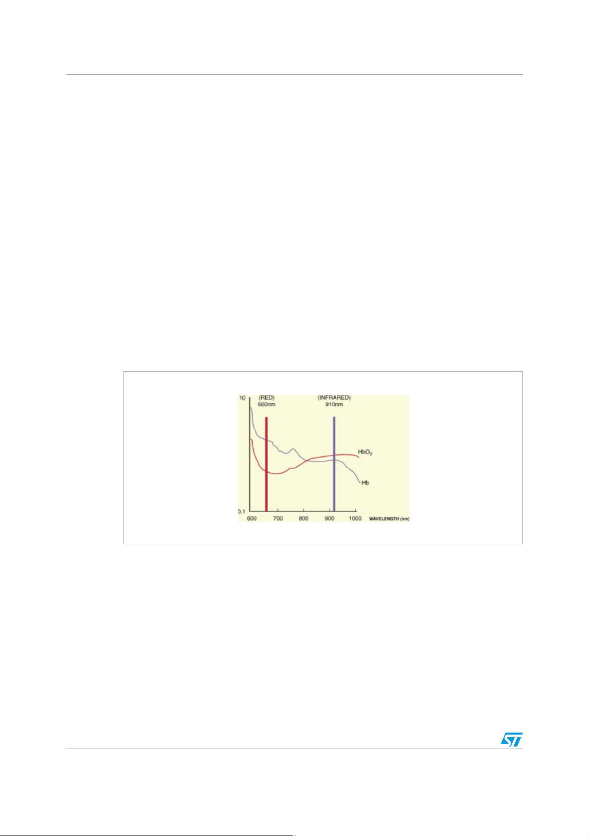
Hemoglobin present in the blood absorbs the light emitted in a particular wavelength range
(see figure below). In this system infrared light can be used with no distinction between
oxyhemoglobin and deoxyhemoglobin.
Figure 1. Light absorption by hemoglobin at different wavelengths
!-V
For this reason, the light which is able to pass through the body at a wavelength of
600-900 nm depends on the quantity of hemoglobin flowing in the blood vessels. Therefore,
since the quantity of hemoglobin at a given time is proportional to the pulse wave at that
time, it is possible to calculate the pulse wave from the transmitted light.
The information from the pulse wave is often used in conjuntion with a three-lead
electrocardiogram (ECG or EKC) with the differential electrodes placed on the thorax. In
fact, from the two measurements, it is possible to calculate the PWTT (pulse wave transit
time) which is the time interval between the R wave peak of the ECG and the positive peak
of the plethysmography (see Figure 2) and can be correlated with blood pressure.
4/12 Doc ID 15467 Rev 1
Page 5

AN2944 Description
Figure 2. Schematic representation of the pulse wave transit time (PWTT)
!-V
Doc ID 15467 Rev 1 5/12
Page 6

Model for creating a plethysmograph AN2944
2 Model for creating a plethysmograph
2.1 Getting started
The construction of the board described in the following sections has been inspired from
other projects already developed in an educational and scientific context [see 1 and 2 in
Section 5: References]. However, in order to prevent accidents, the electrical installation of
the power supply shall be completed in accordance with safety requirements (e.g. by
assuring a satisfactory cross-section of the conductors, by using a fuse, etc.).
2.2 Sensors
The sensor consists of an infrared (800 nm) light emitter diode, the SFH309FA, and an NPN
phototransistor light receiver, the SFH487. The source and detector have to be mounted
side by side on a finger. The light passes through the tissues and the amount of light
captured by the photodetector depends on the quantity of hemoglobin. Timed samples of
the photodetector output allow us to reconstruct the pulse wave. An easy way to mount
these two components to have steady contact with the finger is to use a wooden clothespin
to hold them in a fixed position (see figure below). The infrared filter of the phototransistor
reduces the interference from the fluorescent lights, which exhibit a large AC component in
their output.
Figure 3. Sensing system
2.3 Analog front-end
The analog front-end, used for conditioning the output signal of the phototransistor, is made
of two high-precision rail-to-rail operational amplifiers (TS507) and a programmable shunt
voltage reference (TS3431AILT) with a small number of passive components.
The schematic, shown in Figure 4, consists of an amplification and a filtering block. The
signal received from the phototransistor is high-pass filtered to remove the DC voltage offset
and then amplified by 100 and low-pass filtered with a cutoff frequency of 10 Hz. The power
supply voltage is set to 3.3 V while the VREF signal is set to 1.65 V through the resistors
R12 and R13.
6/12 Doc ID 15467 Rev 1
Page 7

AN2944 Model for creating a plethysmograph
Figure 4. Schematic diagram for the analog front-end plethysmograph
In order to acquire the pulse wave measurement, we suggest connecting the output pin of
the analog board (pin 6 of J6 in figure above) to a digital subsystem where the input of a
microcontroller (e.g. STM32) can be acquired through an A/D converter. The board that
implements the analog front-end and a picture showing the measurement setup are
presented in Figure 5 and 6.
Figure 5. Analog front-end demonstration board
Figure 6. Entire system
!-V
!-V
Doc ID 15467 Rev 1 7/12
Page 8

Bill of material AN2944
3 Bill of material
Table 1. BOM
Item Quantity Value Part reference
1 1 2.2 µ C1
2 1 22 n C2
3 1 2.2 µ C3
4 1 22 n C4
5 1 100 n C6
6 1 10 µ C7
7 1 100 n C9
8 1 CON4 J4
9 1 CON8 J6
10 1 120 Ω R1
11 1 22 kΩ R2
12 1 47 kΩ R3
13 1 10 kΩ R4
14 1 220 kΩ R5
15 1 10 kΩ R6
16 1 47 kΩ R7
17 1 10 kΩ R8
18 1 220 kΩ R9
19 1 2.5 kΩ R10
20 1 3.3 kΩ ±1% R12
21 1 10 kΩ ±1% R13
25 1 TS507ID U1
26 1 TS507ID U2
27 1 TS3431AILT U3
8/12 Doc ID 15467 Rev 1
Page 9

AN2944 Results
4 Results
An example of the pulse wave measurement as recorded by the plethysmograph is shown in
Figure 7. The experiment was carried out on a healthy subject at rest because movement of
the finger causes additional compression of the blood volume which varies the DC
component of the measurement. The peaks in the signals correspond to a maximum in the
blood flow because the greater the voltage, the lesser the light which is received by the
phototransistor and the greater the voltage between the phototransistor collector and
ground.
The time interval between two consecutive peaks can be easily measured by combining the
A/D converter with a triggered timer of the microcontroller which computes this interval.
Figure 7. Five seconds recording by plethysmograph of a healthy subject
Doc ID 15467 Rev 1 9/12
Page 10

References AN2944
5 References
1. The PicoTech project on calculating the heart rate
2. Proceeding of the 2006 IEEE/RSJ International Conference on Intelligent Robots and
Systems (IROS2006), pp. 2620 - 2625 (2006)
10/12 Doc ID 15467 Rev 1
Page 11

AN2944 Revision history
6 Revision history
Table 2. Document revision history
Date Revision Changes
28-Jan-2010 1 Initial release
Doc ID 15467 Rev 1 11/12
Page 12

AN2944
Please Read Carefully:
Information in this document is provided solely in connection with ST products. STMicroelectronics NV and its subsidiaries (“ST”) reserve the
right to make changes, corrections, modifications or improvements, to this document, and the products and services described herein at any
time, without notice.
All ST products are sold pursuant to ST’s terms and conditions of sale.
Purchasers are solely responsible for the choice, selection and use of the ST products and services described herein, and ST assumes no
liability whatsoever relating to the choice, selection or use of the ST products and services described herein.
No license, express or implied, by estoppel or otherwise, to any intellectual property rights is granted under this document. If any part of this
document refers to any third party products or services it shall not be deemed a license grant by ST for the use of such third party products
or services, or any intellectual property contained therein or considered as a warranty covering the use in any manner whatsoever of such
third party products or services or any intellectual property contained therein.
UNLESS OTHERWISE SET FORTH IN ST’S TERMS AND CONDITIONS OF SALE ST DISCLAIMS ANY EXPRESS OR IMPLIED
WARRANTY WITH RESPECT TO THE USE AND/OR SALE OF ST PRODUCTS INCLUDING WITHOUT LIMITATION IMPLIED
WARRANTIES OF MERCHANTABILITY, FITNESS FOR A PARTICULAR PURPOSE (AND THEIR EQUIVALENTS UNDER THE LAWS
OF ANY JURISDICTION), OR INFRINGEMENT OF ANY PATENT, COPYRIGHT OR OTHER INTELLECTUAL PROPERTY RIGHT.
UNLESS EXPRESSLY APPROVED IN WRITING BY AN AUTHORIZED ST REPRESENTATIVE, ST PRODUCTS ARE NOT
RECOMMENDED, AUTHORIZED OR WARRANTED FOR USE IN MILITARY, AIR CRAFT, SPACE, LIFE SAVING, OR LIFE SUSTAINING
APPLICATIONS, NOR IN PRODUCTS OR SYSTEMS WHERE FAILURE OR MALFUNCTION MAY RESULT IN PERSONAL INJURY,
DEATH, OR SEVERE PROPERTY OR ENVIRONMENTAL DAMAGE. ST PRODUCTS WHICH ARE NOT SPECIFIED AS "AUTOMOTIVE
GRADE" MAY ONLY BE USED IN AUTOMOTIVE APPLICATIONS AT USER’S OWN RISK.
Resale of ST products with provisions different from the statements and/or technical features set forth in this document shall immediately void
any warranty granted by ST for the ST product or service described herein and shall not create or extend in any manner whatsoever, any
liability of ST.
ST and the ST logo are trademarks or registered trademarks of ST in various countries.
Information in this document supersedes and replaces all information previously supplied.
The ST logo is a registered trademark of STMicroelectronics. All other names are the property of their respective owners.
© 2010 STMicroelectronics - All rights reserved
STMicroelectronics group of companies
Australia - Belgium - Brazil - Canada - China - Czech Republic - Finland - France - Germany - Hong Kong - India - Israel - Italy - Japan -
Malaysia - Malta - Morocco - Philippines - Singapore - Spain - Sweden - Switzerland - United Kingdom - United States of America
www.st.com
12/12 Doc ID 15467 Rev 1
 Loading...