

Page 1
Clinical Practice Manual
BALANCE
Assessment & Training
Page 2
1 | Clinical Practice Manual
Version Manual: April 2018
The information contained in this manual was partially or fully
taken from the work of several authors and sources not being
original of the company Sensing Future Technologies. This
manual is intended to be a compilation of scientific evidence
applicable to the use of PhysioSensing and i s currently
accessible to anyone. Anyone who reads this manual is able to
find more sources of information on this field or contact the
original authors.
Visit us:
www.physiosensing.net
Page 3
2 | Clinical Practice Manual
Table of contents
1. Balance 3
1.1. Good balance is often taken for granted 3
1.2. What is balance? 3
1.3. Sensory input 3
1.4. Input from the eyes 4
1.5. Input from the muscles and joints 4
1.6. Input from the vestibular system 4
1.7. Integration of sensory input 5
1.8. Processing of conflicting sensory input 5
1.9. Motor output 5
1.10. Motor output to the muscles and joints 6
1.11. Motor output to the eyes 6
1.12. The coordinated balance system 6
2. Assessment protocols 7
2.1. modified Clinical Test of Sensory Interaction on Balance - mCTSIB 7
2.2. Limits of Stability - LOS 9
2.3. Rhythmic Weight Shift - RWS 12
2.4. Weight Bearing/Squat - WBS 14
2.5. Unilateral Stance - US 16
2.6. Fall Risk - FR 17
3. Balance assessment and training applications 20
4. Scientific Evidence 21
5. Bibliography 30
Page 4
3 | Clinical Practice Manual
1. Balance
1.1. Good balance is often taken for granted
Good balance is often taken for granted. Most people don’t find it difficult to walk across a
gravel driveway, transition from walking on a sidewalk to grass, or get out of bed in the
middle of the night without stumbling. However, with impaired balance such activities can be
extremely fatiguing and sometimes dangerous. Symptoms that accompany the unsteadiness
can include dizziness, vertigo, hearing and vision problems, and difficulty with concentration
and memory.
1.2. What is balance?
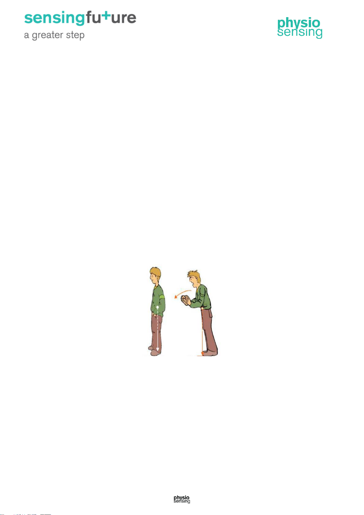
Balance is the ability to maintain the body’s center of mass over its base of support (Figure
1). A properly functioning balance system allows humans to see clearly while moving,
identify orientation with respect to gravity, determine direction and speed of movement, and
make automatic postural adjustments to maintain posture and stability in various conditions
and activities. Balance is achieved and maintained by a complex set of sensorimotor control
systems that include sensory input from vision (sight), proprioception (touch), and the
vestibular system (motion, equilibrium, spatial orientation); integration of that sensory input;
and motor output to the eye and body muscles. Injury, disease, certain drugs, or the aging
process can affect one or more of these components. In addition to the contribution of
sensory information, there may also be psychological factors that impair our sense of
balance.
Figure 1 – Example of the center of mass in balance and outside the base of support (unstable person will fall).
1.3. Sensory input
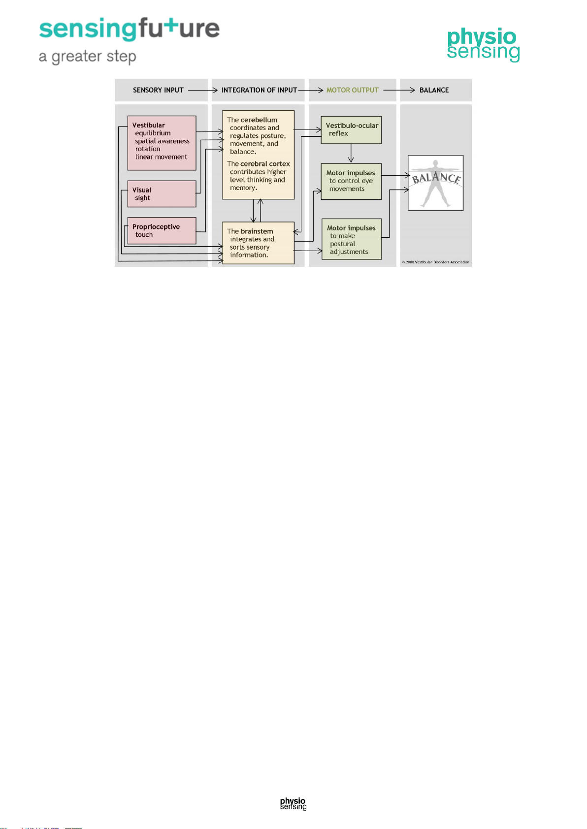
Maintaining balance depends on information received by the brain from three peripheral
sources: eyes, muscles and joints, and vestibular organs (Figure 2). All three of these
information sources send signals to the brain in the form of nerve impulses from special
nerve endings called sensory receptors.
Balance Outside base of support
Page 5
4 | Clinical Practice Manual
Figure 2 – The human balance system: balance is achieved and maintained by a complex set of
sensorimotor control systems.
1.4. Input from the eyes
Sensory receptors in the retina are called rods and cones. Rods are believed to be tuned
better for vision in low light situations (e.g. at night time). Cones help with color vision, and
the finer details of our world. When light strikes the rods and cones, they send impulses to
the brain that provide visual cues identifying how a person is oriented relative to other
objects. For example, as a pedestrian takes a walk along a city street, the surrounding
buildings appear vertically aligned, and each storefront passed first moves into and then
beyond the range of peripheral vision.
1.5. Input from the muscles and joints
Proprioceptive information from the skin, muscles, and joints involves sensory receptors that
are sensitive to stretch or pressure in the surrounding tissues. For example, increased
pressure is felt in the front part of the soles of the feet when a standing person leans
forward. With any movement of the legs, arms, and other body parts, sensory receptors
respond by sending impulses to the brain. Along with other information, these stretch and
pressure cues help our brain determine where our body is in space. The sensory impulses
originating in the neck and ankles are especially important. Proprioceptive cues from the
neck indicate the direction in which the head is turned. Cues from the ankles indicate the
body’s movement or sway relative to both the standing surface (floor or ground) and the
quality of that surface (for example, hard, soft, slippery, or uneven).
1.6. Input from the vestibular system
Sensory information about motion, equilibrium, and spatial orientation is provided by the
vestibular apparatus, which in each ear includes the utricle, saccule, and three semicircular
canals. The utricle and saccule detect gravity (information in a vertical orientation) and linear
movement. The semicircular canals, which detect rotational movement, are located at right
angles to each other and are filled with a fluid called endolymph. When the head rotates in
the direction sensed by a particular canal, the endolymphatic fluid within it lags behind
because of inertia, and exerts pressure against the canal’s sensory receptor. The receptor
Page 6
5 | Clinical Practice Manual
then sends impulses to the brain about movement from the specific canal that is stimulated.
When the vestibular organs on both sides of the head are functioning properly, they send
symmetrical impulses to the brain.
1.7. Integration of sensory input
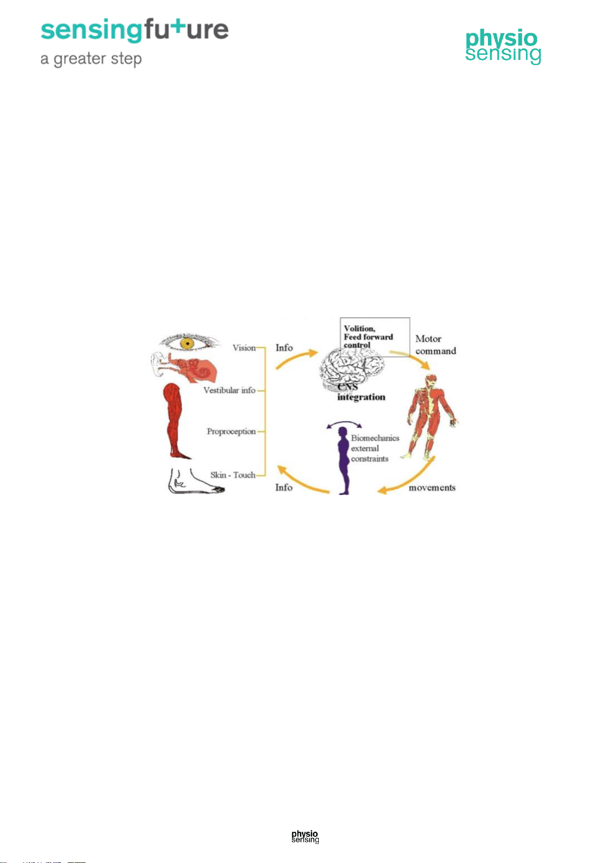
Balance information provided by the peripheral sensory organs – eyes, muscles and joints,
and the two sides of the vestibular system – is sent to the brain stem. There, it is sorted out
and integrated with learned information contributed by the cerebellum (the coordination
center of the brain) and the cerebral cortex (the thinking and memory center). The
cerebellum provides information about automatic movements that have been learned
through repeated exposure to certain motions. For example, by repeatedly practicing serving
a ball, a tennis player learns to optimize balance control during that movement. Contributions
from the cerebral cortex include previously learned information; for example, because icy
sidewalks are slippery, one is required to use a different pattern of movement in order to
safely navigate them, see Figure 3.
Figure 3 – Balance control system.
1.8. Processing of conflicting sensory input
A person can become disoriented if the sensory input received from his or her eyes, muscles
and joints, or vestibular organs sources conflicts with one another. For example, this may
occur when a person is standing next to a bus that is pulling away from the curb. The visual
image of the large rolling bus may create an illusion for the pedestrian that he or she – rather
than the bus is moving. However, at the same time the proprioceptive information from his
muscles and joints indicates that he is not actually moving. Sensory information provided by
the vestibular organs may help override this sensory conflict. In addition, higher level
thinking and memory might compel the person to glance away from the moving bus to look
down in order to seek visual confirmation that his body is not moving relative to the
pavement.
1.9. Motor output
As sensory integration takes place, the brain stem transmits impulses to the muscles that
control movements of the eyes, head and neck, trunk, and legs, thus allowing a person to
both maintain balance and have clear vision while moving.
Page 7
6 | Clinical Practice Manual
1.10. Motor output to the muscles and joints
A baby learns to balance through practice and repetition as impulses sent from the sensory
receptors to the brain stem and then out to the muscles form a new pathway. With repetition,
it becomes easier for these impulses to travel along that nerve pathway – a process called
facilitation – and the baby is able to maintain balance during any activity. Strong evidence
exists suggesting that such synaptic reorganization occurs throughout a person’s lifetime of
adjusting to changing motion environs.
This pathway facilitation is the reason dancers and athletes practice so arduously. Even very
complex movements become nearly automatic over a period of time. This also means that if
a problem with one sensory information input were to develop, the process of facilitation can
help the balance system reset and adapt to achieve a sense of balance again.
For example, when a person is turning cartwheels in a park, impulses transmitted from the
brain stem inform the cerebral cortex that this particular activity is appropriately
accompanied by the sight of the park whirling in circles. With more practice, the brain learns
to interpret a whirling visual field as normal during this type of body rotation. Alternatively,
dancers learn that in order to maintain balance while performing a series of pirouettes, they
must keep their eyes fixed on one spot in the distance as long as possible while rotating their
body.
1.11. Motor output to the eyes
The vestibular system sends motor control signals via the nervous system to the muscles of
the eyes with an automatic function called the vestibulo-ocular reflex (VOR). When the head
is not moving, the number of impulses from the vestibular organs on the right side is equal to
the number of impulses coming from the left side. When the head turns toward the right, the
number of impulses from the right ear increases and the number from the left ear decreases.
The difference in impulses sent from each side controls eye movements and stabilizes the
gaze during active head movements (e.g., while running or watching a hockey game) and
passive head movements (e.g., while sitting in a car that is accelerating or decelerating).
1.12. The coordinated balance system
The human balance system involves a complex set of sensorimotor-control systems. Its
interlacing feedback mechanisms can be disrupted by damage to one or more components
through injury, disease, or the aging process. Impaired balance can be accompanied by
other symptoms such as dizziness, vertigo, vision problems, nausea, fatigue, and
concentration difficulties.
The complexity of the human balance system creates challenges in diagnosing and treating
the underlying cause of imbalance. The crucial integration of information obtained through
the vestibular, visual, and proprioceptive systems means that disorders affecting an
individual system can markedly disrupt a person’s normal sense of balance. Vestibular
dysfunction as a cause of imbalance offers a particularly intricate challenge because of the
vestibular system’s interaction with cognitive functioning, and the degree of influence it has
on the control of eye movements and posture.
Page 8
7 | Clinical Practice Manual
2. Assessment protocols
2.1. modified Clinical Test of Sensory Interaction on Balance mCTSIB
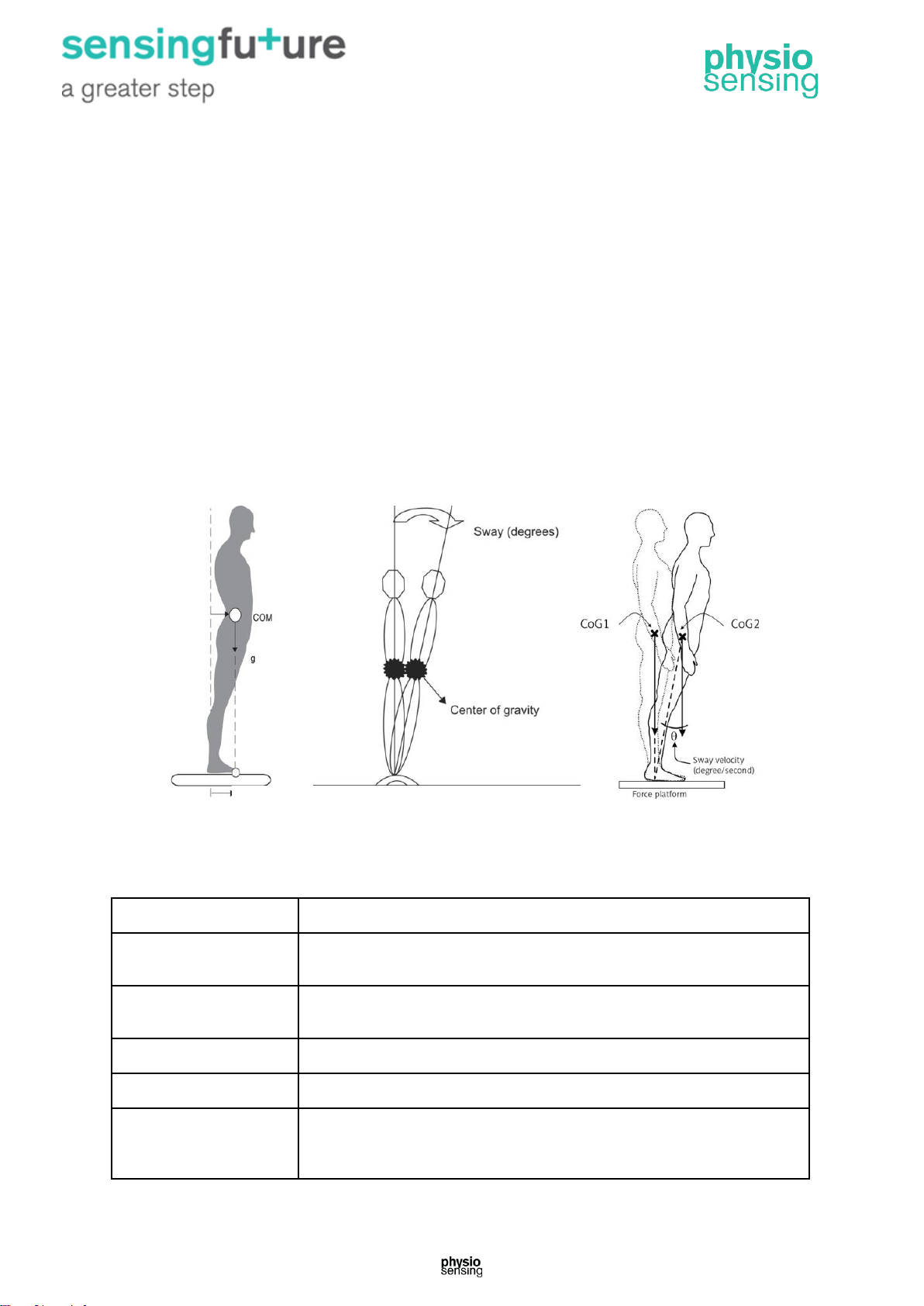
Use the mCTSIB to quantify postural sway velocity (Figure 4) during four sensory conditions:
1. Eyes open firm surface;
2. Eyes closed firm surface;
3. Eyes open unstable surface (foam);
4. Eyes closed unstable surface (foam).
Each condition has three trials of 10 seconds.
The mCTSIB is designed to help the clinician assess the need for further testing in patients
with complaints related to balance dysfunction, and to establish objective baselines for
treatment planning and outcome measurement. The mCTSIB cannot provide impairment
information specific to individual sensory, balance, or motor systems.
Figure 4 – Simplified illustrations of the center of pressure, sway angle and sway velocity (°/s).
At the end of performing all the conditions of the protocol it is possible to save the following
balance data and generate a clinical report (Figure 5).
Data
Description
Trial time
Elapsed time: 10 seconds. In the case of loss of balance, appears the
time until the fall has occurred
Sway velocity
Distance travelled by the center of pressure divided by the test time
(°/s). If a fall occurs the value is 6 °/s
Mean sway velocity
Mean sway velocity for each condition
Composite sway velocity
Mean sway velocity for all 12 trials
COP alignment
Patient’s initial COP position relative to the base of support center at the
beginning of each test, expressed as a percentage of the Limit of
Stability (LOS) and degrees
COP
Page 9
8 | Clinical Practice Manual
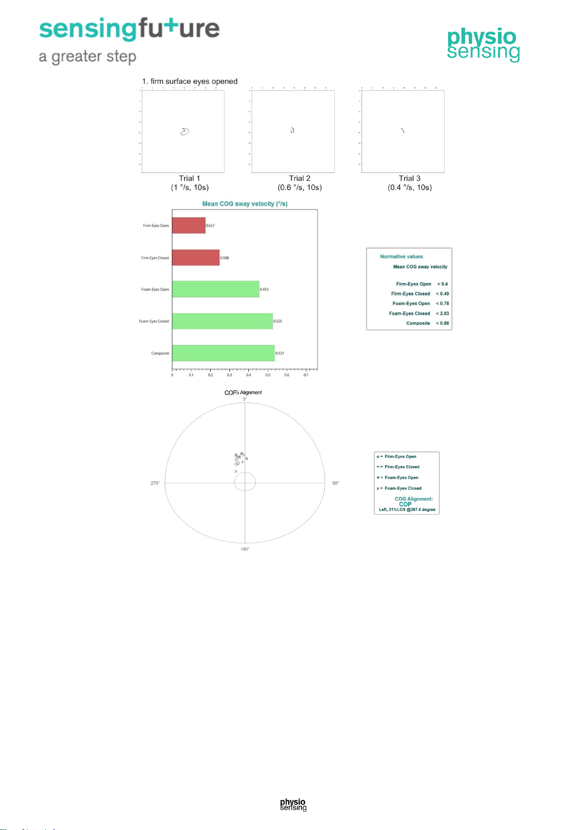
Figure 5 – Example of the results section of the mCTSIB clinical report.
Normal Response (indicated by green): The four conditions of the mCTSIB are designed to
simulate conditions frequently encountered in daily life activities. Normal individuals maintain
their COG near the center of the support base and minimize their sway regardless of the
sensory challenge or condition.
Abnormal Response (indicated by red): For most patients with disequilibrium (with or without
established etiology), the mCTSIB will document the presence of a balance problem and
provide the information required to support further assessments. It also provides focus for
the rehabilitation plan.
Clinical Significance: The mCTSIB can also be used, to a limited extent, to document
progress in a rehabilitation program. Although the results of the mCTSIB can be used to
distinguish normal balance performance versus abnormal balance performance, it cannot be
used to discern the specific patterns of dysfunction. The combination of the mCTSIB and
Page 10
9 | Clinical Practice Manual
LOS forms an objective screening tool for balance problems that can differentiate those
patients who will benefit from a course of rehabilitation from those who require further
diagnostic testing and more advanced balance rehabilitation.
2.2. Limits of Stability - LOS
The LOS protocol quantifies impairments in ability to intentionally displace the COG to the
patient’s stability limits without losing balance. The patient performs the task while viewing a
real-time display of their COP position in relation to targets placed at the center of the base
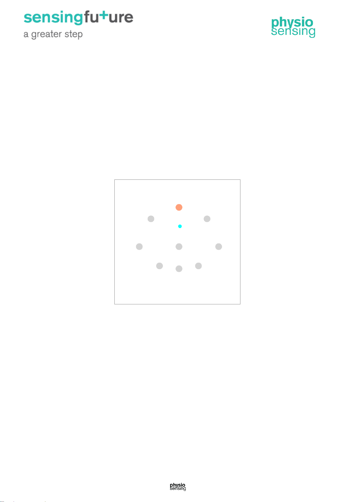
of support and at the stability limits (Figure 6). For each of eight directions, the test
measures movement reaction time, movement velocity, movement distance, and movement
directional control. For each of eight trials, the patient, on command, moves the COP cursor
as quickly and accurately as possible towards a second target located on the LOS perimeter,
set at 100% of the theoretical limits of stability, and then holds a position as close to the
target as possible. The patient is allowed up to 8 seconds to complete each trial.
Figure 6 – Interface of LOS protocol.
This protocol is appropriate for:
• Older adults with mobility impairments;
• Athletes interested in rapid ‘off the block’ reactions and direction changes;
• Patients with peripheral neuropathic disorders: diabetes, polyneuropathy;
• Patients with central nervous system pathologies:
o Degenerative diseases: Parkinson’s disease, multiple sclerosis;
o Acquired paralysis and paresis: stroke, brain injury.
The 8 directions of the protocol are: front, front/right, right, back/right, back, back/left, left and
front/left. The distances of the limits of stability are calculated according to the height of the
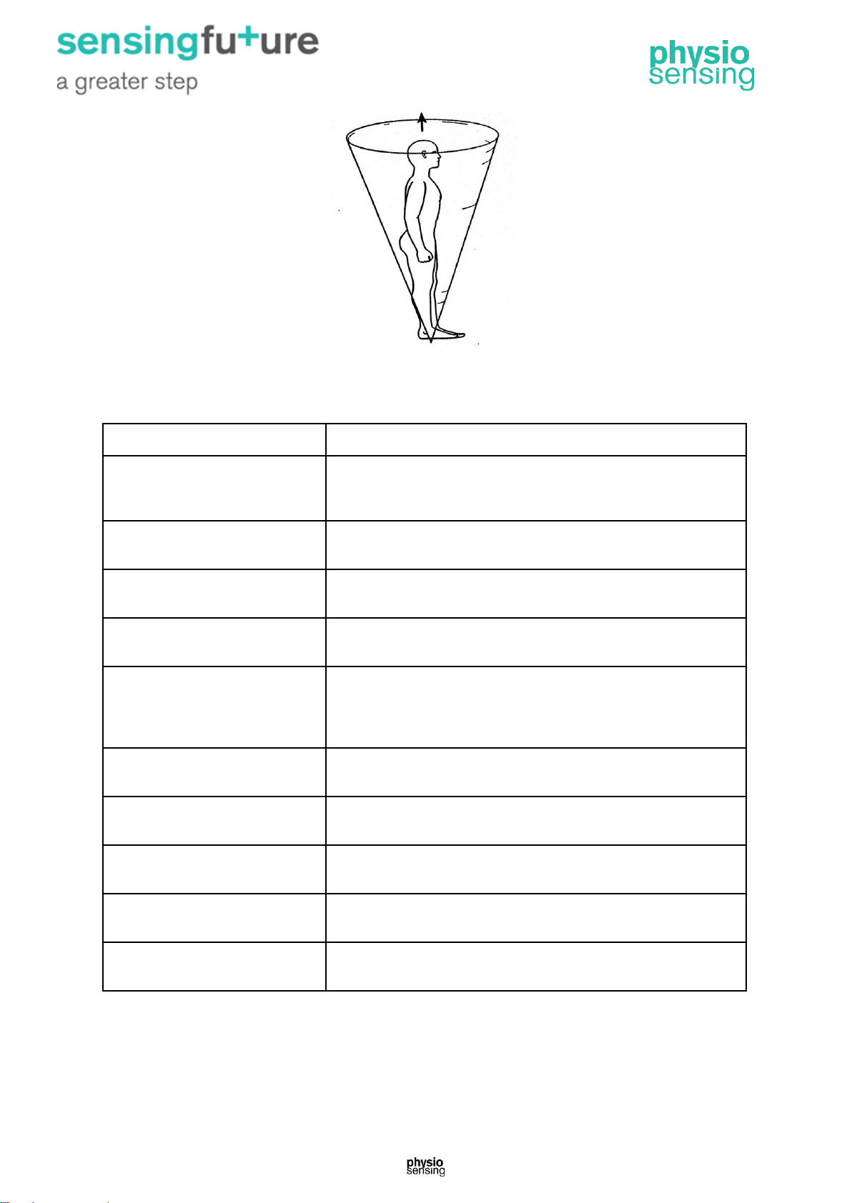
patient's center of gravity. These distances are calculated for 100% LOS, i.e. 8° left, right
and front, and 4.5° back (see Figure 7).
Page 11
10 | Clinical Practice Manual
Figure 7 – Theoretical boundaries of the limits of stability.
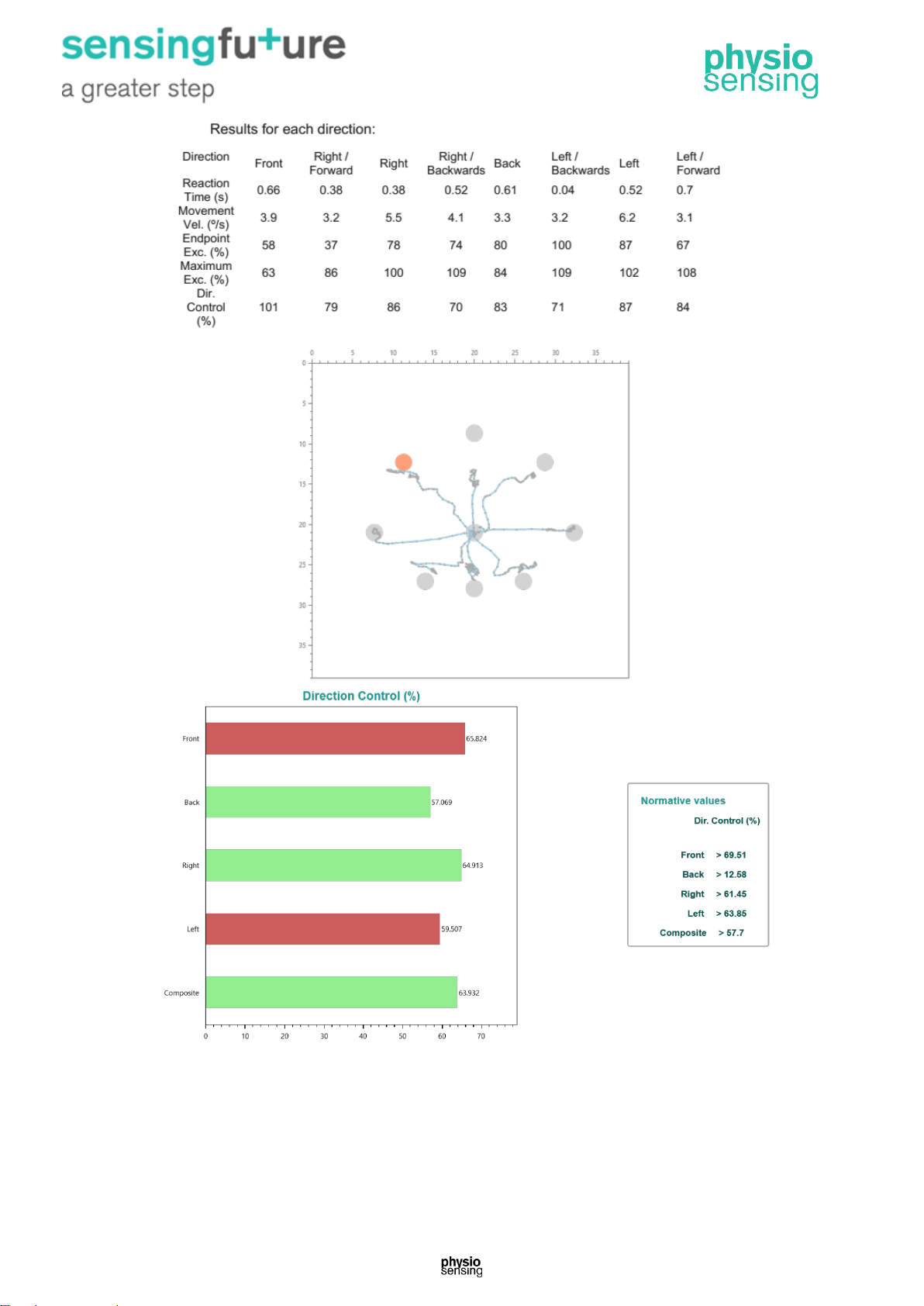
At the end of the protocol appears the trace of the pressure center and the following data
can be saved and exported as a clinical report (Figure 8):
Data
Description
Reaction time
Time between the indication to move (2 seconds after clicking
the Start button Time the color of the point changes) and the
first movement of the patient, in seconds
Mean reaction time per cartesian
axis
Mean reaction time per cartesian axis (front, right, back and left)
Movement velocity
Distance traveled by the center of pressure, between 5% and
95% of the first attempt, divided by 8 seconds (°/s).
Mean movement velocity per
cartesian axis
Mean movement velocity per cartesian axis
Endpoint excursion
Distance from the first attempt to reach the orange dot,
expressed as a percentage of LOS. The end of the first attempt
is considered the point at which the initial movement towards
the goal ceases
Endpoint excursion per cartesian
axis
Mean endpoint excursion per cartesian axis
Maximum excursion
Maximum distance reached during 8 seconds, expressed as a
percentage of LOS
Maximum excursion per
cartesian axis
Mean maximum excursion per cartesian axis
Directional control
Percentage of movement in the intended direction minus offaxis movement during the first attempt
Directional control per cartesian
axis
Mean directional control per cartesian axis
Page 12
11 | Clinical Practice Manual
Figure 8 – Example of the results section for the LOS clinical report.
Normal Response (indicated by green): Performance within age-matched normative range is
indicative of good voluntary motor control and adequate lower extremity proprioception,
range of motion, and strength for mobility.
Page 13
12 | Clinical Practice Manual
Abnormal Response (indicated by red): Abnormal performance is representative of a
voluntary motor impairment. This can be a result of factors including:
• Biomechanics (range, strength);
• Sensory awareness;
• Central movement disorders;
• Perception (fear).
Clinical Significance: Provides information regarding voluntary motor control for safe and
independent function in all activities of daily living (ADLs).
Ability to voluntarily move the COG to positions within the LOS is fundamental to mobility
tasks such as reaching for objects, transitioning from a seated to standing position (or
standing to seated), and walking.
Reaction time delays are commonly associated with difficulties in cognitive processing
and/or motor diseases. Reduced movement velocities are indicative of high level central
nervous system deficits such as Parkinson’s disease and age-related disorders. Inability to
reach targets in single movements, such as reduced endpoint excursions or excessively
larger maximum excursions and poor directional control are indicators of motor control
abnormalities. Excursions may be restricted by biomechanical limitations. Dizzy and/or
unsteady patients and those fearful of falling may artificially restrict their excursions, while
the strength of those with lower extremity weakness may be insufficient to attain and/or
maintain stable target positions.
Limitations in the LOS may correlate to risk for fall or instability during weight shifting
activities such as leaning forward to take objects from a shelf, leaning back for hair washing
in the shower, or opening the refrigerator door. Patients with reduced stability limits in the AP
direction tend to take smaller steps during gait, while laterally reduced limits can lead to
broad-based gaits.
2.3. Rhythmic Weight Shift - RWS
Use the Rhythmic Weight Shift protocol to evaluate the transfer capacity of the center of
pressure rhythmically in the sagittal and anteroposterior plane, at three different velocities:
slow (3s), moderate (2s) and fast (1s). This protocol measures the velocity in the axis and
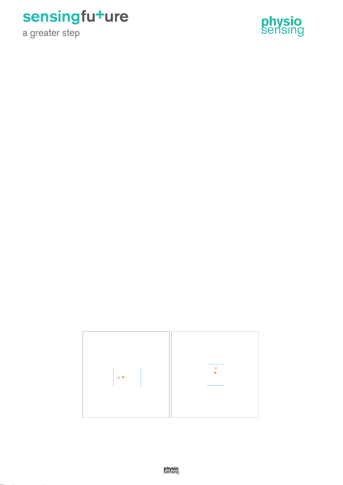
the movement control between two targets at 50% of patient’s LOS (Figure 9).
Figure 9 - Interface of RWS protocol for the two planes: sagittal and anteroposterior.
Page 14
13 | Clinical Practice Manual
During performance of each task, the patient views a real time display of their COP position
relative to a target moving (gray ball) at the desired pace and amplitude. The patient has to
reach the gray ball, which is in motion, and control the load transfer at same pace as the
gray point. The patient must change direction when reaching the blue line, at the same time
of the beep signal. For each direction and pace, the RWS measures movement velocity and
directional control.
Appropriate for patients with:
• Central Nervous System Pathologies:
o Degenerative diseases: Parkinson’s disease, multiple sclerosis;
o Acquired paralysis and paresis: stroke, brain injury;
• Peripheral Neuropathic Disorders:
o Diabetes, polyneuropathy;
• Inability to perform the LOS even after instruction and attempt. The RWS then
becomes the voluntary movement test of choice.
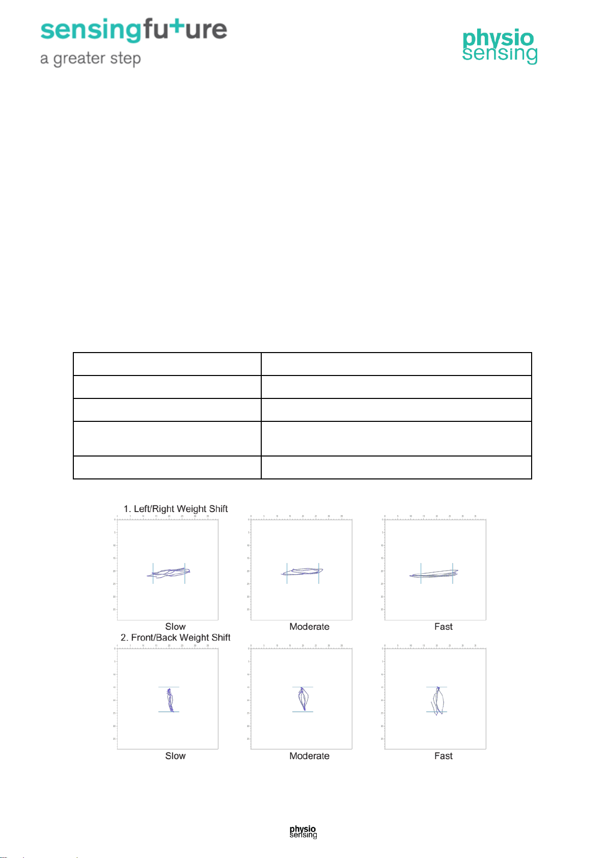
At the end of the protocol the pressure center trace for each of the tests, in the sagittal and
anteroposterior directions, at the three speeds mentioned appears (Figure 10). These
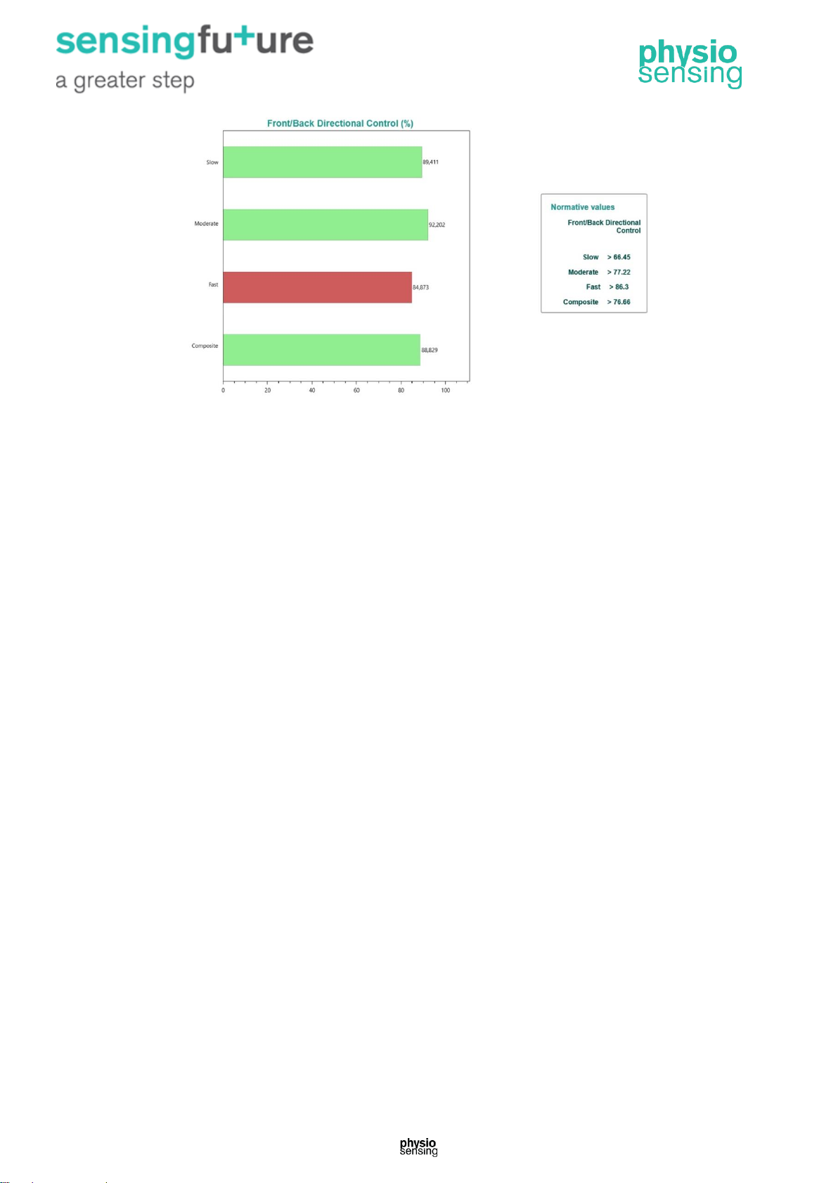
images also appear in the clinical report, such as the following data (Figure 11):
Data
Description
On-axis velocity
COP movement velocity along the specified direction (°/s)
Composite on-axis velocity
Mean velocity at the three speeds
Directional control
Percentage of movement in the desired direction minus
off-axis movement
Composite directional control
Mean directional control at the three speeds
The normative values for each variable are presented next to the results.
Figure 10 – Example of the trace of the COP for the two directions at the three speeds tested.
Page 15
14 | Clinical Practice Manual
Figure 11 – Example of the results section for the RWS clinical report.
Normal Response (indicated by green): Subjects attain the required average velocities set
by the pacing target and cover the full distance between the specified movement boundaries
(50% LOS) with good directional control.
Abnormal Response (indicated by red): Patients with motor disorders disrupting normal
timing of movement control may exhibit slower than normal movement velocities, poor
directional control, or a combination of these two problems.
Clinical Significance: Provides information regarding a patient's effective timing of motor
control for basic safety in performance of activities of daily living (ADLs) and gait weight
transfer.
Normal individuals attain the required average velocities by maintaining the rhythm set by
the pacing target and by covering the full distance between the specified movement
boundaries. At the same time, their movements are straight and well coordinated, with
motions in the off-axis direction being a small percentage of the on-axis motion. Functional
consequences include an inability to meet the timing demands of the environment, such as
crossing the street and stepping onto elevators/escalators. Instability may result when
performing activities that require rapid movement speeds, variability in speeds, or changing
directions. Rhythmic, reciprocal movement patterns are required in many high level athletic
and leisure interests.
2.4. Weight Bearing/Squat - WBS
The WBS protocol quantifies the patient’s ability to perform squats with the knees flexed at
30°, 60°, and 90°, while maintaining equal weight on the two legs (Figure 12). In the erect
position, most body weight is borne through the skeletal system, and relatively less stress is
placed on the knee and hip joints. Increasing depths of squat place greater stress on the
knees and hips, making these positions more sensitive in detecting weight-bearing
abnormalities related to lower extremity musculoskeletal injuries.
Page 16
15 | Clinical Practice Manual
Figure 12 - Interface of WBS protocol with the various angles of knee flexion: 0º, 30º, 60º and 90º.
This protocol allows observation of weight distribution in the sagittal plane with the patient
standing up with different knee flexion angles.
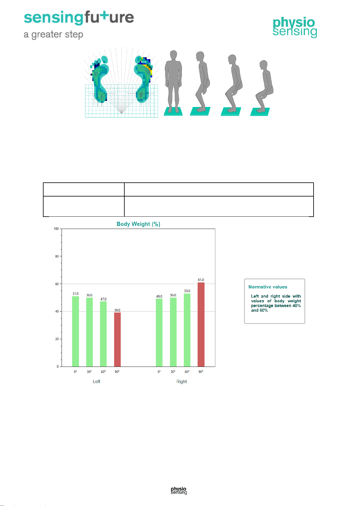
At the end, a graph with the percentage of body weight of the patient on each side (left and
right) with knees straight (at 0°) and with knees bent at 30°, 60° and 90° appears. It is also
possible to save this data and generate a clinical report (Figure 13).
Data
Description
Percentage of body weight
Percentage of weight on each side of the sagittal plane, for each
angle of knee flexion
Figure 13 – Example of the results section for the WBS clinical report.
Normal Response (indicated by green): Normal individuals maintain body weight within ±7%
of equal on the two legs over the full range of squatting positions.
Abnormal Response (indicated by red): Reduced weight bearing on one leg may reflect
sensory (proprioceptive) or strength loss, reduced range of motion, and/or pain. Bending,
stooping and squatting positions substantially increase stress on the ankles and knees, and
may identify weight-bearing differences not detectable in a less challenging (fully erect)
position.
Page 17
16 | Clinical Practice Manual
Clinical Significance: Provides information regarding the symmetry of the patient's voluntary
motor control for basic safety in performance of activities of daily living (ADLs), sports or
leisure activities.
Patients with lower extremity orthopedic injuries may exhibit equal weight bearing in the
erect position, but will bear a preponderance of weight on the uninvolved side during more
stressful squatting positions. Patients with generalized or unilateral weaknesses will
demonstrate impaired motor control for sit-to-stand transitions or an inability to safely
retrieve objects from the floor. In the athletic population, impairments may result in reduced
readiness to move side-to-side or accuracy of weight shift or thrust during squat-to-extend
movements.
2.5. Unilateral Stance - US
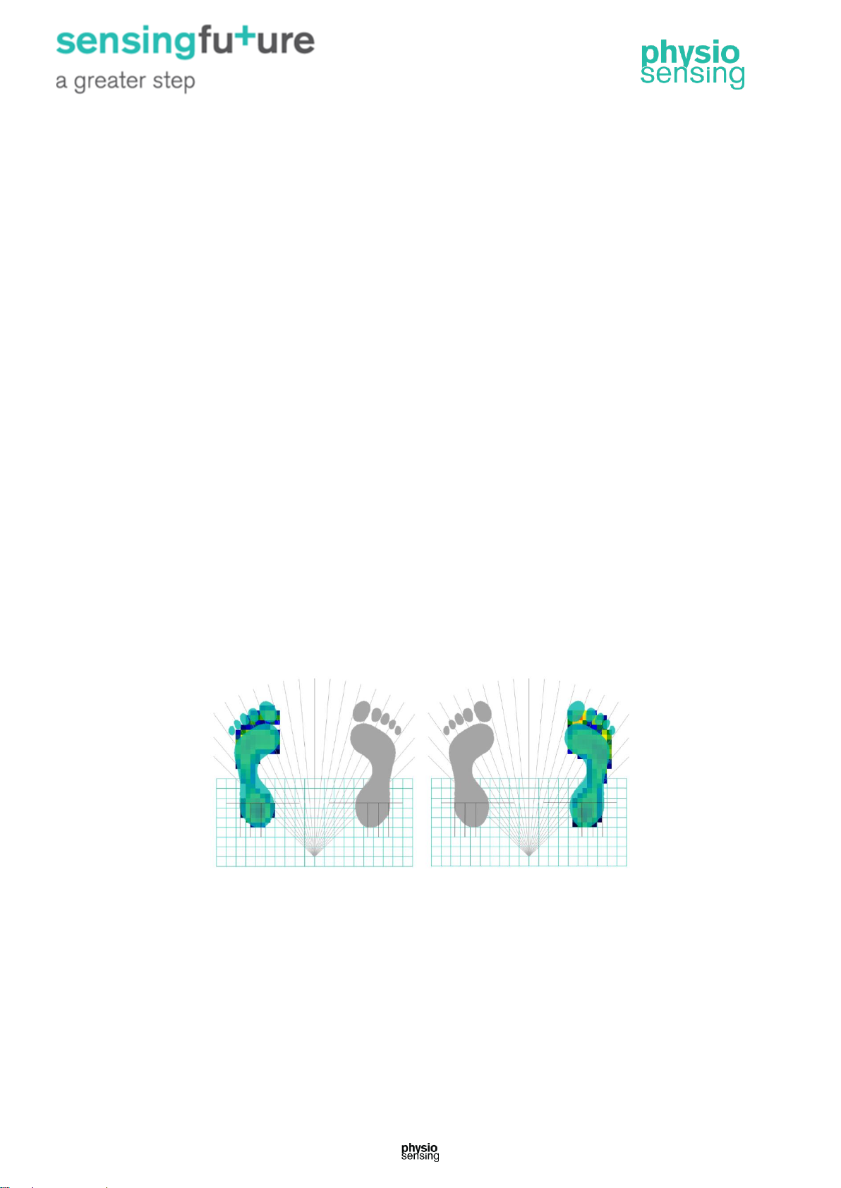
The US assessment quantifies the patient’s ability to maintain postural stability while
standing on one leg at a time with the eyes open and closed (Figure 14). The US enhances
the observational testing of single leg stance performance by providing an objective measure
of patient sway velocity for each of the four task conditions. The length of each trial is ten
seconds.
The US is highly sensitive, but not specific because a large number of independent factors
can impact performance. A partial list of these factors includes lower extremity strength and
weight bearing control, sensory balance control, movement strategies, and prior practice
with the task.
Use this protocol to measure the balance in four conditions: left foot lifted up with eyes open,
left foot lifted up with eyes closed, right foot lifted up with eyes open and right foot lifted up
with eyes closed. This protocol also allows to evaluate the difference of oscillation (in
percentage) between the left and right side with the eyes opened and closed.
Figure 14 - Interface of US protocol, for left and right foot, respectively.
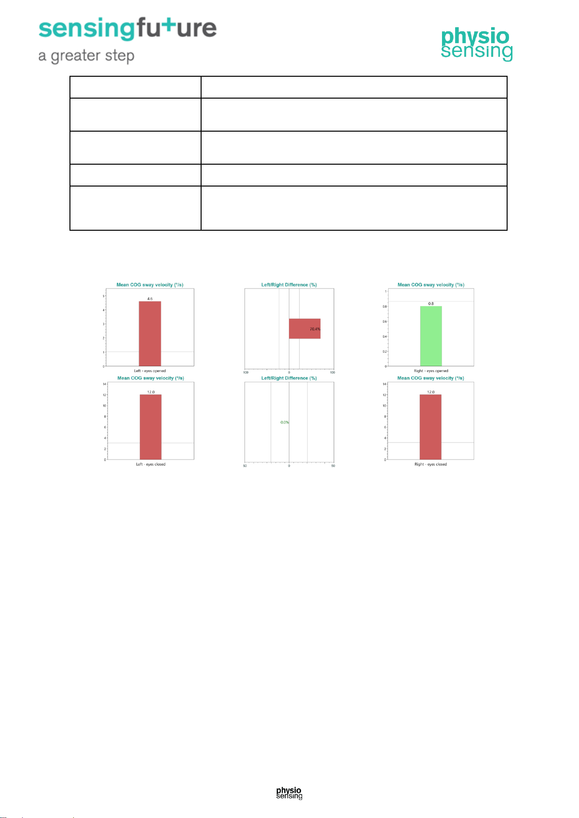
At the end, the following results are presented for each of the indicated conditions, in the
form of graphs (Figure 15).
Page 18
17 | Clinical Practice Manual
Data
Description
Trial time
Elapsed time: 10 seconds. In the case of loss of equilibrium, appears
the time until the fall has occurred
Sway velocity
Distance traveled by the center of pressure divided by the test time
(°/s). If a fall occurs the value is 6 °/s
Mean sway velocity
Mean sway velocity for each condition
Sway velocity difference
Sway velocity difference (in percentage) between left and right side
with eyes open and closed. The bar points to the side with better
performance
In the clinical report can be observed the pressure center trace for each test, as well as the
graphs with the aforementioned data. The normative values for each variable are presented
below the results.
Figure 15 – Example of the results for the US protocol.
Normal individuals have significantly more sway standing on one foot versus two, and even
more sway on one foot with eyes closed. Patients who become unstable may have difficulty
using visual or somatosensory information for balance control, and/or may have
musculoskeletal problems that make it difficult to compensate. Functional consequences are
significant for performance of activities that require single-leg balance, such as donning
pants or hosiery, ascending or descending elevations, or navigating narrow support
surfaces, such as ladders and scaffolding.
2.6. Fall Risk - FR
Falls are a common occurrence among older people, even for those in good health and with
no apparent balance problems. Fall Risk test allows identification of potential fall candidates.
Test results are compared to age-dependent normative data. Scores higher than normative
values suggest further assessment for lower extremity strength, proprioception, and
vestibular or visual deficiencies. The quantification of the patient’s postural sway velocity can
be used to predict risk. Velocity can be described as the speed of an individual's sway as
balance is maintained. Higher velocities, when cues are given to specifically stand as
motionless as possible, are suggestive of postural control deficits. The Fall Risk test protocol
Page 19
18 | Clinical Practice Manual
(Figure 16) is based on research from the University of Dayton (Bigelow KE and Berme N.
2011. ‘Development of a protocol for improving the clinical utility of posturography as a fallrisk screening tool’ J Gerontol A Biol Sci Med Sci 66A: 228–233) and the University of
Jyväskylä in Finland (Pajala, S. et al. 2008. ‘Force platform balance measures as predictors
of indoor and outdoor falls in community-dwelling women aged 63-76 years’ J Gerontol A
Biol Sci Med Sci 63A:171–178).
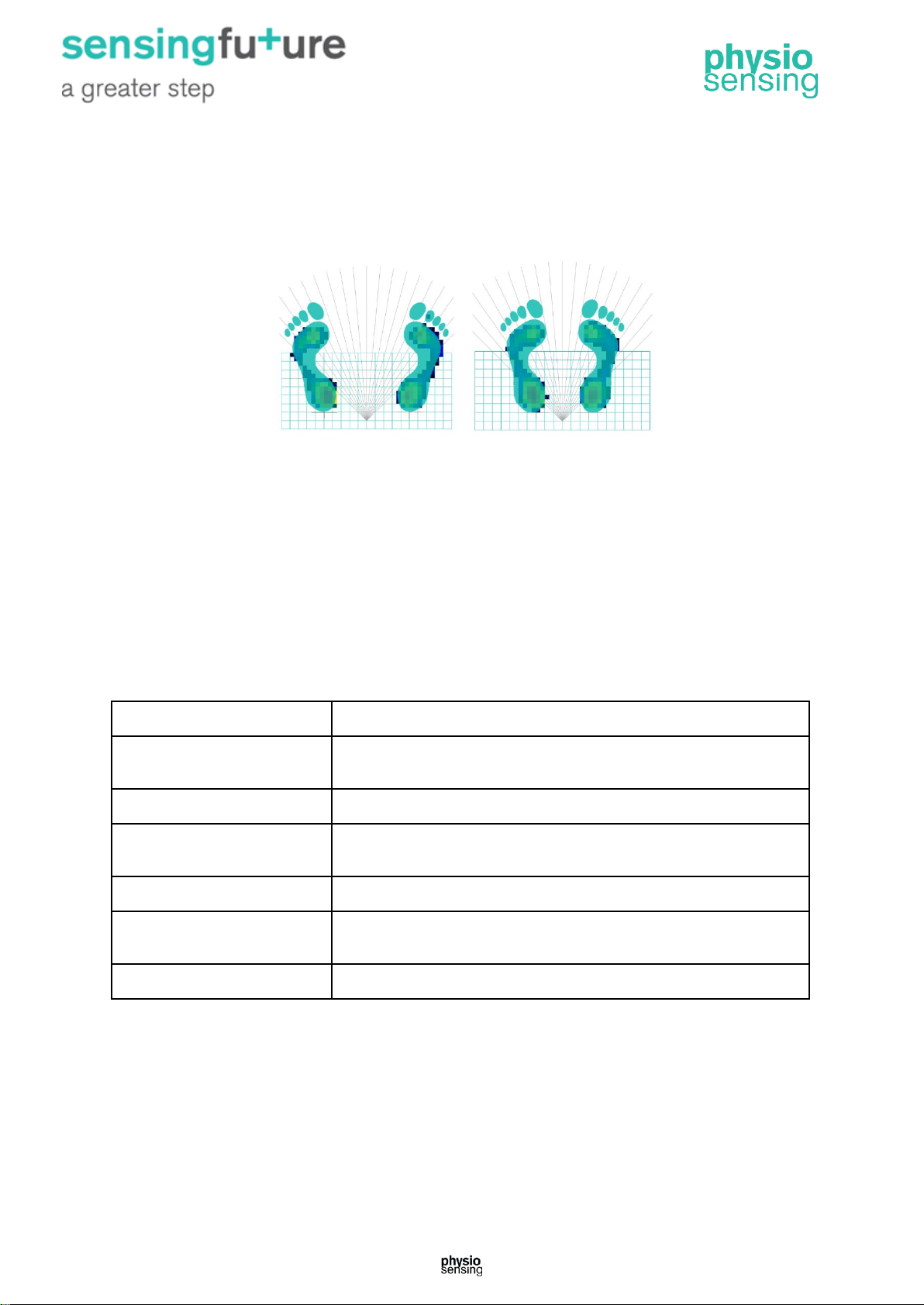
Figure 16 - Interface of Fall Risk protocol, for comfortable and narrow stance.
Use this protocol to measures the static balance in four conditions:
1. Comfortable stance with eyes open;
2. Comfortable stance with eyes closed;
3. Narrow stance with eyes open;
4. Narrow stance with eyes closed.
After performing all the conditions of the protocol, the value of the sway velocity index for
each of the conditions appears. This sequence of tests can provide a fall risk prediction.
At the end of the protocol, it is possible to save the following data and generate a clinical
report.
Data
Description
Velocity
Distance traveled in the sagittal plane divided by the test time, 45
seconds (mm/s)
Composite velocity
Mean velocity for all conditions
Sway velocity index (SVI)
Value based on the velocity and height of the patient, normalized
by the natural logarithm function
Composite SVI
Mean SVI for all conditions
Z-Score
Number of standard deviations of the SVI from the mean value
indicated in the normative values
Composite Z-Score
Mean Z-Score for all conditions
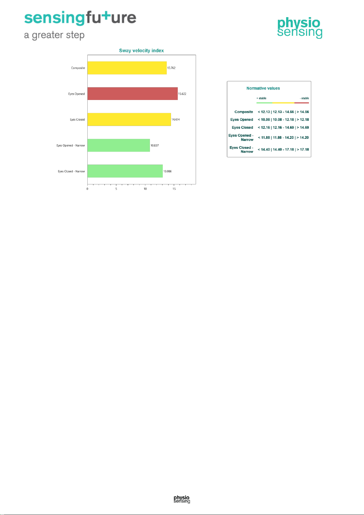
In the clinical report (Figure 17) can be observed the pressure center trace for each
condition. The normative values for each condition are presented next to the results. The
color of the bar will be green, yellow or red depending on the result, higher values will be red
(meaning less stable) and lower values than normative values will be green.
Page 20
19 | Clinical Practice Manual
Figure 17 – Example of the results section for the FR clinical report.
Page 21
20 | Clinical Practice Manual
3. Balance assessment and training applications
PhysioSensing is indicated to assist physiotherapy activities, physical and vestibular
rehabilitation and neurorehabilitation, especially in the early rehabilitation of stroke and
neuromusculoskeletal conditions associated to lower limbs and gait rehabilitation. More
concrete applications provided by medical devices with visual evaluation biofeedback and
balance training are documented by the scientific community:
Neurological
• Stroke or Cerebrovascular Accident (CVA) Patients
• Parkinson Disease
• Amyotrophic Lateral Sclerosis (ALS/ Lou Gehrig’s Disease)
• Multiple Sclerosis
• Spinal Cord or Head Trauma
- Any head or spinal cord injury resulting in loss of balance or ambulatory motor skills.
• Rehabilitation with these patients involves:
- Restoration of effected motor skills by retraining new neural pathways.
- Restoration of kinesthetic sense for proper body positioning to maintain balance and
weight transfer for ambulation.
• PhysioSensing is effective because:
- The instant biofeedback makes it easy for the patient to relate to and repeat the motions.
- Safe controlled environment that allows the patient to progress from non weight bearing to
weight bearing.
- Objectives and reproducible so that progress can be monitored as well as rehab sessions
documented.
Orthopaedics
Lower Extremity Patients:
- ACL’s
- Total Knee
- Total Hip
- Ankle Sprain
- Fractures
- Amputees - weight bearing and balance with the prosthesis
- High Tibial Osteotomies
- Risk for Fall
Spine Patients:
- Ankle, knee and hip movement strategies for improving posture and mechanics.
- Core Stabilization using a gym ball on PhysioSensing.
Upper Extremity Patients:
- Scapular stabilization drills, where the patient uses his arms to load on platform
Geriatric Patients (both neurological and orthopedic):
- Balance and weight shift training as part of rehabilitation from a total hip or knee
replacement, or a stroke.
- Kinesthetic Balance Training.
- Weight bearing.
Sports
Medicine
These patients can work the way either non weight bearing, or full weight bearing. They can
work on weight shifting, proprioceptive and stabilization drills with or without perturbation (with
or without foam). Also, some clinicians are using this type of device for high level agilities.
Vestibular
Disorders
Patients with balance disorder can feel unsteady or dizzy. Either if they are standing, sitting,
or lying down, they might feel as if they are moving, spinning, or floating. When walking, they
might suddenly feel as if they are tipping over.
Everyone has a dizzy spell now and then, but the term “dizziness” can mean different things
to different people. For one person, dizziness might mean a fleeting feeling of faintness, while
for another it could be an intense sensation of spinning (vertigo) that lasts a long time.
The symptoms of a balance disorder might include:
• Dizziness or vertigo (a spinning sensation).
• Falling or feeling as if they are going to fall.
• Staggering when they try to walk.
• Lightheadedness, faintness, or a floating sensation.
• Blurred vision.
• Confusion or disorientation.
Other symptoms might include nausea and vomiting; diarrhea; changes in heart rate and
blood pressure; and fear, anxiety, or panic. Symptoms may come and go over short time
periods or last for a long time and can lead to fatigue and depression.
PhysioSensing can be used to evaluate those conditions.
Page 22
21 | Clinical Manual Practice | March 2018
4. Scientific Evidence
Topic
Subjects
Title
Authors
Cita-
tions
Publication
Key-evidences
Modified
Clinical Test
of Sensory
Interaction on
Balance
Multiple
Sclerosis
Use of Awareness
Through Movement
Improves Balance and
Balance Confidence in
People with Multiple
Sclerosis: A
Randomized Controlled
Study
Stephens, James;
DuShuttle,
Dominique;
Hatcher, Carla;
Shmunes, Jenifer;
Slaninka,
Christine
55
Journal of
neurologic
physical
therapy 2001
25(2)
The ATM (Awareness Through Movement) group exhibited
significantly improved mCTSIB scores indicating an average center
of pressure position closer to theoretical center, had significantly
fewer abnormal mCTSIB tests, and demonstrated improved balance
confidence compared to controls. There was a trend toward
improvement in all other measures in the ATM group compared to
controls.
Benign
paroxysmal
positional
vertigo
Modified clinical test of
sensory interaction on
balance test use for
assessing effectiveness
of Epley maneuver in
benign paroxysmal
positional vertigo
patients' rehabilitation
Yones Lotfi ,
Mohanna
Javanbakht ,
Maryam Sayaf ,
Enayatollah
Bakhshi
0
Aud Vest Res
(2018);27(1):1
2-18.
Meanwhile, it seems that in addition to considering the time criterion
of maintaining the balance in the mCTSIB test, the use of the
amount of sway, especially in patients with BPPV, also provides
more complete information for the specialists in the field of balance
in order to estimate the individual balance problems and the
effectiveness of therapeutic maneuvering. mCTSIB is seems be a
valid test
with simple and quick apply for verifying the
effectiveness of Epley maneuver in BPPV patients.
Attention
Deficit
Hyperactivity
Disorder
Children
Objective Measurement
of the Balance
Dysfunction in Attention
Deficit Hyperactivity
Disorder Children
Yufeng Zang,
Bomei Gu, Qiujin
Qian, Yufeng
Wang
46
Chinese
Journal of
Clinical
Rehabilitation,
May 2002, Vol.
6, No. 9
We found that the sway velocity of center of gravity on conditions,
firm surface with eyes closed, foam pad with eyes open and foam
pad with eyes closed, was significantly higher (P < 0.03) in ADHD
group than in controls. We concluded that the sensory inputs, the
sensory integration, and/or the inhibition of excessive movement are
impaired in ADHD children, which result in the balance dysfunction.
mCTSIB can provide more accurate evaluation on rehabilitation
therapy. Combining CSIS and other methods, e.g., neuroimaging,
could further uncover the mechanisms of inhibition disability of this
group.
Page 23
22 | Clinical Manual Practice | March 2018
Limits of
Stability
Elderly at
different risk of
fall
Effect of a group-based
exercise program on
balance in elderly
Tatjana
Bulat, Stephanie
HartHughes, Shahbaz
Ahmed, Pat
Quigley, Polly
Palacios, Dennis
C Werner,
and Philip Foulis
72
Clin Interv
Aging. 2007
Dec; 2(4):
655–660
We found statistically significant improvement in balance outcomes
as a result of an 8 week group functional balance training class.
LOS composite Reaction Time (RT) and composite movement
velocity (MVL) showed trends toward improvement while LOS
composite end point excursion (EPE), maximum excursion (MXE)
and directional control (DCL) all were significantly improved when
compared to baseline. An eight week group functional balance
training class was safe and effective in improving balance outcomes
in a cohort of elderly veterans at risk for falls.
Multiple
Sclerosis
The Role of Clinical
and Instrumented
Outcome Measures in
Balance Control of
Individuals with Multiple
Sclerosis
Neeta Kanekar
and Alexander S.
Aruin
19
Multiple
Sclerosis
International 2
013;2013:1901
62
Both, the distance of the frst movement towards the target (EPE)
and the maximum distance reached (MXE) by the MS group, were
signifcantly smaller than the distance reached by the HC group.
Posthoc test demonstrated that
for each direction, the MS group had smaller MXE than the HC
group. Specifcally, the MXE for the MS group towards the right side
was smaller as compared to the MXE for the HC group towards the
right side (P = 0.037) and towards the lef side (P = 0.038).
Moreover, irrespective of the direction, the MS group had smaller
MXE than the HC group (P = 0.012). Implementation of both clinical
and instrumented tests of balance is important for the planning and
evaluation of treatment outcomes in balance rehabilitation of people
with MS.
Rhythmic
Weight Shift
Vestibular
Patients
Posturografía estática
con pruebas dinámicas.
Utilidad de los
parámetros
biomecánicos
en la valoración del
paciente vestibular
(Static Posturography
With Dynamic Tests.
Usefulness of
Biomechanical
Parameters in
Assessing Vestibular
Patients)
Ramón Balaguer
García, Salvador
Pitarch Corresa,
José María
Baydal Bertomeu,
María M. Morales
Suárez-Varela
10
Acta
Otorrinolaringo
l Esp.
2012;63:332-
338
In some patients, posturography can detect the negative influence
exerted by a vestibular alteration on postural stability. It does not
replace the classic tests which evaluate the vestibulo-ocular reflex,
but instead provides complementary information, enabling a better
understanding of the functional status of patients with instability.
With the exception of the displacement angle, all biomechanical
parameters of static posturography were able to discriminate
between normal and pathological subjects in our sample. The RWS
test proved useful to discriminate between normal and pathological
subjects in our population. Therefore, vestibular disorders may be
able to significantly alter the voluntary control of rhythmic movement
whilst standing.
Page 24
23 | Clinical Manual Practice | March 2018
Hemiplegic
stroke patients
Effects of balance
training on hemiplegic
stroke patients
Chen IC, Cheng
PT, Chen
CL, Chen
SC, Chung
CY, Yeh TH
111
Chang Gung
Med J. 2002
Sep;25(9):583-
90
For dynamic function training, the patients were instructed to
practice controlling their weight shifts by tracing the moving targets
on the screen in every main
direction while the condition of LOS set at 50%. We found dynamic
balance function showed significant improvements in patient with
visual feedback training when compared with those receiving
conventional therapy only. Patients in the trained group also showed
significant improvements in the self-care ability at 6 months of follow
up.
Weight
Bearing
Squat
Individuals
with Mental
Retardation
Effects of Balance
Training on Individuals
with Mental Retardation
Karen M. Smail
and Michael
Horvat
9
Clinical
Kinesiology:
Journal of the
American
Kinesiotherapy
Association
2005
It is interesting to note that after training, symmetry across left and
right sides for weight distribution (51.9% right and 48.1% left) was
apparent. This demonstrates that individuals with mental retardation
will respond to balance training interventions and supports the
development of specific programs that foster balance related skills.
This study suggests that symmetrical weight distribution can be
corrected with proper training thus resulting in more efficient
movement patterns
Stroke
Impact of Yoga on
Postural Stability in
Stroke
Peter Altenburger
A , Arlene Schmid
A , Marieke Van
Puymbroeck and
Kristine Miller K
3
Int J
Neurorehabilit
ation 2016, 3:1
Postural orientation was measured using the weight bearing squat
assessment. Prior to participation in yoga, significant weight
distribution asymmetry was observed during full upright standing.
Subjects demonstrated greater than 50% improvement in symmetry
alignment for different standing positions.
Unilateral
Stance
Peripheral
Neuropathy
Unipedal Stance
Testing in the
Assessment of
Peripheral Neuropathy
Edward A.
Hurvitz, James K.
Richardson,
Robert A. Werner
45
Arch Phys
Med
Rehabil. 2001
Feb;82(2):198-
204.
Abnormal unipedal stance time (< 45s) identified peripheral
neuropathy with a sensitivity of 83% and a specificity of 71%,
whereas a normal unipedal stance time had a negative predictive
value of 90%. Abnormal unipedal stance time was associated with
an increased risk of having peripheral neuropathy on univariate
analysis. Our results suggest that unipedal stance testing is useful in
detecting peripheral neuropathy.
Lumbar
discectomy
patients with
and without
pain
Comparison of postural
control in unilateral
stance between healthy
controls and lumbar
discectomy patients
with and without pain
Katie Bouche
Veerle Stevens
Dirk Cambier
Jacques
Caemaert Lieven
Danneels
36
Eur Spine J.
2006 Apr;
15(4): 423–
432.
In lumbar discectomy patients, postural sway in unilateral stance is
influenced by age, the presence of pain and the side of former disc
herniation. Long-term following lumbar discectomy, there is no
complete recovery of postural control. In the eyes open condition,
balance of patients with pain was significantly worse than in controls
(P=0.003). In the eyes closed condition, the sway in both groups of
lumbar discectomy patients was significantly worse than in controls
(pain-free P=0.009/painful P<0.001). in stance on the leg of the nonoperated side, postural sway was significantly lower in the eyes
open condition compared to the eyes closed condition.
Page 25
24 | Clinical Manual Practice | March 2018
Fall Risk
Elderly
Postural stability in the
elderly: a comparison
between fallers and
non-fallers
I. MELZER, N.
BENJUYA, J.
KAPLANSKI
474
Age
Ageing. 2004
Nov;33(6):602-
7.
Fallers had significantly higher COP path length, COP velocity and
ML sway in the eyes open condition, compared with non-fallers
(21.6%, 26.3% and 27.5%, respectively). When standing on foam,
fallers had significantly higher elliptical area and ML sway compared
with non-fallers (14.1% and 28.5%, respectively). With eyes closed,
fallers had a significantly higher COP path length, COP velocity,
elliptical area and ML sway (25%, 27%, 34.8% and 30%,
respectively). Multiple regression analysis revealed that those who
had a higher ML sway had a three times higher risk of falling. In
conclusion, there is evidence that simple, safe forceplate
measurement of spontaneous postural sway can identify elderly
individuals at risk of falls and can permit a possible application as a
preliminary screening tool for the risk of falling.
Older adults
Development of a
Protocol for Improving
the Clinical Utility of
Posturography as a
Fall-Risk Screening
Tool
Bigelow
KE, Berme N.
42
J Gerontol A
Biol Sci Med
Sci. 2011
Feb;66(2):228-
33
It was found that the eyes closed comfortable stance testing
condition and its associated model best differentiated recurrent
fallers from nonrecurrent fallers. The current work suggests that
a more comprehensive approach to analyzing COP data where
traditional, fractal, and even demographic information are
considered jointly may be more insightful in differentiating recurrent
fallers from nonrecurrent fallers than examining only a single or few
selected measures.
Older woman
Force Platform Balance
Measures as Predictors
of Indoor and Outdoor
Falls in
CommunityDwelling
Women Aged 63-76
Years
Pajala S, Era
P, Koskenvuo
M, Kaprio
J, Törmäkangas
T, Rantanen T.
184
J Gerontol A
Biol Sci Med
Sci. 2008
Feb;63(2):171-
8.
The participants in the highest COP movement tertile, irrespective of
the balance test, had a two- to fourfold risk for indoor falls compared
to participants in the lowest COP tertile of the test. Thus, our study
accumulates evidence for the relationship between indoor falls and
intrinsic risk factors and shows that the force platform method
serves as a sufficiently sensitive balance measure to identify older
persons who are susceptible to falls due to intrinsic risk factors.
Biofeedback
visual
Stroke
Biofeedback
rehabilitation of posture
and weightbearing
distribution in stroke: a
center of foot pressure
analysis
17
The indices of postural stability, derived from CoP signal analysis,
provided a meaningful approach for studying postural control
recovery. The recovery of symmetrical weight-bearing distribution
due to the audio-visual biofeedback approach was shown by the ML mean CoP values that were reduced only in the SG (Study
Group), at the end of the rehabilitation program, under both visual
conditions.
Elderly woman
Changes in Postural
Balance in Frail Elderly
Women during a 4Week Visual Feedback
Training: A
Randomized Controlled
Trial
Sihvonen
S.E., Sipilä S.
and Era P.A.
195
Gerontology
2004;50:87–95
Our study supports the notion that through enhanced use of
visual feedback, the learning of balance skills can be facilitated. The
participants of the EG (exercise group) showed an improved ability
to move their center of pressure more quickly and accurately
through the different tracks used in testing. The EG
showed improvement after the training period in performance time
and distance of all dynamic force platform balance tests.
Page 26
25 | Clinical Manual Practice | March 2018
Games
Older adults
Effects of an Interactive
Computer Game
Exercise Regimen on
Balance Impairment in
Frail CommunityDwelling Older Adults:
A Randomized
Controlled Trial
Tony Szturm,
Aimee L. Betker,
Zahra Moussavi,
Ankur Desai,
Valerie Goodman
120
Phys
Ther. 2011
Oct;91(10):144
9-62
Findings demonstrated significant improvements in posttreatment
balance performance scores for both groups, and change scores
were significantly greater in the experimental group compared with
the control group. In summary, these findings demonstrated that
graded, dynamic balance exercises on different surfaces could
feasibly be coupled with video game tasks. This coupling, in turn,
resulted in a greater improvement in dynamic standing balance
control compared with the typical exercise program.
Acquired brain
injury
Effectiveness of a Wii
balance board-based
system (eBaViR) for
balance rehabilitation: a
pilot randomized
clinical trial in patients
with acquired brain
injury
José-Antonio GilGómez, Roberto
Lloréns, Mariano
Alcañiz and
Carolina Colomer
328
Journal of
NeuroEngineer
ing and
Rehabilitation2
0118:30
The clinical study presented in this paper suggests that virtual
rehabilitation provided significant improvement in static balance
compared to traditional treatment. atients using eBaViR had a
significant improvement in static balance (p = 0.011
in Berg Balance Scale and p = 0.011 in Anterior Reaches Test).
Regarding dynamic balance, the results showed significant
improvement over time in all these measure. The results suggest
that eBaViR represents a safe and effective alternative to traditional
treatment to improve static balance in the ABI population.
Lower limb
power
rehabilitation
Lower limb power
rehabilitation (LLPR)
using interactive video
game for improvement
of balance function in
older people
Chen P.Y., Wei
S.H., Hsieh
W.L., Cheen
J.R., Chen L.K.
and Kao C.L.
41
Archives of
Gerontology
and Geriatrics
55 (2012)
677–682
For clinical assessments (balance, mobility, and selfconfidence),
exercise group showed significantly better scores. The STS (sit-tostand) movements in video-game-based training mimic real life
situations which may help to transfer the training effects into daily
activities. After six-weeks of training, the video-game-based
exercise group displayed an improvement in lower limb muscle
power, with a percentage change 64%.
Older adults
Games-based
biofeedback training
and the attentional
demands of balance in
older adults
Heiden E.
and Lajoie Y.
38
Aging Clinical
and
Experimental
Research,
2010; Vol. 22,
No. 5-6
Participants in the training group significantly decreased their
reaction time from pre to post testing in a dual task paradigm
compared to a control group. The training group also significantly
increased their scores on the CB&M scale compared to control
participants. Games based balance biofeedback training using a
range of training postures can significantly improve functional
balance in exercise trained older adults by reducing the attentional
demands of postural control.
Neurological
Cerebral palsy
Computerized static
posturographic
assessment after
treatment of equinus
deformity in children
with cerebral palsy
Bourelle S, Berge
B, Gautheron
V, Cottalorda J.
11
J Pediatr
Orthop
B. 2010
May;19(3):211
-20
After treatment of the equinus deformity, the best improvements
were observed for the mCTSIB with the eyes closed on the foam
surface and for the dynamic tests demanding active tasks from the
children, such as weight shifting. After
treatment, mean weight-bearing asymmetry measured by
the Weight Bearing Squat was significantly improved at 30º
of knee flexion. The Balance Master offers the opportunity for
an objective and easy assessment of postural control in
children with cerebral palsy.
Page 27
26 | Clinical Manual Practice | March 2018
Peripheral
Neuropathy
Using Posturography to
Detect Unsteadiness in
13 Patients With
Peripheral Neuropathy:
A Pilot Study
Reid
VA, Adbulhadi
H, Black
KR, Kerrigan
C, Cros D.
16
Neurol Clin
Neurophysiol.
2002;2002(4):
2-8
Posturography showed abnormal sway patterns only in patients who
had EMG abnormalities consistent with large- Žber peripheral
neuropathy. These sway patterns differed signiŽcantly from those of
the control subjects. Posturography may therefore be the test of
choice for assessing functional deŽcits in patients with peripheral
neuropathy—speciŽcally, the deŽcits that increase the risk of
falling.
Chronic
tension-type
headache
Impaired postural
control in patients
affected by tensiontype headache
Giacomini
PG, Alessandrini
M, Evangelista
M, Napolitano
B, Lanciani
R, Camaioni D.
31
Eur J
Pain. 2004
Dec;8(6):579-
83
Spectral analysis of body sway shows a significantly increased body
sway at low and middle frequencies on the antero-posterior (y)
plane and at low frequencies on the lateral (x) plane, in both open
and closed eyes condition. We believe that, in clinical practice,
posturographic results might help not only in the diagnosis of a
postural disturbance, but even more in the follow-up of patients,
revealing any change of the patient postural pattern during and after
the medical and/or rehabilitative treatment.
Parkinson’s
disease
Static posturography in
aging and Parkinson’s
disease
Guntram W.
Ickenstein, Helmut
Ambach, Antonia
Klöditz, Horst
Koch, Stefan
Isenmann, Heinz
Reichmann,
and Tjalf
Ziemssen
30
Front Aging
Neurosci.
2012; 4: 20
The platform Romberg-test with closed eyes detected significant
differences in elderly people and patients with Parkinson’s disease,
which could be objectively quantified with static posturography
testing. Therefore, we highly recommend the use of a standard
platform measurement test in a geriatric setting. Especially the
marked area and mean sway seem to be reliable parameters for
monitoring balance in clinical practice.
Neuro-
ophthalmic
deficits
Postural control after
traumatic brain injury in
patients with neuroophthalmic deficits
Agostini
V, Chiaramello
E, Bredariol
C, Cavallini
C, Knaflitz M.
7
Gait
Posture. 2011
Jun;34(2):248-
53
Using the proposed 10-trial protocol it was possible to clearly
distinguish balance abnormalities of TBI patients with respect to
controls. Moreover, we found that the severity of the residual neuroophthalmic deficit is correlated to the severity of the balance
impairment. This is of paramount importance from a clinical
perspective since it demonstrates that static posturography,
associated to the presented protocol, can be applied to objectively
evaluate the balance performances of a patient enrolled in a
rehabilitation program and assess his/her progresses.
Page 28
27 | Clinical Manual Practice | March 2018
Multiple
sclerosis
Balance Rehabilitation
Unit (BRUTM)
posturography in
relapsingremitting
multiple sclerosis
Natália Kessler;
Maurício Malavasi
Ganança; Cristina
Freitas Ganança;
Fernando Freitas
Ganança; Sabrina
Chiogna Lopes;
Ana Paula Serra;
Heloisa Helena
Caovilla
22
Arq
Neuropsiquiatr
2011;69(3):48
5-490
Posturography was sensitive to identify abnormalities in the balance
of patients with no obvious equilibrium disturbances. In conclusion,
the evaluation of balance control with the posturography of the
Balance Rehabilitation Unit (BRUTM) enables the identifcation of
abnormalities of the sway velocity and confdence ellipse of the
body’s center of pressure distribution area in patients with
relapsingremitting multiple sclerosis.
Stroke
Post-stroke balance
training: Role of force
platform with visual
feedback technique
Srivastava
A., Taly
A.B., Gupta
A., Kumar S.
and Murali T.
124
Journal of the
Neurological
Sciences 287
(2009) 89–93
This study shows that force platform with visual feedback technique
is an effective approach for post-stroke balance training as it results
in better locomotor abilities. The analysis revealed statistically
significant differences for all primary and secondary outcome
measures at follow-up.
Stroke
The effects of balance
training on gait late
after stroke: a
randomized controlled
trial
Yavuzer G., Eser
F., Karakus
D., Karaoglan B.
and Stam H.J.
201
Clinical
Rehabilitation
2006; 20: 960-
969
The difference between before-after change scores of the groups
was significant for pelvic excursion in frontal plane (P=0.039) and
vertical ground reaction force (P=0.030) in favour of experimental
group. Balance training using force platform biofeedback in addition
to a conventional inpatient stroke rehabilitation programme is
beneficial in improving postural control and weight-bearing on the
paretic side while walking late after stroke.
Diabetic
Neuropathy
The Forefoot-toRearfoot Plantar
Pressure Ratio Is
Increased in Severe
Diabetic Neuropathy
and Can Predict Foot
Ulceration
Caselli A., Pham
H., Giurini
J.M., Armstrong
D.G. and Veves
A.
254
Diabetes
Care. 2002
Jun;25(6):1066
-71.
The data suggest that both forefoot and rearfoot peak plantar
pressures are increased in diabetic patients with peripheral
neuropathy, but the F/R peak plantar pressure ratio was significantly
higher only in patients with severe neuropathy, indicating an
imbalance in pressure distribution with increasing degrees of
neuropathy.
Orthopedics
Osteoarthritis
Balance Control and
Knee Osteoarthritis
Severity
Hee-Sang
Kim, Dong Hwan
Yun, Seung Don
Yoo, Dong Hwan
Kim, Yong Seol
Jeong, Jee-Sang
Yun, Dae Gyu
Hwang, Pil Kyo
Jung, and Seong
He Choi.
55
Ann Rehabil
Med. 2011
Oct; 35(5):
701–709
Patients with moderate to severe osteoarthritis (AO) exhibited
signifcantly higher stability indices in all positions than patients with
mild OA. The Fourier index was also higher in patients with
moderate to severe OA than in patients with mild OA. We observed
that moderate to severe OA patients had diminished balance control
compared to mild OA patients and we were able to deduce that a
decrease in muscle strength, proprioception, and increased pain
contributes to postural instability. Quantitative evaluation using force
plates with knee OA patients can be complemented with clinical
balance control tests.
Page 29
28 | Clinical Manual Practice | March 2018
Total hip and
knee
arthroplasty
Force-plate analyses of
balance following a
balance exercise
program during acute
post-operative phase in
individuals with total hip
and knee arthroplasty:
A randomized clinical
trial
Pankaj
Jogi, Aleksandra
Zecevic, Tom J
Overend, Sandi J
Spaulding, and Jo
hn F Kramer
2
SAGE Open
Med. 2016; 4:
205031211667
5097
Improvement in balance ability was found on two test conditions that
involved standing on both limbs, the anterior lean and posterior lean
standing conditions. In conclusion, the addition of three balance
exercises to typical joint ROM and muscle-strengthening exercises
has potential to improve two-limb standing balance in individuals
following THA or TKA as demonstrated by a significant reduction in
the 95% ellipse area.
Rheumatoid
arthritis
Forefoot joint damage,
pain and disability in
rheumatoid arthritis
patients with foot
complaints: the role of
plantar pressure and
gait characteristics
van der Leeden
M., Steultjens
M., Dekker
J.H., Prins A.P.
and Dekker J.
134
Rheumatology
2006;45:465–
469
In this study relationships are identified between foot function on the
one hand and joint damage, pain and disability on the other hand, in
patients with rheumatoid arthritis-related foot complaints. Forefoot
joint damage was significantly correlated with both PP (peak
pressure) and PTI (pressure-time integral) under MTP1 and MTP4.
Gait parameters, i.e. total contact time and duration of heel loading,
were significantly correlated with all disability measures.
Sports
Medicine
Ankle ligament
injury
Balance and Injury in
Elite Australian
Footballers
C. Hrysomallis, P.
McLaughlin, C.
Goodman
88
Int J Sports
Med. 2007
Oct;28(10):844
-7
It was found that low balance ability was a risk factor for injury to the
ankle ligaments. The multivariate analysis revealed that only the
mean balance score of both limbs was a significant independent
predictor of sustaining an ankle ligament injury, Players with low
balance ability had at least twice as many ankle ligaments injuries
as those with average or good balance ability.
Inversion
Ankle Sprains
Intrinsic Risk Factors
for Inversion Ankle
Sprains in Male
Subjects
Willems
TM, Witvrouw
E, Delbaere
K, Mahieu N, De
Bourdeaudhuij
I, De Clercq D.
269
Am J Sports
Med. 2005
Mar;33(3):415-
23
Results of this study show that especially dynamic stabilizers are
compromised in men at risk. We demonstrated that other postural
control parameters could predict an ankle injury. Our results show
that ankle injuries are more common among men with decreased
directional control (limits of stability test). To reduce the amount of
ankle sprains in sports activities, we therefore suggest that balance
and coordination training should be included in prevention
programs.
Vestibular
Disorders
Systemic
vertigo
Body balance in
patients with systemic
vertigo after
rehabilitation exercise
Mraz M, Curzytek
M, Mraz
MA, Gawron
W, Czerwosz
L, Skolimowski T.
26
J Physiol
Pharmacol. 20
07 Nov;58
Suppl 5(Pt
1):427-36
Patients with vertigo underwent a month-long therapy, which
included: exercise of motor-visual coordination on a posturographic
platform and balance exercise, which consisted of repeated visual,
vestibular, and somatosensory stimulation for conscious postural
control. We found a reduction in the range of sways and
improvements in visuomotor coordination, and thus also in balance
control, and in a subjective feeling of the patient’s well-being. Visual
stimulation resulted in an increase of the center-of-foot pressure
index in the forced standing position with biofeedback and in a
decrease of instability.
Page 30
29 | Clinical Manual Practice | March 2018
Parkinson’s
disease
Vestibular rehabilitation
with computerised
dynamic posturography
in patients with
Parkinson’s disease:
Improving balance
impairment
Rossi-Izquierdo
M, Soto-Varela
A, Santos-Pérez
S, Sesar-Ignacio
A, LabellaCaballero
T, Rossi-Izquierdo
M, Soto-Varela
A, Santos-Pérez
S, Sesar-Ignacio
A, LabellaCaballero T
44
Disabil
Rehabil. 2009;
31(23):1907-
16
Rehabilitation was performed on 10 patients using computerised
dynamic posturography (CDP). These exercises involve visual
biofeedback together with sensitive, real-time monitoring of
movement. We found statistically significant improvement in the
sensorial organisation test (SOT) and the limits of stability and
rhythmic weight shift tests measured. we conclude that vestibular
rehabilitation in patients with PD is an effective treatment for
improving the activities of daily life, gait velocity and balance, as well
as reducing the risk of falls. These improvements continue to be
statistically significant 1 year post-treatment.
Fall
Prevention
Older woman
Fall Incidence in Frail
Older Women after
Individualized Visual
Feedback-Based
Balance Training
Sihvonen S, Sipilä
S, Taskinen
S, Era P.
118
Gerontology. 2
004 Nov-
Dec;50(6):411-
6
Our study is one of the first studies to show that specific balance
training can significantly reduce the risk of falling in frail elderly
women living in residential care facilities. Our results also indicate
that the proportion of recurrent falls was significantly smaller in the
EG compared to the CG. In addition, the EG reported decreased
fear of falling and increased physical activity after the training
period. We showed that frail elderly people can benefit from a
carefully targeted training program leading to balance improvements
and a reduced incidence of falls.
Elderly
Effects of Visual
Biofeedback Training
for Fall Prevention in
the Elderly
Kwon-Young
Kang
15
J Phys Ther
Sci. 2013 Nov;
25(11): 1393–
1395.
The individuals in the experiment group selected a biofeedback
program at his or her discretion and exercised for 15 minutes, while
the individuals in the control group exercised one leg while
standing for 15 minutes and then switched legs. The experimental
group showed statistically signifcant changes in weight distribution
index, stability index, and fall index. According to the comparison of
training effects between the two groups, there were statistically
signifcant differences in the variables of stability index and fall index.
Plantar
pressure map
-
Plantar Pressure
Assessment
Margo N. Orlin
and Thomas G.
McPoil
346
Physical
Therapy .
Volume 80 .
Number 4 .
April 2000
Data obtained from a plantar pressure assessment can be used by
the physical therapist in the evaluation and management of adult
and pediatric patients with a wide variety of foot and lower-extremity
disorders associated with the neurological, integumentary, and
musculoskeletal systems.
Page 31
30 | Clinical Manual Practice | March 2018
5. Bibliography
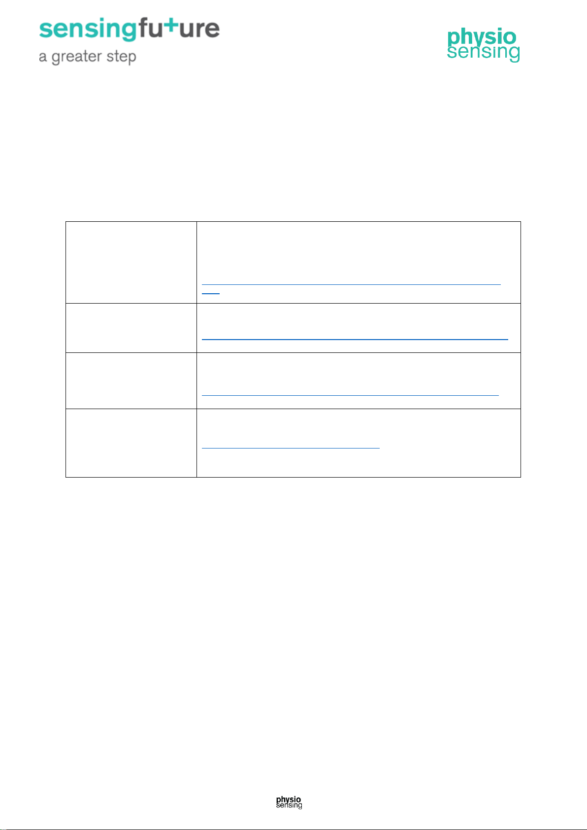
The information contained in this manual was partially or fully taken from the work of several
authors and sources not being original of the company Sensing Future Technologies. This
manual is intended to be a compilation of scientific evidence applicable to the use of
PhysioSensing and is currently accessible to anyone. Anyone who reads this manual is able
to find more sources of information on this field or contact the original authors.
The sources are:
Vestibular Disorders
Association
The Human Balance System—
A Complex Coordination of Central and Peripheral Systems
By The Vestibular Disorders Association, with contributions by Mary Ann Watson, MA,
and F. Owen Black, MD, FACS
http://kernodle.duhs.duke.edu/wp-content/uploads/2013/07/Human_Balance_System-
1.pdf *
Natus
Neurocom International
Objective Quantification of Balance & Mobility (PDF)
http://www.natus.com/index.cfm?page=products_1&crid=271&contentid=397#balmaster *
Biodex
Medical Systems
Biodex Balance System SD- Clinical Resource Manual 945-308 (PDF)
http://www.biodex.com/physical-medicine/applications/fall-risk-screening-conditioning *
National Institute on
Deafness and Other
Communication Disorders
(NIDCD)- USA
Balance Disorders
https://www.nidcd.nih.gov/health/balance-disorders *
*accessed April 2018
 Loading...