

Parks 2100-SX User manual
Medical Electronics, Inc.
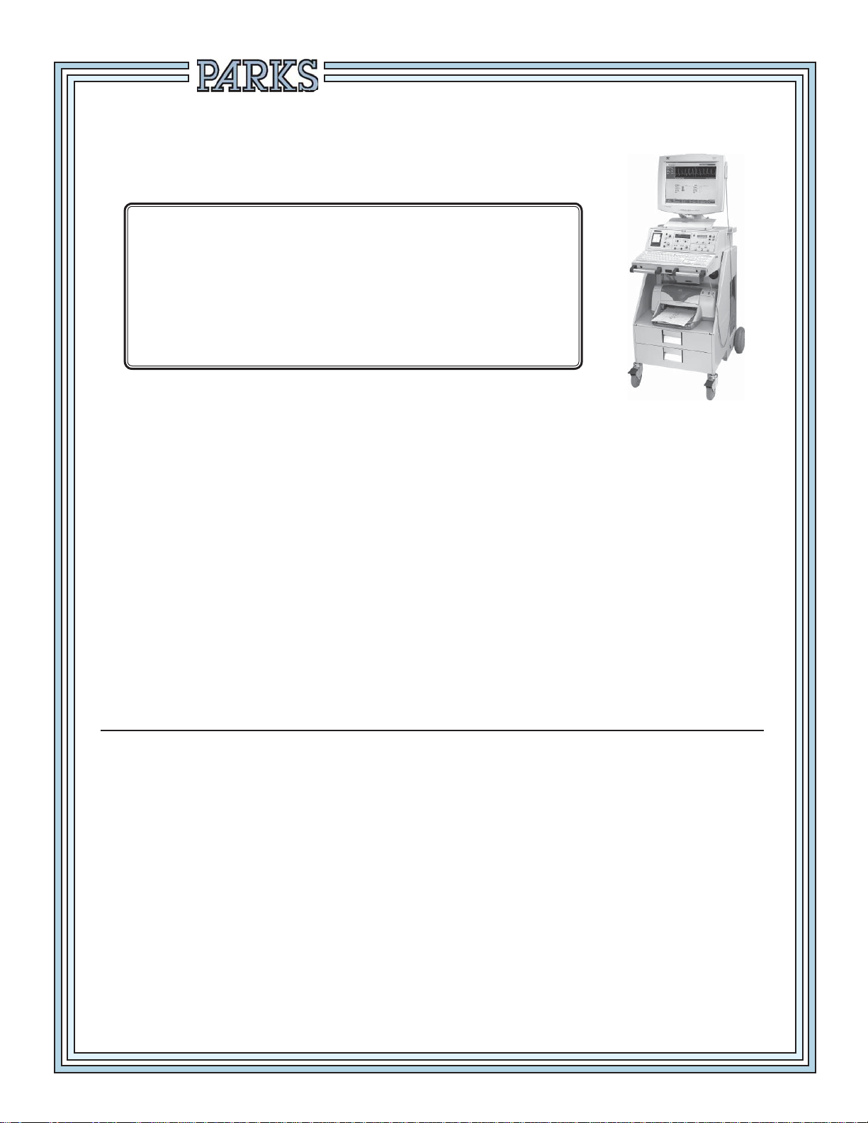
MODEL 2100-SX
CALIBRATION TEST AND
CUFF VOLUME CALIBRATION/VERIFICATION
Notice
This information has been provided to assist you in meeting the accreditation
standards set forth by the Intersocietal Commission for the Accreditation of Vascular
Laboratories.
Parks recommends that you perform the complete fi eld calibration procedure on
your Parks Flo-Lab after every 1000 hours of use or once a year.
To perform both the calibration test and the cuff volume verification/
calibration test, it will be necessary to purchase the calibration test fi xture
(Parks part #80-2100) and─ for units purchased after May 2007─ the 1000 ml
chamber (Parks part #986-3003-25) from the factory.
Please call Parks Medical Electronics, Inc. at 1-888-356-9522, M-F, 7:00-3:30 PM
Pacifi c Time. We will need the model number of your instrument to provide the
correct calibration equipment.

PARKS FLO-LAB
FIELD CALIBRATION
1. Position the Flo-Lab and cart with the back next to a sturdy table or workbench.
2. TURN THE MAIN POWER SWITCH OFF ( ) Located on the lower, right-hand side of the
cart). Set the monitor on the bench, with its cables and connections intact, and remove the six
screws holding the Flo-Lab top cover. Set the cover aside.
3. In the right front corner of the Flo-Lab you will see a grey colored metal box enclosure. Remove
the four screws and take off the box cover. Inside the enclosure there are a set of fi ve plug-in
circuit boards, and one other board mounted behind and at right angles to the others.
4. Carefully remove the left most plug-in circuit card and replace it with the Model 80 calibration
fi xture card. The card to be removed should be marked 8.X near the front. Insert the Model
80 calibration fi xture with the component side of the board facing to the left. Connect the
cable that was furnished with the Model 80 to the jack in the top of the fi xture and to the
EXTERNAL INPUT jack on the back of the Flo-Lab.
(
)
I
5. Turn ON
load the Sonova software. From the opening menu choose NEW STUDY. At the next menu
choose LOWER ARTERIAL. Enter a fi rst and last name and press enter. This will put you into
a PATIENT & STUDY SCREEN. Press escape. Now Choose TEST SELECT, then choose
DOPPLER. On the Model 80 calibration fi xture, turn the POWER switch ON and the CAL.
switch to either CAL A or CAL B position.
the Flo-Lab and computer. The computer will boot into Windows and automatically
6. On the Flo-Lab front panel, press the DOPPLER / ON button. The 8 MHz LED should light up.
Also verify that the OUTPUT FILTER - Hz “28” LED is illuminated. If needed, press the button
below the “28” LED until it illuminates. Next press the MENU button (directly to left of front panel
display) to advance to “5 - EXTERNAL SIGNAL OFF”. Press the UP button (directly to right of
the front panel display) to turn the signal on.
7. Use the POSITION knob on the front panel of the Flo-Lab to move the red cursor position to
the 1st division from the bottom of the display. Turn the SIZE control clockwise until the signal
is maximized. Both channels should display an 8 division signal, plus or minus 10%.
8. Press the 5mm / S button to run the chart recorder. The signal on the chart should be 32 mm
plus or minus 3 mm in amplitude.
9. Press STOP to stop the chart recorder. Press the MENU button to advance to “5 - EXTERNAL
ON”. Press the UP button to turn the signal off. Press DOPPLER / ON. Remove the cable
between the Model 80 calibration fi xture and the EXTERNAL INPUT jack. Switch the Model
80 calibration fi xture power OFF.
10. On the left side of the Flo-Lab front panel, use the POSITION control to center the trace in the
grid on the computer monitor. Turn the Flo-Lab SIZE control counter-clockwise to set SIZE A
and SIZE B both to 85.
11. Start the chart recorder at 5mm / S, then set the switches on the Model 80 calibration fi xture
to “A” and “ON”. After 2 or 3 seconds, set the Model 80 to “B”. Verify that, with the switch on
the Model 80 in position “A”, 5 green LEDs under TOWARD are on; and that in position “B”,
5 red LEDs under AWAY are on. Switch back and forth from “A” to “B” several times, then
STOP the chart recorder to examine it. Press The ( key to “freeze” the trace on the computer
monitor.
12. The upper trace on the monitor will look like a square wave, 8 divisions in amplitude, the lower
trace will be 4 divisions.
PARKS Medical Electronics, Inc. Aloha, Oregon U.S.A.
1 of 3
 Loading...