

Page 1
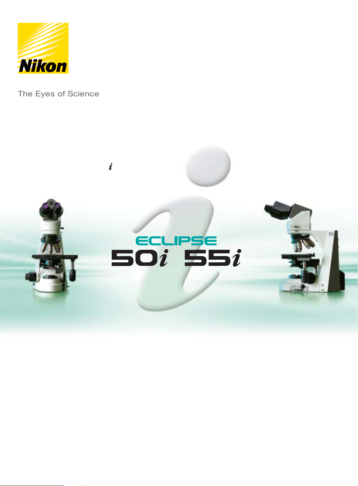
Clinical & Laboratory Microscopes ECLIPSE 50i/55i
/
/
. . . magine microscopy in perfect comfort
ECLIPSE i-Series Website
www.nikon-i.com
Page 2
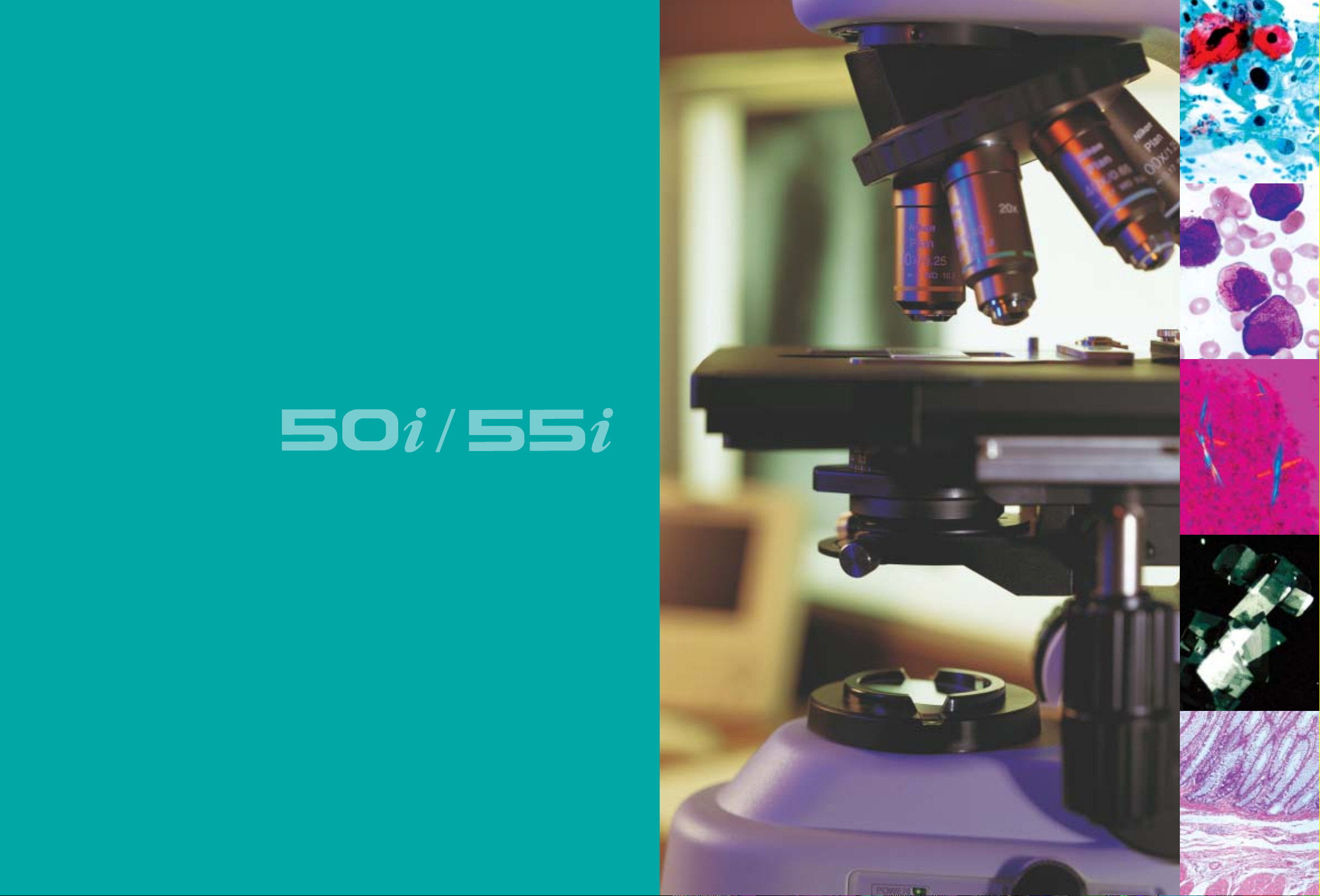
Imagine the ultimate in comfortable microscopy
/
operation; so comfortable, that you would not
even be aware you were carrying out essential
observations!
After carefully listening to customer needs, Nikon
designed the new 50i/55i series of compact,
versatile microscopes that are ideal for
clinical/laboratory inspection and basic research
study.
The ultimate in usability and comfort, the 50i/55i
series incorporates a host of stunning features
that take the stress out of microscopy. A stay-inposition stage handle and tilting/telescoping
ergonomic eyepiece tube ensure ideal viewing
posture, enabling long hours of observations to
be carried out in perfect comfort.
The 50i/55i series is built around Nikon’s tried
and tested infinity optics, the CFI60 system,
which means you not only get the optimum
optical performance, but you also have the
freedom to add various accessories to create the
setup that best suits your purpose.
. . . the ultimate comfort that takes clinical
microscopy to new heights.
— Cool illumination (55i)—LED illumination
generates no heat and maintains the color
temperature even when the brightness is
changed. Also, because this model can run on
batteries, it can be used anywhere. (See page 4)
— Novel ergo stage—the stage handle always
remains in the same position during stage
movement, enabling easy operation with your
hand resting on the desk. (See page 4)
— A digital camera can be mounted on the
ergonomic eyepiece tube by using a DSC port,
enabling easy digital documentation in a
comfortable operating posture. (See page 6)
— The “Ergo-View,” a retrofittable compact
cytodiagnosis unit, enables quick magnification
changeover using a hand switch. (See page 7)
1
1
Page 3
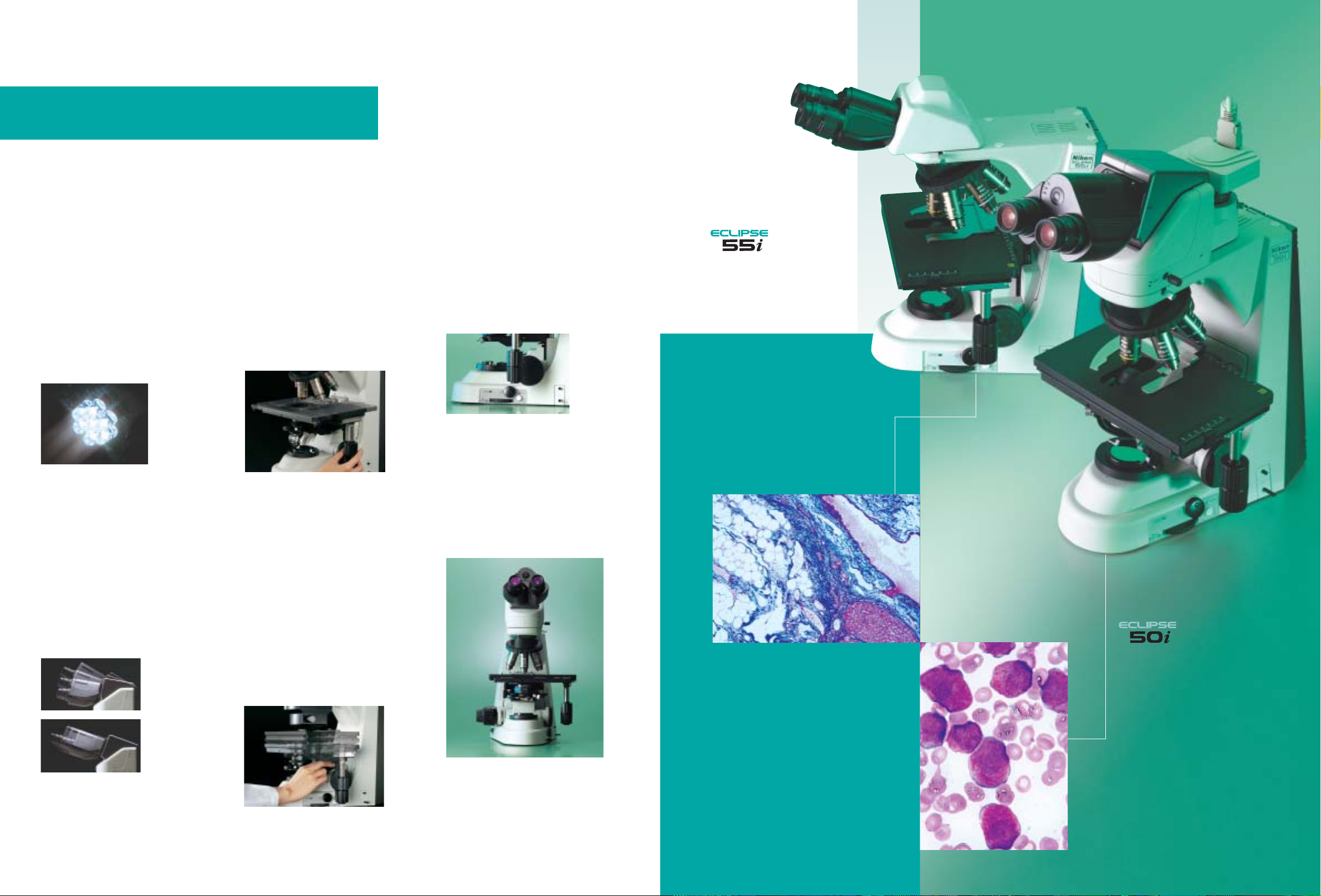
. . . in harmony with you
Cool illumination (55i)
Utilizing white LED illumination, the 55i is
an ideal choice for brightfield applications.
As the color temperature does not change
even when the brightness is altered,
adjusting the color balance filter or voltage
is no longer necessary. And because the
LED illuminator is built into the base,
comprehensive Koehler illumination using a
diaphragm is possible. The illuminator does
not generate heat. Due to its minimal
power consumption, the 55i has an
extremely long lamp lifecycle, and its battery
drive means it can be used anywhere.
New ergonomic tube
The new ergonomic tube can be inclined
from 10° to 30° and the eyepieces can be
extended 40mm. This ensures optimum eye
point and a comfortable viewing posture,
regardless of the operator’s physique or if
intermediate modules are being used. An
optional DSC port for attaching a digital
camera enables users to create a digital
documentation system in comfort. An
optional eye-level riser* can raise the eye
point in 25mm increments.
*The number of risers that can be used at any one time
depends on the intermediate modules being used.
Viewing angle is
adjustable 10°–30°.
Eyepiece length is
extendable up to
40mm.
Refined stage
Stay-in-position stage handle
The stage handle stays at the same position
without interfering with the stage
movement. Because the stage handle and
the focusing knob are always situated close
to each other, users can easily and smoothly
operate the controls with their hand resting
on the desk. The height and torque of the
stage handle are adjustable to further
enhance comfort during operation.
Stage with higher durability and
smoother surface
“Alumite,” a new hardening treatment, has
been applied to the stage surface to increase
durability and smoothness. This facilitates
the smooth exchange of specimens while
preventing the surface from being scratched
by the repeated exchange of slide glasses.
Refocusing stage
The stage can be lowered by pushing the
lever down, and will return to its former
height when the lever is pressed again. This
feature eliminates the need to refocus the
image manually each time the specimen is
changed and the slide oiled, greatly
improving microscopy productivity.
Easy-access controls
Frequently used controls and switches for
adjusting the field diaphragm and
illumination intensity have been
concentrated in the lower part of the
righthand side to minimize the operator’s
hand movements and enable operation
without having to take your eyes off the
specimen.
Rock-solid stability
Utilizing computer-aided engineering (CAE),
Nikon has successfully increased the solidity
of the microscope body and produced a
sleek, modern design. Although compact in
size, the new microscopes boast superb
durability and stability, even during
applications in which they are upgraded with
various attachments.
— With white LED illumination as its
light source, this model is perfect for
brightfield applications.
— No color temperature deviation
even when brightness is changed.
— Cool LED prevents focus
deviations caused by
heat-deformed equipment.
— When Ergo-View cytodiagnosis unit is
installed, it maintains uniform brightness
even if magnifications are changed.
— Long lifecycle illumination consumes
very little power.
— Battery-drive capability using optional
lithium batteries means model can be
used anywhere.
— Bright 6V-30W halogen lamp as light
source.
— ND8 filter is built-in as standard.
— Enables brightfield, darkfield, epi-
fluorescence, phase contrast and simple
polarizing observations.
4
54
Page 4

. . . in harmony with your vision
/
CF160—tried and tested infinity optics
Highly acclaimed optics combine the CF design with infinity optics and utilize a
60mm parfocal distance, resulting in longer working distances and high N.A.’s,
while producing crisp, clear images with minimal flare. The CF160 optics are
perfect for both observations and capturing images with a digital camera, and
they provide a flexible upgrade path to accommodate various accessories to
meet individual applications.
Ergo-View enables quick magnification
switching and marking
The compact Ergo-View cytodiagnosis unit has been developed for
easier and more comfortable cytology examinations. Retrofittable
to the 50i/55i, it can quickly move between 10X and 40X at the
flip of a switch, utilizing a motorized mechanism.
— Compact, sleek design.
— Easily attachable to 50i/55i.
— Fast and accurate motorized magnification changeover with
hand switch.
— Unique quiet, vibration-free mechanism for magnification
change ensures superb parfocality of images and no deviation in
focusing.
— Easy marking while observing the specimen through eyepieces.
— Quick exchange of slides with one hand is possible by using an
optional specimen holder for one slide.
Constant brightness when combined
with the 55i
When the Ergo-View is mounted on the 55i, which utilizes white
LED illumination, light intensity is automatically adjusted in
conjunction with changes in magnification. Constant brightness is
provided throughout the inspection, preventing eyestrain.
. . . in harmony with your hands
DSC port enables digital imaging in comfort
A new optional DSC port can be combined with an ergonomic tilting/telescoping
tube to balance user needs for both digital-image capture and comfortable
viewing. Including a 0.7X lens that is designed to optimize the image to the 2/3inch CCD, the same area as that viewed through the eyepieces can be captured
with a C-mount digital camera. A centering and focus adjustment mechanism is
also provided.
DS-5M-L1 “Digital Sight” standalone digital camera
The standalone design of the DS-5M-L1 allows 5-million-pixel, high-definition
images to be captured without connecting to a personal computer. The cameracontrol unit features a built-in 6.3-inch LCD monitor with excellent resolution,
enabling focusing to be carried out on the screen. Optimal camera settings have
been preprogrammed for each observation method and are selectable from a
menu. Users can take advantage of the camera’s network functions to send or
share images over a network and manage images on a server, saving time and
increasing productivity.
Wide variety of tools
Change between 10X and 40X at the flip of a switch.
10X
40X
Marking is easily performed while observing the image.
Quick-dry ink facilitates fast, clean marking.
Scene menu Count marking Distance measurement between 2 points
76
Page 5

. . . in harmony with your needs
/
. . . and a wide range of accessories
New accessories complement a rich variety of existing accessories.
. . . applying various methodologies
Pathology tests
As the 55i model uses white LED illumination, it can maintain the
same color temperature even if the brightness is changed.
Observations extending to many hours can be carried out in a
natural posture, without physical strain because an ergonomic tube
enables the flexible adjustment of the eye point.
Epi-fluorescence microscopy
A new dedicated turret-type epi-fluorescence illuminator has a
quick-change mechanism combined with a unique filter-lock system
and front-mounted shutter, providing the ultimate in clinical
fluorescence diagnostic microscopy.
2
Simple polarizing/sensitive color-polarizing
microscopy (50i)
Simple polarizing microscopy can be performed easily using
dedicated accessories. Sensitive color-polarizing microscopy enables
uric acid crystals forming inside an organism to be identified by
changing the interference color via a lever. Such microscopy is ideal
for gout tests.
Cytodiagnosis tests
The new Ergo-View cytodiagnosis unit makes magnification switching
and marking faster and easier. When it is combined with the 55i
model, changes in the amount of LED light are aligned with
magnification changes, enabling observation at a constant brightness.
Phase contrast microscopy (50i)
Developed expressly for this technique, our unique Apodized Phase
Contrast objectives enable the detection of minute structures—
previously difficult to detect due to annoying halos—with excellent
contrast and a much wider tonal range. This is ideal for urinary
sediment tests.
Darkfield (50i)
Darkfield microscopy is ideal for observing specimens such as
blood and the minute structures of flagella. A dry- or oil-type
darkfield condenser can be selected.
Epi-fluorescence attachment
This dedicated turret-type epi-illuminator
can accommodate four filter cubes despite
its compact size. Filters or mirrors in the
filter cubes can be easily replaced to create
the desired combination. The attachment’s
glowing display tags make it easy to
recognize filter names in a darkened room.
Ergo-View cytodiagnosis unit
The Ergo-View, a compact cytodiagnosis
unit, can be easily retrofitted to the
microscope. Utilizing a motorized control, it
enables easy switching between 10X and
40X magnifications at the flip of a switch.
Marking operation is also easy, even while
viewing the specimen.
Eyepiece tubes
In addition to the ergonomic tube, which
features a viewing angle of 10°–30° and
eyepieces that can be extended up to
40mm, the following Eclipse series tubes are
also available.
Ergonomic tube
Binocular
tube
Trinocular
tube F
Trinocular
tube T
Eye-level riser
The eye-level riser can raise the eye-point
height by 25mm at one time (up to 100mm*
maximum).
*: The number of risers
that can be used at any
one time depends on the
tube or intermediate
modules being used.
Teaching head (50i)
These teaching heads, which come with a
built-in pointer, facilitate simultaneous
viewing of the same specimen by up to 5
persons without compromising image
brightness.
2-person (side-by-side) version
Double port
Mounted between the main
body and eyepiece tube, the
double port enables
operators to use two CCTV
camera systems
simultaneously or one
CCTV camera and one
digital camera.
Magnification module
The magnification module allows the
intermediate magnification to be changed to
1X, 1.25X, 1.5X or 2X, enabling the
operator to frame the image to be captured
with a digital camera to match the view field
seen through the eyepieces.
DS-5M-L1 Digital Sight digital
camera system
5-megapixel, high-definition images can be
easily captured without connection to a PC
or external monitor. The camera controller
includes a built-in, 6.3-inch LCD monitor
with extremely high resolution, enabling the
operator to focus the image on the
monitor. The “Scene Function” allows the
optimum camera setting for each
observation method to be selected from the
menu.
FX-III series
photomicrographic equipment
The FX series features a direct-projection
system with swing-out prism to ensure fast
exposures and accurate metering.
U-III: 0.1% and 1% spot exposure and 35%
integrated-average measurement modes
H-III: 1% spot and 35% integrated-average
measurement modes
P-III: Manual exposure model
Lithium-ion battery (55i)
The optional lithium-ion battery increases
the mobility of the 55i by freeing it of
dependence on an electrical outlet.
Simple polarizing set and
sensitive color polarizing unit
(50i)
These easily installed options enable
operators to carry out simple
polarizing/sensitive color polarizing
observations.
11
DSC port
The optional DSC port allows a C-mount
digital camera to
be mounted to the
ergonomic tube.
Drawing tube
The observation image and drawing can be
seen together through the eyepieces. When
necessary, 100% of the light can be sent to
the observation port.
Simple polarizing set
Sensitive color polarizing unit
98
Page 6

FF
C-mount Camera
H
F
F
A
B
C
D
A
B
C
D
Y-TV
TV Tube
Y-TB
Binocular
Tube B
Y-TF
Trinocular
Tube FUW
Y-TT
Trinocular
Tube TUW
C-TE
Ergonomic
Binocular Tube
C-TEP
DSC Port
DS AC Adapter
Li-ion
Battery
EN-EL1
Darkfield
* Darkfield*
Condenser
(oil)
Condenser
(dry)
C-C
/Aplanat
Condenser
C-C
AchromatAchromat
Condenser
Swing-out
Condenser
C-C
Abbe
Condenser2-100x
LWD
Achromat
Condenser
C-C
Achromat
Swing-out
Condenser
1-100x
C-C
*
Phase
Condenser
C-C
Low Power
CondenserContrast
Attachment
Epi-fl
Filter Cubes
D
C-SP*
Simple
Polarizer
C-TP
*
Polarizer with
First-order Red
Tint Plate
C-ER Eye-level Riser
C-ISA Intermediate Tube
*
w/Simple Analyzer
C-IA Intermediate Tube*
w/Analyzer
Y-IDP Double Port (45-55 / 0-100, 0-100)
Y-IM Magnification Module
Y-IDT Drawing Tube
Y-THF Teaching Unit Face to Face
*
Y-THS Teaching Unit Side by Side B*
Y-THPS Support for Side by Side
Y-THM Main Teaching Unit
*
Y-THR Teaching Unit Side by Side A
Y-THP Pointer Unit
Y-THA AC Adapter
B
A
CFI60
Objective Lens
J-CY
Cytodiagnostic
Unit
C-HS
Hand Switch
Cable Set
for 55i &
Cytodiagnostic
Unit
C-SR
Mechanical
Stage
C-N
Sextuple
Nosepiece
H H
F
E
CFI
12.5x
CFI
10x
CFI
10x M
CFI
15x
CFI UW
10x
CFI UW
10x M
C-CT
Centering
Telescope
C-FC
Epi-Fl
Collector Lens
Quartz Epi-FI
Collector Lens
C-FC
Epi-Fl
Collector Lens
C-FC
Epi-Fl
Collector Lens
Mercury Lamp
Socket S 100W
Xe Lamp
Socket 75W
Halogen Lamp
Socket 100W/HMX
Hg Lamphouse
HMX-3B
Hg Lamphouse
HMX-4B
Xe Lamphouse
HMX-4
HMX Lamphouse
N
C-SHG1
Power Supply for HG100W
Xenon Power Supply 75W
UN2 Transformer 100W
Camera Mounts
Eyepiece Tubes
Nosepieces
Polarizers
Condensers
Intermediate Modules
Stages / Specimen Holders
Lamphouses
Eyepieces
N
TE-AT Double Lanphouse
Adapter
N
N
N
C-HL2
Specimen Holder Specimen Holder Specimen Holder
(2 slides: Left)
C-HR2
(2 slides: Right)
C-HC1
(1 slide)
GG
G
Y-QT
Quadrocular
Adapter
C
C-CEL Expander Lens
*1: Use the dedicated 0.45X adapter for the double port sub-port.
F
E
F
E
ENG-mount Camera C-mount Camera
ENG-mount
TV Adapter*
1
0.6x
ENG-mount
TV Adapter
Adapter
Relay
Lens
1x
ENG-mount
Zooming
Adapter
Zooming
C-mount
TV Zoom
Lens
Relay
Lens
1x
C-mount
TV Adapter
C-mount TV
Adapter
0.35x
0.38x
0.45x
0.6x
C-mount
TV Adapter A
C-mount
TV Adapter
VM2.5x
C-mount
TV Adapter
VM4x
V-T
Photo
Adapter
FX-III Series
Photomicrographic System
Projection Lens
PLI 2x
PLI 2.5x
PLI 4x
PLI 5x
C-0.7x
DXM Relay Lens
G
System Diagram
*: 50i model.
Built around an optimum digital-imaging platform,
the 80i can visualize weakly fluorescing molecules with
much higher brightness and contrast,
Advanced research microscope
while reducing background noise.
The new Plan Apo VC objectives and unique “fly-eye” optics guarantee uniform
brightness over the whole view field and unparalleled resolution to the
peripheries of the image. These remarkable achievements, which take digital
imaging to new heights, are the result of Nikon’s breakthroughs in optical
technologies and precision engineering.
—Hi S/N Fluorescence System. The universal epi-fluorescence illuminator and
digital-imaging head incorporate Nikon’s unique Hi S/N Fluorescence System,
which employs a “Noise Terminator” to achieve unparalleled contrast with
excellent S/N ratios. An optional “Excitation Balancer” allows specific wavelengths
to be emphasized in multi-stained specimens.
— Digital-Imaging Head. This creates an optimum digital-imaging system that
enables fluorescence imaging with outstanding results. When the DS-5M-L1 digital
camera is mounted to this head, observation data such as magnification and
fluorescence filters in use is automatically detected and can be saved together
with the image file.
— New DIC System. A perfect balance of high resolution and high contrast is
possible, even at low magnifications. Three types of DIC prisms are available:
standard, high-contrast, and high-resolution.
Nikon offers various options to best suit your digital-imaging needs
An all-in-one microscope
that can be operated by mouse
Digital microscope
The COOLSCOPE is revolutionary in every respect, from its looks
to its functionality and ease of use. After loading the slide glass
preparation onto the tray, users need only click the mouse to
operate the microscope. Observations, image recording and
networking can all be done with a single unit of the COOLSCOPE—
no need for a PC. The popular image of a microscope has
undergone a major metamorphosis.
— Motorized operation enables brightfield observations through the
monitor’s GUI.
— Simultaneous display of both enlargements (micro) of specimen
portions and whole image (macro) on a single screen.
— The point of interest can be easily moved to the center by simply
clicking the point in the micro image. In the macro image, the
selected point is enlarged and displayed in the micro image.
— Instant recall of previous observations can be achieved by clicking
the numbered button.
— Remote operation* and viewing of images is possible through
networked computers anywhere.
Please visit our dedicated website, where you will find
extensive information on the uses and operation of the
COOLSCOPE.
* Some operations are limited
www.coolscope.com
1110
Page 7

NIKON CORPORATION
Yokohama Plant
NIKON CORPORATION
Instruments Company
ISO 9001
Accredited by the
Dutch Council for
Accreditation
ISO 14001
/
/
I
O
FIL
200
330
60
404
208
60
I
O
FIL
486
330
200
208
Specifications
Main body 50i 55i
Magnification 10–1500X
Optical system CFI60 Infinity Optical System
Coarse/fine focusing Fine: 0.1mm per rotation, Coarse: 13.8mm per rotation, Minimum reading: 1 micron
Illumination 6V-30W halogen lamp White LED array
Built-in filter ND8 LA60 color balance filter
Eyepiece tube Binocular tube B (for F.O.V. 22mm)
Eyepiece lens 10X (F.O.V.
Nosepiece Sextuple nosepiece
Stage Super-hard Alumite coated surface, Stay-in-position stage handle, Stage handle height and tension adjustable
Condenser focusing stroke 27mm
Intermediate accessories Epi-fluorescence illuminator (4 filter positions), Magnification module, Eye-level riser, Double port
Observation method Brightfield, Epi-fluorescence
Coarse motion torque adjustable, Refocusing function (with stopper)
100-240V (worldwide voltage)
AC adapter (100-240V) or optional lithium battery (7.4V)
Trinocular tube "F" UW (for F.O.V. 22mm/25mm, observation/photo: 100/0, 0/100)
Trinocular tube "T" UW (for F.O.V. 22mm/25mm, observation/photo: 100/0, 20/80, 0/100)
Ergonomic binocular tube (for F.O.V. 22mm, inclination:10-30°, extension: 40mm); DSC port: 100/0, 50/50 (optional)
:
22mm), 10X M photo mask (F.O.V.: 25mm), 12.5X (F.O.V.:16mm), 15X (F.O.V.:14.5mm),
UW 10X (F.O.V.:25mm), UW 10X M photo mask (F.O.V.:25mm)
Ergo-View cytodiagnostic unit (Motorized changeover 10X to 40X with hand switch, Stamp type marking
— Automatic illumination adjustment with Ergo-View
Rectangular 159mm X 243mm surface stage, 78mm X 54mm cross travel (x-y movement)
1-slide or 2-slide specimen holder available (option)
Teaching head —
Darkfield, Phase contrast, Simple polarizing —
Dimensional Diagrams
Configured with binocular tube B Configured with ergonomic tube
Nikon has reduced the amount of chromium, cadmium and lead used in the Eclipse-i Series
to an absolute minimum to diminish its environmental impact.
Specifications and equipment are subject to change without any notice or obligation
on the part of the manufacturer. December 2003. ©2003 NIKON CORPORATION
WARNING
* Monitor images are simulated.
Company names and product names appearing in this brochure are their registered trademarks or trademarks.
NIKON INSTECH CO., LTD.
Parale Mitsui Bldg.,8, Higashida-cho, Kawasaki-ku,
Kawasaki, Kanagawa 210-0005, Japan
phone: +81-44-223-2167 fax: +81-44-223-2182
www.nikon-instruments.jp/eng/
www.i-series.com
NIKON INSTRUMENTS (SHANGHAI) CO., LTD.
CHINA phone: +86-21-5058-5055 fax: +86-21-5058-5060
NIKON SINGAPORE PTE LTD
SINGAPORE phone: +65-6559-3618 fax: +65-6559-3668
NIKON MALAYSIA SDN. BHD.
MALAYSIA phone: +60-3-78763887 fax: +60-3-78763387
TO ENSURE CORRECT USAGE, READ THE CORRESPONDING MANUALS
CAREFULLY BEFORE USING YOUR EQUIPMENT.
NIKON INSTRUMENTS EUROPE B.V.
P.O. Box 222, 1170 AE Badhoevedorp, The Netherlands
phone: +31-20-44-96-222 fax: +31-20-44-96-298
http://www.nikon-instruments.com/
NIKON FRANCE S.A.
FRANCE phone: +33-1-45-16-45-16 fax: +33-1-45-16-00-33
NIKON GMBH
GERMANY phone: +49-211-9414-0 fax: +49-211-9414-322
NIKON INSTRUMENTS S.p.A.
ITALY phone: + 39-55-3009601 fax: + 39-55-300993
NIKON AG
SWITZERLAND phone: +41-43-277-2860 fax: +41-43-277-2861
NIKON UK LTD.
UNITED KINGDOM phone: +44-20-8541-4440 fax: +44-20-8541-4584
Please contact Nikon for a handy pamphlet listing compatible
accessories, including objectives and epi-fluorescence filters.
NIKON INSTRUMENTS INC.
1300 Walt Whitman Road, Melville, N.Y. 11747-3064, U.S.A.
phone: +1-631-547-8500; +1-800-52-NIKON (within the U.S.A.only) fax: +1-631-547-0306
http://www.nikonusa.com/
NIKON CANADA INC.
CANADA phone: +1-905-625-9910 fax: +1-905-625-0103
Unit: mm
Microscopy images courtesy of :
1 Nihon University Itabashi Hospital
2 Naoyuki Miyokawa, M.D., Ph.D., Associate
Professor, Dept. of Surgical Pathology,
Asahikawa Medical College Hospital.
Printed in Japan (0312-13)T
NIKON CORPORATION
Fuji Bldg., 2-3, Marunouchi 3-chome, Chiyoda-ku,
Tokyo 100-8331, Japan
www.nikon.com/
Code No. 2CE-MSHH-1
This brochure is printed on recycled paper made from 40% used material.
En
 Loading...