

Page 1

Instruction Manual
The LOWER and UPPER MRD was specifically designed for
either the knee or shoulder joints to be used to help pre and
post-surgical patients early in their rehabilitation when
forces that affect the healing tissues need to be controlled.
However, it can do much more which this manual will reveal.
The MRD’S can not only be used for pre and post surgery as
mentioned, but also with traumatic injuries involving the
upper and lower extremity, kinetic chain of joints. Because
of the synergistic and integrated nature of the nervous
system, the MRD’s will impact the tone of muscles and the
length of their myofascia that affect the position and motion
of the shoulder, elbow, wrist, and hand with the UPPER MRD
and the hip, knee, ankle, and foot with the LOWER MRD.
The UPPER AND LOWER MRD’s are “open kinetic” chain
devices a term used to describe a phase in the way the
human body moves against the vertical forces of gravity and
ground reaction force (GRF).
Simply when one bone is moved within a kinetic chain of
joints, it creates some kind of movement or effect in
adjacent, nearby joints along with affecting the muscles,
tendons, fascia, and ligaments attached at the joints.
A kinetic chain is considered “open” when either the hand or
foot is free to move and not affected by GRF. Conversely, a
kinetic chain is considered “closed” when the hand or foot is
fixed against the ground and reacting to GRF.
Page 2

In function, the human body has to be able to do both. But
with open kinetic chain movement, it is much easier to
control the linear and rotational forces on a joint which is a
necessity in the early healing phase of injured tissues or
post surgery. The closed kinetic chain, on the other hand,
where the hand and foot react to GRF, the movement will
occur at multiple joints and at multiple joint axes. This
simultaneous movement occurring at more than one joint or
segment makes it much more difficult to control the linear
and rotational forces. Therefore, the UPPER AND LOWER
MRD’S are tools that should be used in the early
rehabilitation phase after surgery or trauma.
In describing functional, human movement like walking, it is
in fact a continuum, a sequence of open and closed kinetic
chain activity. The UPPER AND LOWER MRD’S, therefore,
have their place in this continuum of movement that
ultimately leads to a more stable and optimum outcome
from either surgery or trauma as the patient becomes more
aggressive and functional in their movements requiring a
response to GRF.
The UPPER AND LOWER MRD’S can be used either in the
clinic or more importantly in the patient’s home environment
to begin the process of changing the patient’s muscle
memory or “myomemory” and remodeling scar tissue vital in
preventing “arthrofibrosis.” Therefore, the MRD’s are an
important key to a patient’s recovery and return to function
because as we all know “motion is life, motion is freedom!”
When I mention changing “myomemory” I am referring to the
ability of a patient to reproduce a particular movement
without intervention of the conscious part of the brain.
Page 3

In short, turn your client’s muscles on for a specific
movement or skill to “autopilot.” That can only occur with
active, patient participation and practice and altering visual,
vestibular, and proprioceptive sensory input.
Considering that approximately 90% of all neuronal activity
of the human brain is somehow involved with vision and the
eyes, the movement of the eyes is the key to transforming
the neuronal patterns, the muscle and myofascial patterns
that ultimately create posture and movement.
Simply, myomemory is the process of reorganizing and
rewiring a patient’s brain-muscle-myofascial connection.
When a patient practices a new movement over and over
again in conjunction with eye movements while using the
MRD’S, they are literally decreasing the resistance within
their own CNS to create a new neuronal or muscle pathway
that will ultimately alter the position and motion of an
isolated joint and the integrated posture and movement.
This is referred to as “neuroplasticity,” a process that
involves adaptive structural and functional changes to the
brain and its output.
Over time, our posture and movement and the muscle and
myofascial patterns that create it can become dysfunctional
altering the position and motion of a joint from the ideal
eventually causing pathology. That is why surgical
intervention in many cases is required. But, the surgery only
addresses the joint dysfunction and pathology. It does not
alter the muscle and myofascial memory that caused it.
Page 4

The UPPER AND LOWER MRD’S can be useful tools in
altering dysfunctional muscle and myofascial memory by
creating new neuronal, muscle pathways.
With the transformation of muscle and myofascial memory, a
patient’s posture and movement will also change that will
better prepare the client for weight bearing and closed
kinetic chain, functional activities. Let’s get started to
transforming your client’s “myomemory” and improving their
function.
“KEY POINTS TO REMEMBER”
•WHEN POSITIONING A CLIENT TO PRACTICE A MOVEMENT
ON EITHER THE UPPER OR LOWER MRD, MAKE SURE THE
AXIS OF MOTION FOR THE PRIMARY JOINT WORKING IS
ALIGNED WITH AXIS OF MOTION FOR EITHER MRD.
•POSTURE WHEN USING THE UPPER AND LOWER MRD’S IS
IMPORTANT TO A SUCCESSFUL OUTCOME. MAKE SURE
THE CLIENT IS IN A “NEUTRAL” STANDING OR SITTING
POSTURE. THAT IS WHEN SITTING, THEIR WEIGHT IS
EQUALLY DISTRIBUTED BETWEEN BOTH “SITS” BONES OF
THE PELVIC GIRDLE AND THEY DEMONSTRATE A SLIGHT
LORDOSIS OF THE LUMBOSACRAL SPINE.
IN STANDING,THEIR WEIGHT SHOULD BE EQUALLY
DISTRIBUTED BETWEEN BOTH FEET FEELING EQUAL
PRESSURE BETWEEN THE BALL OF LITTLE AND BIG TOES
AND INSIDE AND OUTSIDE OF BOTH HEELS. FEET ARE
PARALLEL TO EACH OTHER AND STRAIGHT AHEAD. THE
HEAD, FACE, AND PELVIC AND SHOULDER GIRDLES ARE
STRAIGHT AHEAD WITHOUT ROTATION. THE CHIN IS
PARALLEL WITH THE FLOOR.
Page 5

•WHEN BEGINNING A PROGRAM, INSTRUCT YOUR PATIENT
HOW TO “READ” THEIR PAIN. LET THEM KNOW THEY MAY
INITIALLY EXPERIENCE PAIN WITH MOVEMENT, BUT THE
PAIN WILL GO AWAY ONCE THEY STOP THE MOVEMENT.
THAT IS “DYSFUNCTIONAL PAIN” AND IS EXPECTED
BECAUSE THEY ARE WORKING TISSUES IN A NEW AND
REMODELED WAY.
•IF THEY WORK THE TISSUES AND EXPERIENCE PAIN AND
THE PAIN PERSISTS FOR HOURS AFTER DOING THE
MOVEMENT, THAT IS MOST LIKLEY PAIN FROM
INFLAMMATION. THE IDEA IS TO WORK THE HEALING
TISSUES IN THE NEW, REMODELED WAY WITHOUT
CAUSING INFLAMMATION. THIS SIMPLY IS NOT A
SITUATION OF “NO PAIN, NO GAIN.”
•WHEN WORKING WITH “DYSFUNCTIONAL PAIN,” WORK UP
TO THE POINT OF FIRST EXPERIENCING PAIN AND/OR
STRETCH. DO NOT TRY TO WORK THROUGH THE PAIN. IF
THE PAIN IS DYSFUNCTIONAL, THEY WILL BE ABLE TO
GRADUALLY GAIN MORE AND MORE RANGE OF MOTION
(ROM) WITHOUT INCREASING INFLAMMATION AND THEIR
LEVEL OF PAIN. IF SLIGHT PAIN IS EXPERIENCED, WORK
UP TO THE POINT WHERE FIRST EXPERIENCING A PULL OR
STRETCH.
•THE TIME HOLDING THE END OF A MOVEMENT DEPENDS
ON THE TISSUE YOU ARE TRYING TO ISOLATE. FOR
MUSCLE, HOLD A POSITION FOR UP TO A MAXIMUM OF 30
SECONDS WHEREAS LONGER WILL LENGTHEN THE
CONNECTIVE TISSUE (MYOFASCIA). IN THE EXERCISE
DESCRIPTIONS, A MAXIMUM HOLD TIME OF 30 SECONDS IS
USED.
Page 6

•INITIALLY, HOLD THE POSITION FOR 5 SECONDS AND
GRADUALLY INCREASE THE TIME OF HOLD.
•THE FREQUENCY OF DOING THE MOVEMENT VARIES WITH
EACH PATIENT. HOWEVER, IT IS BETTER TO SPREAD THE
REPETITIONS DONE THROUGHOUT THE DAY RATHER THAN
DOING ONE TO TWO INTENSE SESSIONS. IF THE CLIENT IS
USING THE MRD AT HOME FOR EXAMPLE, DOING A
PRACTICE OF A MOVEMENT EVERY OTHER HOUR WHILE
MONITORING THEIR PAIN, SWELLING, AND SKIN
TEMPERATURE RESPONSE IS PREFERRED.
•HAVE CLIENTS DO 3-12 REPETITIONS EACH SESSION.
•REMEMBER THE BUILT IN GONIOMETER TO DOCUMENT
INITIAL JOINT RANGE OF MOTION (ROM) AND PROGRESS.
•REMEMBER YOU CAN INCREASE THE INTENSITY OF
EXERCISE BY THE HOLDING TIME, REPETITIONS, ROM, AND
THE “VARIABLE RESISTANCE” CAPABILITY OF THE UPPER
AND LOWER MRD’S.
•WITH THE LOWER MRD, THE SIMPLE MOVEMENT
PRACTICED IS WITH KNEE EXTENSION AND FLEXION IN
THE SAGITTAL PLANE. BY CHANGING THE SENSORY INPUT
WITH THE POSITION OF THE EYES, HEAD AND NECK, AND
FOOT AND ANKLE, YOU CAN AFFECT THE TENSION, TONE
OF MUSCLES AND LENGTH OF THE MYOFASCIA THAT
ULTIMATELY ALTER THE POSITION AND MOTION OF A
JOINT IN ALL THREE PLANES.
Page 7

•WITH THE UPPER MRD, THE SIMPLE MOVEMENTS
PRACTI
THE FRONTAL PLANE,
THE SAGITTAL PLANE, SHOULDER HORIZONTAL
ABDUCTION/ADDUCTI
ROTATION/EXTERNAL ROTATION IN THE TRANSVERSE
PLANE.
•BY CHANGING EYE AND HEAD
CONJUNCTI
WILL ADDRESS THE POSITION AND MOTION OF THE
SHOULDER JOINT IN ALL THREE PLANES.
•ALL M
CLOSED TO LIMIT THE AFFECT OF REFLEXES REFERRED TO
AS THE “VESTI
CED ARE SHOULDER ABDUCTION/ADDUCTIO
SHOULD
ON AND SHOULDER IN
ON WITH THESE SHOULDER MO
OVEMENTS WILL BE PE
BULAR-OCULAR
ER FLEXION/EXTENSION IN
TERNAL
AND NECK POSITIONS, IN
VEMENTS, YOU
RFORMED WITH THE EYES
REFLEXES.” THESE ARE EYE
N IN
MOVEM
COUNTERING WITH THE MOVEMENT OF THE HEAD.
ENTS THAT REFLEXIVELY STABILI
ZE THE GAZE BY
Page 8
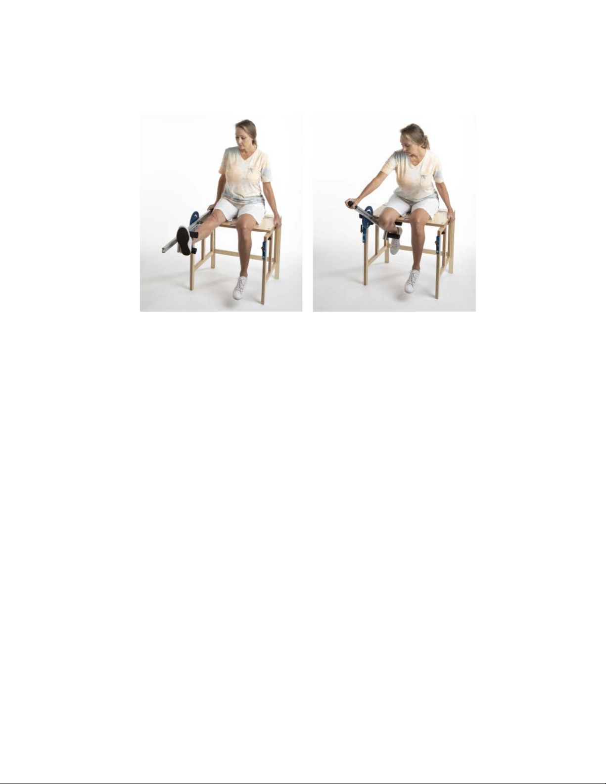
“THE LOWER MRD PROGRAM”
Progression of use:
1) Have client familiarize th
by extendi
using the acti
control of their movem
2) Extend the leg up at the knee
the LO
dysfunctional pain and/or stretch. Flex the leg back into
knee flexi
Repeat as much as needed to familiarize client with the
MRD.
ng and flexing their knee in the sagittal pl
ve-assist handle.
WER MRD’s arm to wh
on where first experiencing pain and/
emselves with the LOWER MRD
Emphasize that they are in
ent.
into knee extension with
ere first experiencing
or stretch.
ane
Page 9

3) SAGITTAL PLANE MOTION:
Begin working the knee in the sagittal plane now altering
the visual and proprioceptive input. During the knee flexion
and extension movement, have the client keep their chin
parallel with the floor and close their eyes. Move eyes out to
the R and down and flex the R toes down toward the plantar
surface of foot. With the LOWER MRD’S active-assist handle,
have client move into knee flexion until first experiencing
dysfunctional pain and/or stretch and hold. Initially, hold for
5 seconds and relax. Gradually increase the hold time to a
maximum of 30 seconds. Relax toes of R foot and eyes R
and down. During the knee extension phase, have the client
move eyes out to the R and up and extend toes of R foot up
toward the front of the knee. With the LOWER MRD’S active
assist handle, bring R knee into knee extension until first
experiencing dysfunctional pain and hold. Initially, hold for 5
seconds and gradually increase the hold time to a maximum
of 30 seconds. Relax toes of R foot and eyes R and up. Do
this sequence as just described of R knee flexion and
extension 3-12 times.
The L knee is done in the same manner, but eyes are closed
and moving out to the L up with L knee and toe extension
and moving out to L and down with L knee and toe flexion.
You can have the client do this flexion and extension
movement with both knees. If there is a significant fear of
moving their involved leg, start with their uninvolved limb
and then do the involved limb.
Page 10

KEY POINTS TO REMEMBER FOR SAGITTAL PLANE
MOTION:
•ALIGNMENT AND POSTURE IS IMPORTANT TO A
SUCCESSFUL OUTCOME.
•EYES ARE CLOSED AND MOVE WITHOUT STRAIN.
EYES MOVE OUT TO THE SAME SIDE AND DOWN AS
EXTREMITY MOVING WITH TOE AND KNEE FLEXION
AND OUT TO THE SAME SIDE AND UP AS EXTREMITY
MOVING WITH TOE AND KNEE EXTENSION.
4) FRONTAL PLANE MOTION:
As the client gains confidence and has less fear of
movement, begin frontal plane motion that involves
tilting or side bending of the head and neck 15-30º as
well as plantarflexion and dorsiflexion of the foot and
ankle.
Have the client close their eyes and keeping their head
and face straight ahead, position their head and neck
so it is tilted or side bent toward their R shoulder and
move their eyes out to the R and up. Point the R foot
and ankle down away from the front of the knee into
plantarflexion and with the active-assist handle, have
the client flex their R knee to where first experiencing
pain and/or stretch and hold. Initially hold for 5 seconds
and gradually increase to a maximum of 30 seconds.
Relax head and neck tilt R, eyes R and up, R foot and
ankle plantarflexion, and R knee flexion.
Page 11

Bring head and neck to vertical and straight ahead and
close eyes. Move eyes out to the R and down. Have
client bring R foot up toward front of knee into
dorsiflexion and with the MRD active-assist arm, extend
the R leg up into extension until first experiencing pain
and/or stretch and hold. Hold this extended position of
the R knee initially for 5 seconds and up to a maximum
of 30 seconds. Repeat this cycle of R knee flexion and
extension 3-12 times.
L knee flexion is done in the same manner with eyes
closed and side bend or tilt of head and neck toward L,
eyes move out to the L and up, and foot and ankle in
plantar flexion. With L knee extension, head and neck is
brought to vertical and straight ahead and eyes closed.
Have client move eyes out to the L and down and
dorsiflex their L foot and ankle.
KEY POINTS TO REMEMBER FOR FRONTAL PLANE
MOTION:
•ALIGNMENT AND POSTURE IS IMPORTANT TO A
SUCCESSFUL OUTCOME.
•EYES ARE CLOSED AND MOVE OUT TO SAME SIDE
AND UP AS DOING THE KNEE FLEXION AND OUT TO
THE SAME SIDE AND DOWN AS DOING THE KNEE
EXTENSION.
•HEAD AND NECK IS TILTED ABOUT 15-30º TOWARD
SAME SIDE AS FLEXING KNEE AND VERTICAL AND
STRAIGHT AHEAD WHEN EXTENDING KNEE.
•FOOT AND ANKLE IS PLANTARFLEXED WITH KNEE
FLEXION AND DORSIFLEXED WITH KNEE EXTENSION.
Page 12

5) FRONTAL/TRANSVERSE PLANE MOTION:
Involves turning head and neck with eyes closed and
eye movement. Have client close their eyes and
keeping their chin parallel with floor, turn their head
and neck toward R shoulder. Move eyes out to the R
and down. With the MRD active-assist arm, have the
client flex or bend their R knee back to where first
experiencing pain and/or stretch and hold. Hold initially
for 5 seconds and gradually increase to a maximum of
30 seconds. Now, have client extend or straighten the R
knee up to where pain is first experienced and/or
stretch and hold. Again, hold initially for 5 seconds and
gradually increase to a maximum of 30 seconds. Relax
head and neck rotation R, eyes R and down, and R knee.
Repeat 3-12 times.
Keeping the client’s eyes closed and their chin parallel with
the floor, have client turn their head and neck to the L and
move eyes out to the R and up. With the MRD active-assist
arm, have the client flex or bend their R knee to where first
experiencing pain and/or a stretch and hold. Hold initially for
5 seconds and gradually increase to a maximum of 30
seconds. Now, have client extend or straighten the R knee
to just where pain and/or stretch is first experienced and
hold. Again, hold initially for 5 seconds and gradually
increase to a maximum of 30 seconds. Relax head and neck
rotation L, eyes R and up, and R knee. Repeat 3-12 times.
The L leg is done in the same manner. First eyes are closed,
head and neck is turned toward L shoulder, and eyes L and
down while doing L knee flexion and extension movements.
This is followed by eyes are closed, head and neck turned to
R shoulder, and eyes L and up while doing L knee flexion
and extension movements.
Page 13

KEY POINTS TO REMEMBER FOR FRONTAL/
TRANSVERSE PLANE MOTION:
•ALIGNMENT AND POSTURE IS IMPORTANT TO A
SUCCESSFUL OUTCOME.
•EYES ARE CLOSED AND LOOK OUT TO THE SAME SIDE
AS KNEE FLEXION AND EXTENSION MOVEMENT.
•HEAD AND NECK IS TURNED TOWARD THE SAME SIDE
AS KNEE FLEXION AND EXTENSION MOVEMENT WITH
EYES LOOKING DOWN.
•HEAD AND NECK IS TURNED TOWARD OPPOSITE SIDE
OF KNEE FLEXION AND EXTENSION MOVEMENT WITH
EYES LOOKING UP.
6) SAGITTAL/TRANSVERSE PLANE MOTION:
Involves turning the head and neck as well as inversion
and eversion of the foot with eyes closed. Keeping chin
parallel with the floor, have the client turn their head
and neck toward the R shoulder and move eyes out to
the L and down. Turn R foot outward (eversion) away
from midline of body. With the MRD active-assist arm,
have the client flex their R knee back to where first
experiencing dysfunctional pain and/or stretch and hold.
Initially hold for 5 seconds gradually increasing to a
maximum of 30 seconds.
Now, have the client extend or straighten the R knee
to where first experiencing dysfunctional pain and/or
stretch is experienced and hold. Hold initially for 5
seconds gradually increasing to a maximum of 30
seconds. Relax head and neck rotation R, eyes L and
down, R knee, and R foot. Repeat 3-12 times.
Page 14

Keeping eyes closed and chin parallel with the floor,
turn head and neck toward the L shoulder and move
eyes out to the L and up. Turn R foot inward (inversion)
toward the midline of body. With the MRD active-assist
arm, have the client flex their R knee back to where
first experiencing dysfunctional pain and/or stretch and
hold. Hold initially for a 5 seconds gradually increasing
to a maximum of 30 seconds.
Now, have the client extend or straighten the R knee up
to where experiencing dysfunctional pain and/or stretch
is first experienced and hold. Initially hold for a count of
5 seconds and gradually increase to a maximum of 30
seconds. Relax head and neck rotation L, eyes L and
up, R knee, and R foot. Repeat 3-12 times.
The L leg is done in the same manner. First eyes are
closed, head and neck is turned toward L shoulder, and
eyes look out to the R and down while doing L knee
flexion and extension movements. This is followed by
eyes are closed, head and neck turned toward R
shoulder, and eyes look out to the R and up while doing
L knee flexion and extension movements.
KEY POINTS TO REMEMBER SAGITTAL/TRANSVERSE
PLANE MOTION:
•ALIGNMENT AND POSTURE IS IMPORTANT TO A
SUCCESSFUL OUTCOME.
•EYES ARE CLOSED AND MOVE OUT TO THE OPPOSITE
SIDE OF EXTREMITY MOVING.
•WITH FOOT EVERSION AND KNEE FLEXION AND
EXTENSION MOVEMENTS, NECK ROTATION IS TOWARD
THE SAME SIDE AS EXTREMITY MOVING WITH EYES
DOWN.
•WITH FOOT INVERSION AND KNEE FLEXION AND
EXTENSION MOVEMENTS, NECK ROTATION IS TO THE
OPPOSITE SIDE AS EXTREMITY MOVING AND EYES UP.
 Loading...