

Page 1
Medical
BioSphere
®
Instructions for use
Instructions d'utilisation
Gebrauchsanweisungen
Instrucciones de utilización
Istruzioni per l’uso
Instruções de utilização
Gebruiksaanwijzing
Brugsanvisning
Bruksanvisning
Käyttöohjeet
Instruksjoner for bruk
Οδηγίες χρήσης
Kullanma talimatları
Pokyny pro použití
Instrukcja stosowania
Instrucţiuni de utilizare
Инструкции за употреба
Használati útmutató
Lietošanas norādījumi
Naudojimo instrukcijos
Návod na použitie
Kasutusjuhised
Инструкции по применению
0459 - 2000
Z 1691 rev E 06/12
730044002/A
Page 2

English.......................................................................................................3
Français (French)
Deutsch (German)
Español (Spanish)
Italiano (Italian)
Português (Portuguese)
Nederlands (Dutch)
Dansk (Danish)
.........................................................................5
.......................................................................7
........................................................................9
.........................................................................11
.....................................................13
................................................................15
...........................................................................17
Svenska (Swedish)
Suomi (Finnish)
..........................................................................21
Norsk (Norwegian)
Ελληνικά (Greek)
Türkçe (Turkish)
Čeština (Czech)
Polski (Polish)
........................................................................28
...........................................................................30
..............................................................................32
Română (Romanian)
...................................................................19
.................................................................23
...................................................................25
.............................................................34
Български (Bulgarian)
Magyar (Hungarian)
Latviešu (Latvian)
............................................................39
....................................................................41
Lietuviškai (Lithuanian)
Slovenčina (Slovak)
Eesti (Estonian)
Русский (Russian)
...............................................................45
..........................................................................47
...................................................................49
.....................................................36
..................................................43
Page 3

ENGLISH
DESCRIPTION
Embosphere® Microspheres are biocompatible, hydrophilic,
non-abs orbable, precise ly calibrated acr ylic polymer
microspheres impregnated with porcine gelatin and are
available in a large range of sizes and concentrations.
HOW SUPPLIED
20-ml pre filled syringe w ith a standard Lu erlock tip,
individually packaged in blister tray sealed by a Tyvek® peelaway lid. Plastic screw cap and plunger. Elastomer three-skirt
plunger joint.
Contents: 1 ml or 2ml of microspheres in sterile, pyrogen-free
0.9% NaCl solution.
INDICATIONS
Embosphere Microspheres are designed to occlude blood
vessels, for therapeutic or preoperative purposes, in the
following procedures:
- Embolisation of hypervascular tumours and processes,
including uterine fibroids, meningiomas,etc.
- Embolisation of arteriovenous malformations.
- Haemostatic embolisation.
40-120 µm microspheres are more specifically designed for
embolisation of meningiomas and liver tumours.
CONTRAINDICATIONS
- Patients unable to tolerate vascular occlusion procedures.
- Vascular anatomy precluding correct catheter placement.
- Feedin g arteries to o small to acce pt the select ed
microspheres.
- Presence or suspicion of vasospasm.
- Presence of distal arteries directly supplying cranial nerves.
- Presence of patent extra-to-intracranial anastomoses.
- High-flow arteriovenous shunts or with a diameter greater
than the selected microspheres.
- Pulmonary embolism.
- Severe atherosclerosis.
- Patients with known allergy to gelatin.
40-120 µm and 10 0-300 µm microspher es a re n ot
recommended for use in the bronchial circulation.
POTENTIAL COMPLICATIONS
Vascular embolisation is a high-risk procedure. Complications
may occur at any time during or after the procedure, and may
include, but are not limited to, the following:
- Stroke or cerebral infarction
- Occlusion of vessels in healthy territories
- Vascular rupture and haemorrhage
- Neurological deficits
- Infection or haematoma at the injection site
- Allergic reaction, cutaneous irritations
- Transient pain and fever
- Vasospasm
- Death
- Ischemia at an undesirable location, including ischemic
stroke, ischemic infarction (including myocardial infarction),
and tissue necrosis
- Blindness, hearing loss, loss of smell, and/or paralysis
- Additional information is found in the Warnings section
CAUTION
DO NOT USE THIS PREFILLED SYRINGE TO DIRECTLY
INJECT EMBOSPHERE MICROSPHERES. THIS IS A
“RESERVOIR”SYRINGE.PLEASE REFERTO INSTRUCTIONS
PARAGRAPH.
Embosphere Microspheres must only be used by specialist
physicians trained in vascular embolisation procedures. The
size and quantity of microspheres must be carefully selected
according to the lesion to be treated, entirely under the
physician’s responsibility. Only the physician can decide the
most appropriate time to stop the injection of microspheres.
Do not use if blister tray, peel-away film, screw cap or syringe
are damaged. This is a disposable product. Discard opened
syringes after use. All procedures mus t be perfo rmed
according to an aseptic technique.
For singlepatient use only - Contents supplied sterile
Do not reuse, reprocess, or resterilize. Reusing, reprocessing
or resterilizing may compromise the structural integrity of the
device and or lead to device failure, which in turn may result
in patient injury, illness or death. Reusing, reprocessing or
resterilizing may also create a risk of contamination of the
device and or cause patient infection or cross infection
including, but not limited to, the transmission of infectious
disease(s) from one patient to another. Contamination of the
device may lead to injury, illness or death of the patient.
WARNINGS
• Embosphere Microspheres contain gelatin of porcine origin,
and therefore, could cause an immune reaction in patients
who are hypersensitive to collagen or gelatin. Careful
consideration should be given prior to using this product in
patients who are suspected to be allergic to injections
containing gelatin stabilizers.
• Studies have shown that Embosphere Microspheres do not
form aggregates, and, as a result, penetrate deeper into the
vasculature as compared to similarlysized PVA particles. Care
must be taken to choo se larger sized Embosph ere
Microspheres when embolizing arteriovenous malformations
with large shunts to avoid passage of the spheres into the
pulmonary or coronary circulation.
• Some of the Embosphere Microspheres may be slightly
outside of the range, so the physician should be sure to
careful ly s elect the size of Embosph ere Microsphere s
according to the size of the target vessels at the desired level
of occlusion in the vasculature and after consideration of the
arterio venous an giographic appe arance. Embosph ere
Microspheres size should be selected toprevent passage from
artery to vein.
• Because of the significant complications of misembolization,
extreme caution should be used for any procedures involving
the extracranial circulation encompassing the head and neck,
and the physician should carefully weigh the potential benefits
of using emb olization agai nst the risks and pote ntial
complications of the procedure. These complications can
include blindness, hearing loss, loss of smell, paralysis and
death.
• Serious radiation induced skin injury may occurto the patient
due to long periods of fluoroscopic exposure, large patient
diameter, angled x-ra y pro jections, and multiple ima ge
recording runs or radiographs. Refer to your facility’s clinical
protocol to ensure the proper radiation dose is applied for each
specific type of procedure performed. Physicians should
monitor patients that may be at risk.
• Onset of radiation-induced injury to the patient may be
delayed. Patients should be counseled on potential radiation
side effects and whom they should contact if they show
symptoms.
• Pay careful attention for signs of mistargeted embolization.
During injection carefully monitor patient vital signs to include
SaO
(e.g. hypoxia, CNS changes). Consider terminating the
2
procedure, investigating for possible shunting, or increasing
microsphere size if any signs of mistargeting occur or patient
symptoms develop.
• Consider upsizing the microspheres if angiographic evidence
of embolization does notquickly appear evident during injection
of the microspheres.
Warningsabout use of small microspheres
• Careful consideration should be given whenever use is
contemplated of embolic agents that are smaller in diameter
than the resolution capability of your imaging equipment. The
presence of arteriov enous anastomoses, branch vessels
leading away from the target area or emergent vessels not
3
Page 4

evident pri or to emb olization can lead to mista rgeted
S
TERILIZE
2
2
STERILE
embolization and severe complications.
• Microspheres smaller than 100 microns will generally
migrate distal to anastomotic feeders and therefore are more
likely to terminate circulation to distal tissue. Greater potential
of ischem ic injury resul ts from use of small er sized
microspher es and con sideration must be given to the
consequence of this injury prior to embolization. The potential
consequences include, swelling, necrosis, paralysis, abscess
and/or stronger post embolization syndrome.
• Post embolization swelling may result in ischemia to tissue
adjacent to target area. Care must be given to avoid ischemia
intolerant, nontargeted tissue such as nervous tissue.
INSTRUCTIONS
• Position the catheter at the desired site and perform
baseline angiography to evaluate the blood supply of the
lesion.
• Embosphere Microspheres are available in a range of sizes.
Because of the potential for misembolization and the inherent
variability in sphere sizes, the physician should be sure to
careful ly s elect the size of Embosph ere Microsphere s
according to the size of the target vessels at the desired level
of occlusion in the vasculature.
• Carefully select the size of microspheres according to the
size of the vess els identified and the ca theter used.
Embosphere Microspheres are flexible particles that support
temporary compression by 20 to 30% to facilitate passage
through microca theters. Studie s h ave shown a dire ct
correlation between the size of microspheres and the size of
occluded vessels.
• Inspect packaging and syringe before use to ensure that
they are intact. The external surface of the syringe is sterile.
• Unscrew the cap of the Embosphere Microsphere prefilled
syringe and gently draw contrast agent directly into the
reservoir syringe.
• The ideal suspension is usually obtained with a mixture of
50% contrast agent and 50% saline solution. To obtain a
homogeneous suspen sion of Embosp here Mi crospheres,
gently invert the 20-ml syringe several times. Contrast agent
and 0.9% NaCl solution can be added in thesame proportions
to obtain a more diluted suspension.
• Do n ot use the 20-ml prefilled syringe to inject
Embosphere Microspheres through the catheter!
• Remove all air from the syringe and connect it to one hub of
the three-way stopcock.
• Draw up the suspension using a small syringe (1 to 3 cc)
connected to another hub of the three-way stopcock. Avoid
back and forth movements to reducethe risk of introducing air
into the system. Check that the desired quan tity a nd
concentration of microspheres are used.
• Remove all air from the syringe.
• Screw the syringe onto the hub of the catheter, using the
male Luer-lock connector of the stopcock.
• Open stopcock to connect the injection syringe with the
catheter.
• Under continuous flu oroscopic control, slowly infuse
microspheres into the blood stream. Always inject under free
flow conditions. Reflux of microspheres can induce immediate
ischaemia of healthy tissues or vessels.
• Continue infusion until the desired devascularisation is
obtained. Studies have shown that Embosphere Microspheres
penetrate more distally into the lesion than PVA particles of
similar size. Reduction of thearterial blood supply to the lesion
is therefore more progressive.
• At the end of the infusion, remove the catheter while
maintaining gentle aspiration to avoid dislodging any residual
microspheres still inside the catheter, thenclose the three-way
stopcock.
• Remove the catheter.
• Discard any remaining Embosphere Microspheres and the
used syringes.
• Do not use the 20-ml prefilled syringe to inject
Embosphere microspheres through the catheter!
• Remove all air from the syringe and connect it to one hub of
the three-way stopcock.
• Draw up the suspension using a small syringe (1 to 3 cc)
connected to another hub of the three-way stopcock. Avoid
back and forth movements to reducethe risk of introducing air
into the system. Check that the desired quan tity a nd
concentration of microspheres are used.
• Remove all air from the syringe.
• Screw the syringe onto the hub of the catheter, using the
male Luer-lock connector of the stopcock.
• Open stopcock to connect the injection syringe with the
catheter.
• Under co ntinuous fluor oscopic control , slo wly infuse
microspheres into the blood stream. Always inject under free
flow conditions. Reflux of particles can induce immediate
ischaemia of healthy tissues or vessels.
• Continue infusion until the desired devascularisation is
obtained. Studies have shown that Embosphere microspheres
penetrate more distally into the lesion than PVA particles of
similar size. Reduction of thearterial blood supply to the lesion
is therefore more progressive.
• At the end of the infusion, remove the catheter while
maintaining gentle aspiration to avoid dislodging any residual
microspheres still inside theca theter, thenclose the three-way
stopcock.
• Remove the catheter.
• Discard any remaining Embosphere microspheres and the
used syringes.
CONSERVATIONAND STORAGE
Embosphere Microspheres must be stored in a cool, dry, dark
place in their original syringe and packaging. Use by the date
indicated on the labels of the outer box and blister’s pack. Do
not freeze.
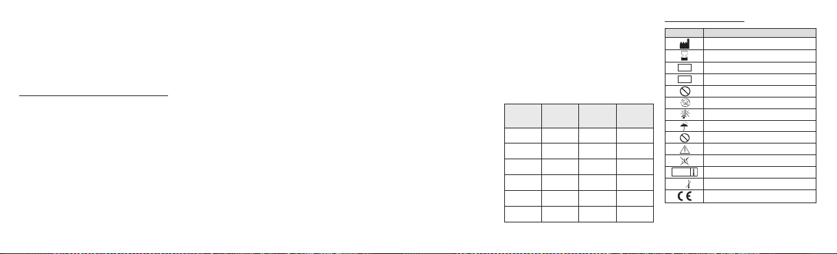
Size Range
Color Code 1 ml 2 ml
(µm)
40-120 Orange S110GH S120GH
100-300 Yellow S210GH S220GH
300-500 Blue S410GH S420GH
500-700 Red S610GH S620GH
700-900 Green S810GH S820GH
900-1200 Purple S1010GH S1020GH
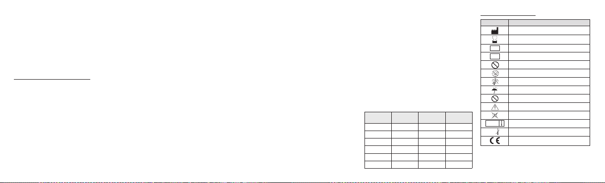
Information on packaging:
Symbol
Designation
Manufacturer: Name & Address
Use by date: year-month
Batch code
LOT
Catalogue number
REF
Do not resterilize
Do not use if package is damaged
Keep away from sunlight
Keep dry
Do not re-use
Caution - Refer to Instructions For Use
Non-pyrogenic
Sterilized using steam
Lower limit of temperature
0°C
EC mark logo - Notified body identification : 0459
All serious or life threatening adverse events or d eaths
associated with use of Embosphere Microspheres should be
reported to the device manufacturer.
4
Page 5

FRANÇAIS
DESCRIPTION
Les Microsphères Embosphe re
biocompatibles, hydrophiles, non résorbables, calibrées avec
précision, en polymère acrylique et imprégnées de gélatine
porcine. Elles sont disponibles dans une large gamme de
tailles et de concentrations.
PRÉSENTATION
Seringue pré -remplie de 20ml avec embout Lue r-lock
standard, conditionnée individuellement sous blister rigide
scellé par un film pelable en Tyvek
en plastique. Joint de piston à trois collerettes en élastomère.
Contenu : 1 ml ou 2 ml de microsphères dans une solution de
NaCl 0,9 % apyrogène et stérile.
INDICATIONS
Les Microsphères Embosphere sont conçues pour occlure les
vaissea ux sangui ns, à des fins théra peutiques ou
préopératoires, dans le cadre des procédures suivantes :
- Embolisation de tumeurs et processus hyper vasculaires,
dont fibromes utérins, méningiomes, etc.
- Embolisation de malformations artério-veineuses.
- Embolisation pour hémostase.
Les microsphères de 40-120 µm sont plus spécialement
conçues pour l'embolisation des méningiomes et des tumeurs
du foie.
®
sont des microsphères
®
. Bouchon à vis et piston
CONTRE-INDICATIONS
- Patients intolérants aux procédures d'occlusion vasculaire.
- Anatomie vasculaire empêchant lamise en place correcte du
cathéter.
- Artère s nourr icières trop pe tites po ur rece voir les
microsphères sélectionnées.
- Présence ou suspicion de vasospasme.
- Présence d'artères distales irriguant directement les nerfs
crâniens.
- Présence d'anastomoses extra-intracrâniennes manifestes.
- Shunt artério-veineux à débit élevé ou avec un diamètre
supérieur aux microsphères sélectionnées.
- Embolie pulmonaire.
- Athérosclérose sévère.
- Antécédents d'allergie à la gélatine.
L'utilisation des microsphères de 40-120 µm et de 100-300
µm est déconseillée dans le réseau bronchique.
COMPLICATIONSPOTENTIELLES
L'embolisation vasculaire est une procédure à haut risque.
Des complications peuvent se produire à tout moment, durant
ou après l'intervention, et peuvent inclure notamment :
- Accident vasculaire cérébral ou infarctus cérébral
- Occlusion de vaisseaux irriguant des territoires sains
- Rupture vasculaire et hémorragie
- Déficits neurologiques
- Infection ou hématome au point de ponction
- Réaction allergique, irritation cutanée
- Douleur et fièvre transitoires
- Vasospasme
- Décès
- Ischémie à un endroit non souhaité, y compris accident
ischémique cérébral, infarctus ischémique (dont l’infarctus du
myocarde), et nécrose tissulaire
- Cécité, perte auditive, perte de l'odorat et/ou paralysie
- Informations supplémentaires disponibles dans la section
Avertissements
MISE EN GARDE
NE PAS UTILISER DIRECTEMENT CETTE SERINGUE PRÉREMPLIE POUR INJECTER LES MICROSPHÈRES
EMBOSPHERE. ILS'AGIT D'UNE SERINGUE« RÉSERVOIR ».
SE REPORTER AU PARAGRAPHEINSTRUCTIONS.
Les Micro sphères Embosp here do ivent ê tre util isées
exclusivement par des médecins spécialisés, formés aux
procédures d'embolisation vasculaire. La taille et la quantité
de microsphères doivent êtrechoisies avec soin en fonction de
la lésion à traiter, ce qui est entièrement de la responsabilité
du médecin. Seul le médecin peut décider du moment le plus
approprié pour arrêter l'injection des microsphères.
Ne pas utiliser si le blister, le film pelable, le bouchon à vis ou
la seringue est endommagé.Il s'agit d'un produit jetable.Toute
seringue entamée doit être jetée après utilisation. Toutes les
procédures doivent être effe ctuées selon une technique
aseptique.
Pour unusage unique-Contenu fourni stérile
Ne pas réutiliser, retraiter ou restériliser. La réutilisation, le
retraiteme nt ou la restéri lisation peuvent compromettr e
l'intégrité structurelle du dispositif et/ou conduire à une
défaillance du dispositif, pouvant entraîner une lésion, une
maladie ou le décès du patient.La réutilisation, le retraitement
ou la rest érilisation risquent également de générer une
contamination du dispositif et/ou de causer une infection ou
une infection croisée chez le patient, y compris notamment, la
transmission de maladies infectieuses d'un patient à l'autre.
La contamination du dispositif peut entraîner une lésion, une
maladie ou le décès du patient.
AVERTISSEMENTS
• Les Microsphères Embosphere contiennent de la gélatine
d'origine porcine et peuvent, par conséquent, entraîner une
réaction immunitaire chez les patients hypersensibles au
collagène ou à la gélatine. Une mûre réflexion est nécessaire
avant d'utiliser ce produit chez les pa tients pour qui on
suspecte une allergie aux injections contenant des stabilisants
en gélatine.
• Des études ont montré que les Microsphères Embosphere
ne formen t pas d'agr égats et p énètrent donc plus
profondément dans le système vasculaire que des particules
de PVA de taille similaire. Veiller à choisir des Microsphères
Embosphere de plus grande taille pour l'embolisation de
malformations artério-veineuses avec de grands shunts, pour
éviter le passage des micr osphères dans la circulation
pulmonaire ou coronaire.
• Il est possible que certaines Microsphères Embosphere soit
légèrement hors de la gamme de taille, par conséquent, le
médecin devra s'assurer de sélectionner avec soin la taille
des Microsphères Embosphere, en fonction de la taille des
vaisseaux au niveau désiré d’occlusion dans le système
vasculaire et après considération de l'aspect angiographique
artério-veineux. La taille des Microsphères Embosphere doit
être choisie de façon à éviter leur passage de l'artère à la
veine.
• Du fait des complications importantes liées à une mauvaise
embolisation, une prudence extrême doit être appliquée pour
de quelconques inte rventions impliquant une circu lation
extracrânienne englobant la tête et le cou et le médecin doit
sérieusement peser les bienfaits potentiels du recours à
l'embolisation par rapport aux risques et aux complications
potenti els de l a pr océdure. Ces complica tions peuvent
comprendre la cécité, une perte auditive,une perte de l'odorat,
la paralysie et la mort. Ces complications peuvent inclure :
cécité, perte auditive, perte de l'odorat, paralysie et décès.
• Le patient peut développer de graves lésions cutanées
inhéren tes à l’irradi ation du fait d e longues péri odes
d'exposition à l'angiographie, de patients de forte corpulence,
d’incidences obliques, de séries répétées d'enregistrement
d'images ou de radiographies multiples. Se reporter au
protocole clinique de votre établissement pour vous assurer
que la dose d'irradiation correcte est utilisée pour chaque type
de procédure réalisée. Les médecins doivent surveiller les
patients qui peuvent présenter un risque.
• L’apparitionde lésions par irradiation chez lepatient peut être
retardée. Les patients doiven t être infor més de s effet s
potentiels des rayons, de ce qu'il faut rechercher et de la
personne à contacter en cas d'apparition de symptômes.
• Apporter une attention particulière aux signes d'embolisation
mal ciblée. Durant l'injection, suivre attentivement les signes
5
Page 6
vitaux du patient, tels que le
STERILIZE
2
2
STERILE
changements du SNC). Envisager d'arrêter la procédure, en
cherchant un shunt éventuel, ou s'orienter vers une taille de
microsph ères s upérieure si de quelconques sign es de
mauvais ciblage se produisent ou que les symptômes du
patient s'aggravent.
• Envisager d'utiliser unetaille des microsphères supérieure si
l'angiographie ne démontre pas rapidement une embolisation
évidente pendant l'injection des microsphères.
Avertissements relatifs à l'utilisationdes petitesmicrosphères
• Une attention toute particulière doit être apportée lorsque les
emboles ontun diamètre inférieur à la capacité de résolution de
votre équipement d'imagerie. La présence d'anastomoses
artério-veineuses, de vaisseaux ramifiés conduisant hors de la
zone cible ou de vaisseaux émergents non évidents, peuvent
conduire à une embolisation mal ciblée et à des complications
graves.
• Des microsphères de moins de 100 microns effectueront
généralement une migration d istale vers les sources
anastomotiques et sont ainsi susceptibles d'emboliser un tissu
distal. L'utilisation de microsphères de taille plus petite peut
conduire à un risque plus élevé de lésion ischémique et les
conséquences de cette lésion doivent être prises en compte
avant l'embolisation. Les conséquen ces potentielles
comprennent le gonflement, la nécrose, la paralysie, un abcès
et/ou un syndrome post-embolisation plus fort.
• Un gonflementpost embolisation peut conduire àune ischémie
du tissu adjacent à la zone cible. Il faut prendre soin d'éviter
l'ischémie d'untissu intolérant, non ciblétel que letissu nerveux.
SaO
(par ex. l'hypoxie, les
2
INSTRUCTIONS
• Positionner le cathéter à l'endroit souhaité et pratiquer un
bilan angiographique de référence pour évaluer le réseau
vasculaire de la lésion.
• Les Micro sphères Embosp here sont dispon ibles en
différentes tailles. Du fait du risque de mauvaise embolisation
et de la variabilité inhérente aux tailles des sphères, le
médecin doit s’assurer qu’il sélectionne avec soin la taille des
Microsphères Embosph ere en fonctio n de la taille des
vaisseaux ciblés au niveau souhaité de l’occlusion du système
vasculaire.
• Sélectionner avec soin la tailledes microsphères en fonction
de la taille des vaisseaux identifiés et du cathéter utilisé. Les
Microsphères Embosphere sont desmicrosphères flexibles qui
admettent une compression temporaire de 20 à 30 % pour
faciliter leur passage à travers desmicrocathéters. Des études
ont montré une corrél ation directe entre la taille des
microsphères et la taille des vaisseaux occlus.
• Avant utilisation, inspecter l'emballage et la seringue pour
s'assurer qu'ils sont intègres. La surface externe de la
seringue est stérile.
• Dévisse r le bouc hon de la s eringue pré-rem plie
Microsphères Embosphere et aspirerdoucement du produit de
contraste directement dans la seringue réservoir.
• La suspension idéale est généralement obtenue avec un
mélange de 50 % de produit de contraste et 50 % de sérum
physiol ogique. Pour obteni r une mise en su spension
homogèn e d es M icrosphères Embo sphere, renv erser
doucement la seringue de 20 ml à plusieurs reprises. Il est
possible d'ajouter du produit de contraste et du sérum
physiologique (NaCl à 0,9 %) dans les mêmes proportions
pour obtenir une suspension plus diluée.
• Ne pas utiliser la seringue pré-remplie de 20 ml pour
injecter les Microsphères Embosphere par le cathéter !
• Purger tout l'air de la seringueet la raccorder à une embase
du robinet trois voies.
• Aspirer la suspension en utilisant une petite seringue (1 à 3
ml) raccordée à une autre embase du robinet à trois voies.
Éviter les mouvements de va-et-vient pour réduire le risque
d'introduction d'air dans le système. Vérifier que la quantité et
la concentration de microsphères souhaitées sont utilisées.
• Purger tout l'air de la seringue.
• Visser la seringue sur l'embase du cathéter, en utilisant le
raccord Luer-lock mâle du robinet.
• Ouvrir le robinet pour raccorder la seringue d'injection au
cathéter.
• Sous contrôle angiographiquecontinu, injecter lentement les
microsphères dans le flux sanguin. Toujours injecter dans des
conditions de débit libre. Le reflux de microsphères peut
provoquer une ischémie immédiate de vaisseaux ou de tissus
sains.
• Continuer l'injection jusqu'à ce que la dévascularisation
souhaitée soit obtenue. Des études ont montré que les
Microsphères Embosphere pénètrent plus profondément dans
la lésion que les particules de PVA de taille similaire. La
réduction de l'irrig ation artérielle de la lésion est par
conséquent plus progressive.
• Dès que l'injection est terminée, retirer le cathéter tout en
maintenant une légère aspiration pour éviter de déloger les
microsphères résiduelles présentes à l'intérieur du cathéter,
puis fermer le robinet à trois voies.
• Retirer le cathéter.
• Jeter toutes Microsphères Embosphere restantes et les
seringues utilisées.
CONSERVATIONET STOCKAGE
Les Microsphères Embosphere doivent être conservées dans
un endroit sec, à l'abri dela lumière et de la chaleur, dans leur
seringue et leur emballage d'origine. Utiliser avant la date
indiquée sur les étiquettes de la boîte extérieure et du blister.
Ne pas congeler.
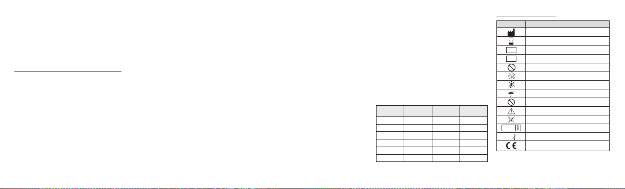
Gamme de
Code couleur 1 ml 2 ml
tailles (µm)
40-120 Orange S110GH S120GH
100-300 Jaune S210GH S220GH
300-500 Bleu S410GH S420GH
500-700 Rouge S610GH S620GH
700-900 Vert S810GH S820GH
900-1200 Violet S1010GH S1020GH
Informations sur l'emballage :
Symbole
Designation
Fabricant : nom et adresse
Date limite d'utilisation : année-mois
Numéro de lot
LOT
Référence catalogue
REF
Ne pas restériliser
Ne pas utiliser si l'emballage est endommagé
Conserver à l'abri de la lumière du soleil
Tenir au sec
Ne pas réutiliser
Attention - Consulter les instructions d'utilisation
Apyrogène
Stérilisé à la vapeur
Limite inférieure de température
0°C
Logo du marquage CE - Indentification de l'organisme
Tous les événements indésirables graves ou menaçant la vie,
ou entrainant la mort, liés à l'utilisation des Microsphères
Embosphere doivent être signalés au fabricant du dispositif.
notifié : 0459
6
Page 7

DEUTSCH
BESCHREIBUNG
®
Mikrosphären sind biokompatible, hydrophile,
Embosphere
nicht resorbierbare, präzise kalibrierte Mi krosphären aus
einem Acryl-Polymer, die mit Schweinegelatine imprägniert
sind. Sie sind in vi elen ver schiedenen Größ en und
Konzentrationen erhältlich.
LIEFERZUSTAND
Vorgefüllte 20-ml-Spritze mit normgerechter Luer-Lock-Spitze,
einzeln verpack t in einer Blistersc hale und mit einem
abziehbaren Tyvek
Kolben aus Kunststoff. Kolbendichtung mit drei Lippen aus
Elastomer.
Inhalt: 1 ml oder 2 ml Mikrosphären in steriler, pyrogen-freier
Kochsalzlösung (0,9% NaCl).
INDIKATIONEN
Embosphere Mikrosphären sindzur Okklusion von Blutgefäßen
zu therapeutischen oder präoperativen Zwecken bei den
folgenden Eingriffen vorgesehen:
- Embolisation von hypervaskulären Tumoren und Prozessen,
einschließlich Uterus myomatosus, Meningiomen usw.
- Embolisation von arteriovenösen Fehlbildungen.
- Embolisation zur Blutungsstillung.
Die Mikrosphären von 40-120 µm sind insbesondere für die
Embolisation vonMeningiomen und Lebertumorenvorgesehen.
®
-Deckel versiegelt. Schraubverschluss und
KONTRAINDIKATIONEN
- Patienten , die einen Eingriff zur Gefäßo kklusion nicht
verkraften würden.
- Gefäßanatomie, die keine sachgemäße Katheterplatzierung
zulässt.
- Zu kleiner Durchmesser der zuführenden Arterien für die
gewählten Mikrosphären.
- Bekannter oder vermuteter Vasospasmus.
- Vorliegen von distalen Arterien, die Schädelnerven direkt
versorgen.
- Vorliegen von durchgängigen Anastomosen von extrakranial
nach intrakranial.
- Arteriovenöse Shunts mit hoher Durchflussrate oder einem
Durchmesser über dem der gewählten Mikrosphären.
- Lungenembolie.
- Schwere Atherosklerose.
- Patienten mit bekannter Allergie auf Gelatine.
Die Mikrosphären von 40-120 µm und 100-300 µm werden
nicht für den Einsatz im Bronchialkreislauf empfohlen.
POTENZIELLE KOMPLIKATIONEN
Bei einer Gefäßembolisation handelt es sich um einen
riskanten Eingriff. Komplikationen können jederzeit während
des Eingriffs oder danach eintreten. Insbesondere sind die
folgenden zu nennen:
- Schlaganfall bzw. Hirninfarkt
- Okklusion von Gefäßen in gesunden Bereichen
- Gefäßruptur und Blutung
- Neurologische Ausfälle
- Infektion oder Hämatom an der Injektionsstelle
- Allergische Reaktion, Hautreizungen
- Vorübergehende Schmerzen und Fieber
- Vasospasmus
- Tod
- Ischämie an einer unerwünschten Stelle, einschli eßlich
ischämischer Schlaganfall, ischämischer Infarkt(einschließlich
Herzinfarkt) und Gewebenekrose
- Blindheit, Gehörverlust, Verlust des Geruchssinns und/oder
Lähmung
- Weitere Informationen befinden sich unter „Warnungen“
VORSICHT
DIESE VORGEFÜLLTE SPRITZE DARFNICHT ZUR DIREKTEN
INJEKTION VON EMBOSPHERE MIKROSPHÄREN
VERWENDET WERDEN. ES HANDELT SICH UM EINE
VORRATSSPRITZE. NÄHERES IST DEM ABSCHNITT
„ANLEITUNG“ ZU ENTNEHMEN.
Embosphere Mikrosphären dürfen nur von Fachärzten mit
einer Ausbildung in Gefäßembolisationseingriffen verwendet
werden. Größ e un d Me nge der Mikrosphär en m üssen
sorgfäl tig entsp rechend der zu behan delnden L äsion
ausgewä hlt werden. Diese Auswa hl liegt ganz in der
Verantwortung des Arztes. Nur der Arzt kann entscheiden,
wann die Injektion der Mikrosphären gestoppt werden sollte.
Das Produk t darf nich t verwend et werden , wenn die
Schalenverpackung, dera bziehbareFilm, der Schraubverschluss
oder die Spritze sichtbare Schäden aufweist. Dies ist ein
Einwegprodukt. Offene Spritzen nach Gebrauch entsorgen. Alle
Eingriffe sind unter aseptischen Bedingungen durchzuführen.
Nur zumGebrauch beieinem einzelnen Patienten–Inhalt
ist im Lieferzustandsteril
Nicht wiederverwenden, wiederaufbereiten oder resterilisieren.
Eine Wiederv erwendung, Wi ederaufbereitung o der
Resterili sation kann die struktu relle Unversehrthei t d es
Produktes gefährden und/oder zum Versagen des Produktes
führen, was seinerseits Verletzungen, Erkrankungen oder den
Tod des Patienten z ur Folge haben kann. Eine
Wiederverwendung, Wiederaufbereitung oder Resterilisation
kann darüber hinaus ein Kontaminationsrisiko für das Produkt
darstellen und/oder eine Infektion oder Kreuzinfektion des
Patienten verursachen,darunter insbesondere die Übertragung
von Infektionskrankheiten von einem Patienten auf einen
anderen. EineKontamination des Produkteskann Verletzungen,
Erkrankungen oder den Tod des Patienten zur Folge haben.
WARNUNGEN
• Embosphere Mikrosphären enthalten Gelatine, die von
Schweinen stammt und daher bei Patienten, die überempfindlich
aufKollagenoder Gelatinereagieren, eineImmunreaktionauslösen
kann. Vor der Anwendung dieses Produktes bei Patienten, die
vermutlich allergisch auf Injektionen mit Gelatinestabilisatoren
reagieren,empfiehltsich eine sorgfältigeAbwägung.
• Studien zufolge bilden Embosphere Mikrosphären keine
Anhäufungen und können daher im Vergleich zu PVA-Partikeln
ähnlicher Größe tiefer in das Gefäßsystem vordringen. Bei der
Embolisation von arteriovenösen Fehlbildungen mit großen
Shunts muss darauf geachtet werden, größere Embosphere
Mikrosphären auszuwählen, um zu verhindern, dass diese in
den Lungen- oder Herzkreislauf gelangen können.
• Einige Embosphere Mikrosphären können etwas außerhalb
des angegebenen Größenbereichs liegen. Der Arzt muss daher
die Größe der Embosphere Mikrosphären sorgfältig auf die
Größe der Zielgefäße der zu okk ludierenden Ebene des
Gefäßsy stems a bstimmen und d as arte riovenöse
angiographische Erscheinungsbild in Betracht ziehen. Die
Größe der EmbosphereMikrosphären ist so zu wählen, dassein
Durchtritt aus der Arterie in eine Vene ausgeschlossen ist.
•Da mit einer Fehlembolisation erhebliche Komplikationen
verbunden sind, muss bei Eingriffen mit Einfluss auf die
extrakraniale Durchblutung von Kopf und Nacken mit äußerster
Vorsicht vorgegangen werden. Der Arzt muss den potenziellen
Nutzen der Embolisation sorgfältig gegen die Risiken und
potenziellen Komplikationen des Eingriffs abwägen. Zu diesen
Komplikationen gehören u.a. Blindheit, Gehörverlust, Verlust
des Geruchssinns, Lähmung und Tod.
• Der Patient kann durch lang andauernde Durchleuchtung,
großenKörperumfang, Röntgenaufnahmen inSchrägansicht sowie
mehrfache Röntgenaufzeichnungen bzw. -aufnahmen schwere
strahlungsbedingte Hautverletzungen erleiden. Zur Einhaltung der
richtigen Strahlungsdosis für jeden Eingriffstyp wird auf das
klinische Protokoll der jeweiligen Einrichtung verwiesen. Der Arzt
muss gefährdetePatienten entsprechend überwachen.
• Strahlungsbedingte Verletzungen des Patienten können mit
Verzögerung auftreten. Der Patient ist über die potenziellen
Nebenwirkungen der Strahlung aufzuklären und darüber zu
informieren, an wen er sich wenden kann, falls Symptome
auftreten.
• Es muss sor gfältig auf Anzeichen einer fehlgeleiteten
Embolisation geachtet werden. Während der Injektion sind die
7
Page 8
Vitalzeichen des Patienten einschließlich
STERILIZE
2
2
STERILE
Veränderungen des ZNS) zu überwachen. Falls Zeichen einer
Fehlembolisation auftreten oder der Patient Symptome zeigt,
sind ein Abbruch des Eingriffs, eine Untersuchung auf
möglich es Shun ting od er eine S teigerung der
Mikrosphärengröße in Betracht zu ziehen.
• Falls da s An giographieb ild bei der In jektion der
Mikrosphären nicht rasch Anzeichen einer Embolisation zeigt,
sind größere Mikrosphären in Betracht zu ziehen.
Warnungen zur Anwendung von kleinen Mikrosphären
• Embolisationsm ittel, deren Durchmesser unterha lb des
Auflösu ngsvermögens der zur Verfü gung stehen den
bildgebenden Verfahren liegt, dürfen nur nach sorgfältiger
Abwägun g angewen det werde n. Wen n a rteriovenöse
Anastomosen, aus dem Zielbereich abführende Gefäßzweige
oder hervortretende Gefäße, die vor der Embolisation nicht
sichtbar waren, vorliegen, kann es zu Fehlembolisationen und
schwerwiegenden Komplikationen kommen.
• Mikrosp hären unterhalb v on 1 00 µm migrieren im
Allgemeinen distal zu anastomotischen Zuflüssen, sodass die
Wahrscheinlichkeit einer Unterbrechung der Blutversorgung
zum distalen Gewebe höher liegt. Die A nwendung von
kleiner en Mikros phären is t mit größere n potenzi ellen
ischämi schen Verletzu ngen verbun den, w eshalb die
Konsequ enzen einer derartigen Verl etzung vor d er
Embolisation abzuwägen sind. Die potenziellen Konsequenzen
sind: Schwellung, Nekrose, Lähmung, Abszess und/oder
stärker ausgeprägtes Nachembolisationssyndrom.
• Schwellungen nach der Embolisation können eine Ischämie
SaO
(z. B. Hypoxie,
2
von Geweben in der Nähe des Zielgebiets auslösen. Nicht zu
behandelndes Gewebe, das keine Ischämie toleriert, wie z. B.
Nervengewebe, ist sorgfältig zu umgehen.
ANLEITUNG
• Den Katheter an der vorgesehenen Stelle platzieren und ein
Ausgangsangiogramm aufnehmen, um die Blutzufuhr zur
Läsion zu bewerten.
• Embosphere Mikrosphären sind in verschiedenen Größen
erhältl ich. A ufgrund des Ri sikos eines Sch eiterns der
Embolis ation und der un vermeidliche n Variabilität der
Sphärengröße muss der Arzt darauf achten, die Größe der
Embosphere Mikrosphä ren ents prechend der G röße der
Zielgef äße auf der gewün schten Versc hlussebene im
Gefäßsystem auszuwählen.
• Die Größe der Mikrosphären sorgfältig anhand der Größe der
identifizierte n Gefäße und des verwend eten Katheters
auswählen.Embosphere Mikrosphärensind flexible Partikel,die
sich zur leichteren Passage durc h einen Mik rokatheter
kurzfristigum 20 bis 30%komprimieren lassen.Studien zufolge
besteht ein direkter Zusammenhang zwischen der Größe der
Mikrosphären und der Größe der okkludierten Gefäße.
• Verpackung und Spritze vor Gebrauch inspizieren, um
sicherzustellen, dass sie intakt sind. Die Außenoberfläche der
Spritze ist steril.
• Den Verschluss von der vorgefüllten Embosphere Spritze
abschrauben und vorsichtig Kontrast mittel d irekt in die
Vorratsspritze aufziehen.
• Die ideale Susp ension wird normalerweis e mit ein er
Mischung aus 50% Kontrastmittel und 50% Kochsalzlösung
erzielt. Um eine homogene Suspension der Embosphere
Mikrosphären zu erzielen, die 20-ml-Spritze vorsichtig einige
Male umdrehen. Kontrastmittel und 0,9-%ige NaCl-Lösung
können zu gleichen Teilen zugegeben werden, um die
Suspension weiter zu verdünnen.
• Keinesfalls die vorgefüllte 20-ml-Spritze verwenden,
um dieEmbosphereMikrosphären durch den Katheter zu
injizieren!
• Die Spritze restlos entlüften und an einem Ansatz des DreiWege-Absperrhahns anschließen.
• Die Suspension mithilfe einer kleinen(1 bis 3 ml) Spritze, die
an einem anderen Ansatz des Drei-Wege-Absperrhahns
angeschlossen ist, aufziehen . Hin- und Herbewegungen
vermeiden, um das Risiko zu senken, dass Luft in das System
gelangt. Überprüfen,dass die Mikrosphären der vorgesehenen
Größe und Konzentration verwendet werden.
• Die Spritze restlos entlüften.
• Die Spritze auf den Ansatz des Katheters schrauben, indem
der männ liche Luer-Lock -Anschluss de s A bsperrhahns
verwendet wird.
• Den Absperrhahn öffnen, um die Injektionsspritze mit dem
Katheter zu verbinden.
• Die Mikrosphären unter ständiger Durchleuchtung langsam
in den Blutstrom infundieren. Die Injektion muss stets unter
freiem Fluss erfolgen. Bei einem Reflux der Mikrosphären
kann es zu einer sofortigen Ischämie von gesunden Geweben
bzw. Gefäßen kommen.
• Die Infus ion forts etzen, bis die vorg esehene
Devasku larisation err eicht ist. Studie n z ufolge dringe n
Embosphere Mikrosphären im Vergleich zu PVA-Partikeln
ähnlicher Größe tiefer in das Gefäßsystem vor. Die Reduktion
der arteriellen Blutzufuhr zur Läsion erfolgtdaher progressiver.
• Am Ende derInfusion den Katheter entfernen und gleichzeitig
weiterhin vorsicht ig aspir ieren, um zu verhindern , dass
eventuell im Katheter verbleibende Mikrosphären disloziert
werden. Anschließend den Drei-Wege-Absperrhahn schließen.
• Den Katheter entfernen.
• Eventuell zurückbleibende Embosphere Mikrosphären und
die benutzten Spritzen entsorgen.
AUFBEWAHRUNG UND LAGERUNG
Embosphere Mikrosphären müssen kühl, trocken und dunkel
in der Origin alspritze und Originalverpackung aufbewahrt
werden. Das Produkt muss vor dem auf dem Etikett der
Außenschachtel und der Blisterverpackung angegebenen
Datum verwendet werden. Nicht einfrieren.
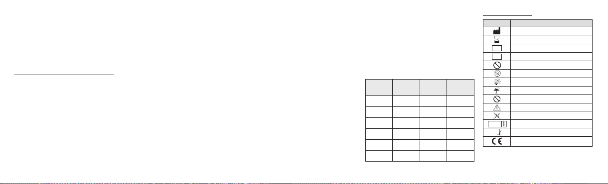
Größenbereich
Farbcode 1 ml 2 ml
(µm)
40-120 Orange S110GH S120GH
100-300 Gelb S210GH S220GH
300-500 Blau S410GH S420GH
500-700 Rot S610GH S620GH
700-900 Grün S810GH S820GH
900-1200 Violett S1010GH S1020GH
Angaben auf der Verpackung:
Symbol
Bezeichnung
Hersteller: Name und Adresse
Verwendbarkeitsdatum: Jahr-Monat
Chargenbezeichnung
LOT
Bestellnummer
REF
Nicht resterilisieren
Inhalt bei beschädigter Verpackung nicht verwenden
Vor Sonnenlicht schützen
Vor Nässe schützen
Nicht zur Wiederverwendung
Vorsicht – Siehe Gebrauchsanweisung
Nicht pyrogen
Sterilisation mittels Dampf
Untere Temperaturgrenze
0°C
CE-Kennzeichnung - Angabe der Benannten Stelle:
All e s chwe rwie gend en o der le bens bedr ohli chen
unerwünschten Ereignisse oder Todesfälle in Zusammenhang
mit der Anwendung von Embosphere Mikrosphären müssen
dem Hersteller des Produktes gemeldet werden.
0459
8
Page 9

ESPAÑOL
DESCRIPCIÓN
Las Microesferas Embosphere
acrílico, calibradas con precisión, no absorbibles, hidrófilas y
biocompatibles,que están impregnadas con gelatinaporcina, y
están disponibles en una amplia gama de tamaños y
concentraciones.
PRESENTACIÓN
Jeringa precargada de 20 ml con cono Luer-lock estándar, en
un envase individual consistente en un blíster sellado con una
película Tyvek
plástico. Junta del émbolo de tres faldas,de elastómero.
Contenido: 1 ml o 2 ml demicroesferas en solución de NaCl al
0,9%, apirógena y estéril.
INDICACIONES
Las Microesferas Embosphere están diseña das para la
oclusión con fines terapéuticos o preoperatorios de vasos
sanguíneos en los procedimientos siguientes:
- Emboliza ción d e proc esos y tumo res hi pervasculares,
incluidos fibroides uterinos,meningiomas, etc.
- Embolización de malformaciones arterio-venosas.
- Embolización hemostática.
Las microesferas de 40 a 120 µm están diseñadas más
específicamente para la embolización de meningiomas y
tumores al hígado.
®
son microesferas de polímero
®
desprendible. Émbolo y obturador con roscade
CONTRAINDICACIONES
- Pacientes intolerantes a los procedimientos de oclusión
vascular.
- Imposibilidad de colocar correctamente el catéter a causa de
la anatomía vascular.
- Arterias nutrientes demasiado pequeñas para aceptar las
microesferas seleccionadas.
- Presencia o sospecha de vasoespasmo.
- Presencia de arterias distales que riegan directamente
nervios craneales.
- Presencia de anastomosis extra-intracraneal patentes.
- shunts arterio-venosas de altoflujo o de diámetro mayor que
el de las microesferas seleccionadas.
- Embolia pulmonar.
- Aterosclerosis grave.
- Pacientes con alergia conocida a la gelatina.
Las microesferas de 40 a 120 µm y 100 a 300 µm no están
recomendadas para utilizarse en la circulación bronquial.
POSIBLES COMPLICACIONES
La embolización vascular es un procedimiento de alto riesgo.
Pueden presentarse complicaciones en cualquier momento,
tanto durante el procedimiento como después de él, y estas
pueden incluir,entre otras, las siguientes:
- Accidente vascular cerebral o infarto cerebral
- Oclusión de vasos en territorios sanos
- Rotura de vasos y hemorragia
- Déficits neurológicos
- Infección o hematoma en el sitio de la inyección
- Reacción alérgica, irritaciones cutáneas
- Dolor y fiebre transitorias
- Vasoespasmo
- Muerte
- Isquemia en un lugar no deseado, incluido ictus isquémico,
infarto isquémico (incluido infarto de miocardio) y necrosis
tisular
- Ceguera, pérdida auditiva,pérdida del olfato y parálisis
- Informac ión ad icional se enc uentra en la sección
Advertencias
ADVERTENCIA
NO UTILICE ESTA JERINGA PRECARGADA PARA INYECTAR
DIRECTAMENTE LAS MICROESFERAS EMBOSPHERE. ESTA
ES UNAJERINGA «RESERVORIO».CONSULTEEL APARTADO
DE INSTRUCCIONES.
Las Microesf eras Embos phere debe rán ser utili zadas
únicamente por médicos especializad os y formados en
procedimientosde embolización. Eltamaño de las microesferas
y su cantidad deben ser seleccionados cuidadosamente de
acuerdo con la lesión que se va a tratar, quedando dicha
selección enteramente bajo la responsabilidad del médico.
Únicamente el médico puede decidir el momento idóneo para
detener la inyección de microesferas.
No utilice este producto si el blíster, lapelícula desprendible, el
obturador con rosca o la jeringa están dañados. Este es un
producto desechable. Deseche las jeringas abiertas después
de su uso. Todos los procedimientos deben realizarse de
acuerdo con una técnica aséptica.
Para uso en un único paciente solamente - El contenido
se suministra estéril
No reutilice, reprocese ni reesterilice este producto. Su
reutilización,reprocesamiento o reesterilización podríanafectar
a la integridad estructural del dispositivo o provocar su fallo, lo
que a su vez podría ocasionar lesión, enfermedad o la muerte
del paciente. Su reutilización, reprocesamie nto o
reesterilización podría n constitu ir tambié n un riesgo de
contaminación del dispositivo o causar infecciónen el paciente
o infección cruzada, incluido, entre otras cosas,la transmisión
de enfermedades infecciosas de un paciente a otro. La
contamina ción del di spositivo puede prov ocar lesió n,
enfermedad o la muerte del paciente.
ADVERTENCIAS
• Las Microesferas Embosphere contienen gelatina de origen
porcino y, por lo tanto, podrían provocar un a reacción
inmunitaria en pacientes conhipersensibilidad al colágeno o la
gelatina. Debe considerarse previa y detenidamente el uso de
este producto en pacientes que se sospecheque son alérgicos
a inyecciones que contienen estabilizantes de gelatina.
• Estudios realizados han mostrado que las Microesferas
Embosphere no forman agregado s y, por consiguiente,
penetran más pr ofundamente en la va sculatura en
comparación con partículas de alcohol polivinílico (PVA) de
tamaño pare cido. Debe te nerse cui dado para e legir
Microesferas Embosphere de mayor tamaño al embolizar
malformaciones arterio-venosas con shunts grandes para
evitar el paso de las esferas a la circulación pulmonar o
coronaria.
• Algunas de las Microesferas Embosphere podrían estar
ligeramente fuera de la gama.Por consiguiente, tras considerar
la apariencia angiográfica arterio-venosa, el médico debe
asegurarse de seleccionar cuidadosamente el tamaño de las
Microesferas Embosphere de acuerdo con el tamaño de los
vasos diana y el nivel de oclusión deseado en la vasculatura.El
tamaño de lasMicroesferas Embosphere debe seleccionarse de
modo que se impida el paso de la arteria a la vena.
• Debido alas importantes complicacionesde una embolización
fallida, se d ebe proceder con sumo cuidad o en los
procedimientos que implican la circulación extracraneal que
abarca la cabeza y el cuello, y el médico debe sopesar
detenidam ente los ben eficios po tenciales de aplicar
embolización frente a los riesgos y las posibles complicaciones
del procedim iento. Estas complicacion es p ueden incluir
ceguera, pérdida auditiva,pérdida del olfato , parálisisy muerte.
• El paciente puedesufrir una lesión cutánea seria inducidapor
la radiación como consecuencia de largos períod os de
exposición radioscópica, un paciente de diámetro grande,
proyecciones radiográficasen ángulo y varias series de registro
de imágenes o radiografías. Consulte el protocolo clínico de su
centro para asegurar que se aplique la dosis de radiación
correcta a cada tipo específico de procedimiento realizado. Los
médicos deben monitorizar a los pacientes que puedan correr
riesgo.
• La lesión inducida por radiación en el paciente podría ser de
aparición diferida. Debe advertirse a los pacientes de los
posibles efectossecundarios de la radiacióne indicarles a quién
deben contactar si presentan síntomas.
• Preste mucha atención a los signos de una embolización de
9
Page 10
regiones n o diana. Duran te la inye cción, monit orice
STERILIZE
2
2
STERILE
cuidadosamente las constantesvitales del paciente, incluido la
SaO
(por ejemplo, hipoxia, cambios en el sistema nervioso
2
central). Si hay signos de embolización de regiones no diana o
si el pacien te presenta síntomas, considere termi nar el
procedimiento, investigar la presencia de posibles shunts o
aumentar el tamaño de las microesferas.
• Si durantela inyección de las microesferas no aparecen clara
y rápidam ente señales ang iográficas de e mbolización,
considere aumentar el tamaño de las microesferas.
Advertenciassobre el uso de pequeñas
• Siempre quese contemple el usode medios embólicos deun
diámetro más pequeño que la capacidad de resolución del
equipo de imágenes, deberá considerarse detenidamente
dicha opción. La presencia de anastomosis arterio-venosas,
vasos ramificados hacia fuera del área diana o va sos
emergentes no evidentes antes de la embolización puede
derivar en una e mbolización de region es no diana y
complicaciones graves.
• Las micro esferas de tamaño in ferior a 100 micras
generalme nte migran distalmente a las tribu tarias
anastomóticas y, por lo tanto, tienen más probabilidad de
interrumpir la circulación al tejido distal. Existe una mayor
probabilidad delesión isquémica con eluso de microesferas de
tamaño más pequeño, ydeben considerarse las consecuencias
de esta lesión antes de la emboli zación. Las posibles
consecuencias incluyenhinchazón, necrosis, parálisis, absceso
y síndrome posembolización más intenso.
• La hinchazón posembolización podría provocar isquemia en
microesferas
el tejido adyacente alárea diana. Deben tomarse precauciones
para evitar la isquemia de tejido no tolerante y no deseado,
como es el caso del tejido nervioso.
INSTRUCCIONES
• Coloque el catéter en el sitio deseado y realice una
angiografía inicial para evaluar el riego sanguíneo de la lesión.
• Las Microesferas Embosphere están disponibles en una
gama de tamaños. Debido al potencial de embolización fallida
y la variabilidad inherente de los tamaños de las esferas, el
médico debe asegurarse de seleccionar cuidadosamente el
tamaño de las Microesferas Embosphere según el tamaño de
los vasos diana en el nivel deseado de oclusión de la
vasculatura.
• Seleccione cuidadosamente el tamaño de las microesferas
de acuerdo con el tamaño de los vasos identificados y del
catéter utiliz ado. La s Mi croesferas E mbosphere son
microesferas flexibles que toleranuna compresión temporaldel
20 al 30% para facilitar el paso a través de microcatéteres.
Estudios realizados hanmostrado una correlación directa entre
el tamaño de las microesferas y el tamaño de los vasos
ocluidos.
• Examine el envase y la jeringa antes de utilizarlos para
asegurar que estén intactos. La superficieexterna de la jeringa
es estéril.
• Desenrosque el obturador de la jeringa precargada de
Microesferas Embosphere e introduzca suavemente medio de
contraste directamente en la jeringa reservorio.
• La suspensión ideal se suele obtener con una mezcla de
50% de medio de contraste y 50% de solución salina. Para
obtener una susp ensión homo génea de Micro esferas
Embosphere, invierta suavemente la jeringa de 20 ml varias
veces. Para obtener una suspensión más diluida, se puede
añadir medio de contraste y soluciónde NaCl al 0,9% en igual
proporción.
¡No utilice la jeringaprecargada de 20 ml para inyectar
•
Microesferas Embosphere a través del catéter!
• Retire todo el aire de la jeringa y conéctela a un conector de
la llave de tres vías.
• Aspire la suspensión con una jeringuilla(1 a 3 ml) conectada
a otro conector de la llave de tres vías. Evite los movimientos
de avance y retroceso para reducir el riesgo de introducir aire
en el sistema. Compruebe que se utilice la cantidad y la
concentración deseadas de microesferas.
• Extraiga todo el aire de la jeringa.
• Enrosque la jeringa en el conector del catéter, usando el
conector Luer-lock macho de la llave.
• Abra la llave para conectar la jeringa de inyección con el
catéter.
• Bajo control fluoroscópico continuo, infunda lentamente
microesferas en el fluj o sanguíneo. Inyecte siempre en
condiciones de flujo libre. El reflujo de microesferas puede
inducir isquemia inmediata en tejidos o vasos sanos.
• Continúe con la infusi ón h asta que se obtenga la
desvascularización deseada.Estudios realizados han mostrado
que las Microesferas Embosphere penetran más distalmente
en la lesión que las partículas de alcohol polivinílico (PVA) de
tamaño parecido.La reducción del riego de sangre arterial a la
lesión es por lo tanto más progresiva.
• Al finalde la infusión, retireel catéter al tiempo que mantiene
una suave aspiración para no desalojar ninguna de las
microesferas residuales que todavía quede en el interior del
catéter,y luego cierre la llave de tres vías.
• Retire el catéter.
• Deseche las microesferas Embosphere restantes y las
jeringas utilizadas.
CONSERVACIÓNY ALMACENAMIENTO
Las Microesferas Embospheredeben almacenarse en un lugar
fresco, seco y oscuro, en su jeringa y envase originales.
Utilícelas antes de la fecha indicada en las etiquetas de la caja
y del blíster.No las congele.
Gama de
tamaños
(µm)
Código de
color
1 ml 2 ml
40-120 Naranja S110GH S120GH
100-300 Amarillo S210GH S220GH
300-500 Azul S410GH S420GH
500-700 Rojo S610GH S620GH
700-900 Verde S810GH S820GH
900-1200 Violeta S1010GH S1020GH
Información enel envase:
Símbolo
Designación
Fabricante: Nombre y dirección
Utilizar antes del: año-mes
Código de lote
LOT
Número de catálogo
REF
No reesterilizar
No utilizar si el envase está dañado
Mantener al abrigo de la luz del sol
Mantener seco
No reutilizar
Atención - Consultar las Instrucciones de uso
Apirógeno
Esterilizado utilizando vapor
Límite inferior de temperatura
0°C
Logotipo de la marca CE - Identificación del organismo
Todos los eventos adversos serios o potencialmente mortales o
muertes asociadas con el uso de las Microesferas Embosphere
deben notificarse al fabricante del dispositivo.
notificado: 0459
10
Page 11

ITALIANO
DESCRIZIONE
Le Microsfere Embosphere
acrilico calibrate con precisione, biocompatibili, idrofile, non
riassorbibili e impregnate di gelatina porcina. Sono disponibili
in una vasta gamma di misure e concentrazioni.
CONFEZIONAMENTO
Siringa da 20 ml con punta Luer Lock standard preriempita,
confezionata singolarmente in blister sigillato con pellicola a
strappo in Tyvek
Guarnizione dello stantuffo in elastomero, con tre ner vature.
Contenuto: 1 ml o 2 ml di microsfere in soluzione fisiologica
(0,9% di NaCl) apirogena sterile.
INDICAZIONI
Le Microsfere Embosphere sono previste per l’occlusione dei
vasi sanguigni a scopi terapeutici o preoperatori nell’ambito
delle seguenti procedure:
- embolizzazione di tumori e processi ipervascolari, inclusi
fibromi uterini, meningiomi, ecc.
- embolizzazione di malformazioni arterovenose
- embolizzazione a scopo di emostasi
Le mic rosfere da 40-120 µm son o con cepite più
specificamente per l’embolizzazione dei meningiomi e dei
tumori epatici.
®
sono microsfere in polimero
®
. Tappo a vite in plastica e stantuffo.
CONTROINDICAZIONI
- Pazienti non in grado di tollerare procedure di occlusione
vascolare.
- Anatomia vascolare in grado di precludere il corretto
posizionamento del catetere.
- Arterie affluenti troppo piccole per accogliere le microsfere
selezionate.
- Presenza o sospetto di vasospasmo.
- Presenza di arterie distali che irrorano direttamente i nervi
cranici.
- Presenza di anastomosi extra-intracraniche pervie.
- Shunt arteriovenosi con flusso elevato o con un diametro
superiore a quello delle microsfere selezionate.
- Embolia polmonare.
- Aterosclerosi grave.
- Pazienti con allergia nota alla gelatina.
Le microsfere da 40-120 µm e da 100-300 µm non sono
consigliate per l’uso nella circolazione bronchiale.
POTENZIALI COMPLICANZE
L’embolizzaz ione vascolare è una procedu ra al tamente
rischiosa. Le complicanze che possono verificarsi in qualsiasi
momento durante o dopo la procedura includono, senza
limitazioni, le seguenti:
- ictus o infarto cerebrale
- occlusione di vasi di aree sane
- rottura del vaso ed emorragia
- deficit neurologici
- infezione o ematoma in corrispondenza del sito di iniezione
- reazione allergica, irritazioni cutanee
- dolore e febbre transitori
- vasospasmo
- decesso
- ischemia in un sito indesiderato, inclusi ictus ischemico,
infarto ischemico (compreso infarto miocardico) e necrosi
tissutale
- cecità, perditadell’udito, perdita dell’odorato e/o paralisi
- ulteriori informazioni si trovano nella sezione Avvertenze
ATTENZIONE
NON USARE LA SIRINGA PRERIEMPITA PER INIETTARE
DIRETTAMENTE LE MICROSFERE EMBOSPHERE. LA
PRESENTE È SOLAMENTE UNA SIRINGA DI
“PREPARAZIONE”. CONSULTARE ILPARAGRAFO RELATIVO
ALLE ISTRUZIONI PER L’USO.
Le Microsfere Embos phere devono essere usate
esclusivamente da medici esperti e debitamente addestrati
nelle procedure di embolizzazione vascolare. La misura e la
quantità di microsfere vanno attentamente selezionate in base
alla lesione da trattare; la responsabilità di questa selezione
spetta esclusivamente al medico. Solo il medico, inoltre, può
decidere il momento più opportuno per concludere l’iniezione
delle microsfere.
Non utilizzare il prodotto se la confezione blister, la pellicola a
strappo, il tappo a vite o la siringa presentano danni. Il
presente è un prodotto monouso. Dopo l'uso, gettare le
siringhe aperte.Tutte le procedure devono essereeseguite con
tecniche asettiche.
Esclusivamente per l’uso in un singolo paziente Prodottosterile
Non riutilizzare, rigenerare né risterilizzare. Il riutilizzo, il
ricondi zionamento o la ri sterilizzazio ne p ossono
compromettere l'integrità strutturale delprodotto e/o causarne
il malfunzionamento che, a sua volta, può provocare lesioni,
malattie o la mo rte del paziente. Inoltre, il riu tilizzo, il
ricondizionamento o la risterilizzazione possono determinare il
rischio di contaminazione del prodotto e/o causare infezioni al
paziente o infezioni crociate tra pazienti, inclusa, tra le altre,la
trasmissione di malattie infettive da un paziente all'altro. La
contaminazione del prodotto può causare lesioni, malattie o la
morte del paziente.
AVVERTENZE
• Le Microsfere Embosphere contengono gelatina di origine
porcina e potrebbero quindi causare una reazione immunitaria
nei pazienti con ipersensibilità al collagene o alla gelatina.
Prima di usare questo prodotto, è necessario valutar e
attentamente i pazienti con sospetta allergia ai materiali
iniettabili contenenti gelatine stabilizzanti.
• Gli studi condotti hanno dimostrato che le Microsfere
Embosphere non formano aggregati e penetrano quindi a
maggiori prof ondità nel sistema vascolare rispett o alle
partice lle in PVA di dimen sioni si mili. Nel cas o di
embolizzazione di malformazioni arteriovenose con shunt di
grandi dimensioni è quindi necessario selezionare Microsfere
Embosphere di misura maggiore, allo scopo di evitarne il
passaggio nella circolazione polmonare o coronarica.
• È possibile che le misure di alcune delle Microsfere
Embosphere siano leggermente fuori gamma; il medico deve
quindi accertarsi di selezionare con cautela le Microsfere
Embosphere della misura idonea in base alle dimensioni dei
vasi interessati al livellodi occlusione previsto; questa selezione
deve inoltre tenere in considerazione l’aspetto arteriovenoso
desunto da lle immagini angio grafiche. La misura dell e
Microsfere Embosp here de ve esse re tale da evitarn e il
passaggio da arteria a vena.
• A causa delle gravi complicanze di un’embolizzazione
sbagliata, è necessario prestare estrema cautela in tutte le
procedure che coinvolgono la circolazione extracranica del
capo e del collo; il medico deve inoltre ponderare attentamente
i possibili benefici dell’embolizzazione a fronte dei rischi e delle
potenziali complicanze della procedura. Queste complicanze
possono i ncludere cecit à, perdita d ell’udito, perdi ta
dell’odorato, paralisi e morte.
• Lunghi period i di esposi zione del paziente ai sistemi
fluoroscopici, una grossa corporatura, proiezioni radiografiche
inclinate e sessioni di imaging o radiografie multiple possono
provocare lesioni cutanee indotte dalle radiazioni. Attenersi al
protocollo in vigore pressola struttura sanitaria di appartenenza
per accertarsi di erogare la corretta dose di radiazioni per
ciascun tipo specifico di procedura eseguita. I medici devono
monitorare i pazienti potenzialmente a rischio.
• L’insorgenza delle lesioni indotte da radiazioni può avere
luogo in fase postoperatoria. I pazienti devono essere a
conoscenza dei potenziali effetti collaterali delle radiazioni e
devono sapere a chi rivolgersi in caso di sintomi.
• Prestare molta attenzione a eventualisegni di embolizzazione
fuori dal vaso interessato. Durante l’iniezione, monitorare
11
Page 12
attentamente i segni vitali del paziente, inclusa la saturazione
STERILIZE
2
2
STERILE
di ossigeno nel sangue (per rilevare, ad esempio, ipossia o
variazioni a carico del sistema nervoso centrale). Se si
riscontrano segni di embolizzazione fuori dal vaso interessato
o se il paziente sviluppa sintomi, considerare l’opportunità di
terminare la procedura, di investigare la possibile presenza di
shunting o di aumentare le dimensioni delle microsfere.
• Se l’evidenza angiografica di embolizzazione non diventa
rapidamente evidente durante l’iniezione delle microsfere,
prendere in considerazione ilpassagg ioa microsfere di misura
superiore.
Avvertenze relative all’uso di
• È necessario ponderare attentamente l’uso di agenti per
emboliz zazione con diametro inf eriore alla capacità di
risoluzione dell’apparecchiatura di imaging in dotazione. La
presenza di anastomosi arteriovenose, di diramazioni laterali
che si dipartono dall’area interessata o di vasi affluenti non
evident i prima de ll’embolizza zione po ssono pr ovocare
embolizzazioni in aree non previste e gravi complicanze.
• Le microsfere di misura inferiore a 100 micron migrano
generalment e in direzi one di stale rispetto agli affluenti
anastomotici ed è quindi più probabile che blocchino la
circolazione a tessuto distale. Una maggiore probabilità di
lesioni ischemiche deriva quindi dall’uso delle microsfere di
misure più p iccole: è pe rtanto nece ssario ponde rare
attentamente le conseguenze di questotipo di lesione prima di
procedere all’emb olizzazione. Le potenziali conseguenze
includono tumefazione, necrosi, paralisi, ascesso e/o una
sindrome post-embolizzazione più grave.
microsfere
• La tum efazione pos t-embolizzaz ione p uò pro vocare
ischemia ai tessuti adiacenti all’area interessata. È necessario
fare attenzione a evitare tessuti non in grado di tollerare
l’ischemia e non direttamente interessati dalla procedura,
come il tessuto nervoso.
ISTRUZIONI
• Posizionare il catetere in corrispondenza del sito desiderato
ed eseguire un’angiografia al basale per valutare l’apporto
ematico alla lesione.
• Le Microsfere Embosp here sono disponibili in varie
dimensi oni. A causa della possi bilità di errori
nell’embolizzazione e dell’inerente variabilità della dimensione
delle sfere, il medico deve essere sicuro di selezionare
accuratamente la grandezza delle Microsfere Embosphere in
base alla dimensione dei vasi da trattare nel livello desiderato
di occlusione nella vascolatura.
• Selezionare con cura le microsfere della misura idonea in
base alle dimensioni dei vasi identificati e del catetere usato.
Le Microsfere Embosphere sono particelle flessibili che
supportando una compressione temporanea compresa tra il
20 e il 3 0% per agevola rne il passagg io attr averso i
microcateteri. Gli studi hanno dimostrato una correlazione
diretta tra la misura delle microsfere e le dimensioni dei vasi
occlusi.
• Prima dell’uso, esaminare la confezione e la siringa per
accertarsi che siano intatte. La superficie esterna della siringa
è sterile.
• Svitare il tappo della siringa di Microsfere Embosphere
preriempita e aspirare delicatamente il mezzo di contrasto
direttamente nella siringa di preparazione.
• La sospensione ideale si ottiene generalmente aggiungendo
una miscela al 50% di mezzo di contrasto e al 50% di
soluzione fisiologica. Per ottenere una sospensione omogenea
di Mic rosfere Emb osphere, cap ovolgere de licatamente
svariat e vo lte l a si ringa da 20 ml. P er ot tenere una
sospensione più diluita, è possibile aggiungere mezzo di
contrasto e soluzione fisiologica (0,9% di NaCl) in proporzioni
uguali.
• Non usare la siringa preriempita da 20 ml per iniettare
le Microsfere Embosphere attraverso il catetere.
• Espellere tutta l’aria dalla siringa e collegarla a uno dei
raccordi del rubinetto a tre vie.
• Aspirare la sospensione usando una piccola siringa (da 1 a
3 ml) collegata a un altro raccordo del rubinetto a tre vie.
Evitare movimenti avanti e indietro per ridurre il rischio di
introdurre aria nel sistema. Accertarsi di usare la quantità e la
concentrazione desiderata di microsfere.
• Espellere tutta l’aria dalla siringa.
• Avvitare la siringa al connettore del catetere usando il
raccordo Luer Lock maschio del rubinetto.
• Aprire il rubinetto per collegare la siringa di iniezione al
catetere.
• Sotto os servazione f luoroscopica contin ua, infondere
lentamente le microsfere nel flusso sanguigno. Eseguire
sempre l’iniezione in condizioni di flusso libero. Il riflusso di
microsfere può provocare l’ischemia immediata di tessuti o
vasi sani.
• Continuare l’infusione fino a ottenere la devascolarizzazione
desiderata. Gli studi hanno dimostrato che le Microsfere
Embosphere hanno una maggiore penetrazione distale nella
lesione rispetto alle particelle in PVA di dimensioni simili. La
riduzione dell’apporto di sangue arterioso alla lesione avviene
quindi in modo più progressivo.
• Alla fine dell’infusione, rimuovere il catetere continuando ad
aspirare delicata mente p er evit are di libera re even tuali
microsfere residue ancora presenti nel catetere , quindi
chiudere il rubinetto a tre vie.
• Rimuovere il catetere.
• Alla conclusione della procedura, le siringhe usate e le
Microsfere Embosphere residue devono essere gettate.
CONSERVAZIONE ESCADENZA
Le microsfere Embosphere devono essere conser vate in un
luogo fresco, asciutto e buio nelle siringhe e nelle confezioni
originali. Vanno usate entro la data indicata sull’etichetta della
scatola esterna e della confezione blister. Non congelarle.
Gamma di
misure (µm)
Codifica
cromatica
40-120 Arancio S110GH S120GH
100-300 Giallo S210GH S220GH
300-500 Azzurro S410GH S420GH
500-700 Rosso S610GH S620GH
700-900 Verde S810GH S820GH
900-1200 Viola S1010GH S1020GH
1 ml 2 ml
Informazioni sulleconfezioni
Símbolo
Significato
Produttore: nome e indirizzo
Data di scadenza: anno-mese
Codice del lotto
LOT
Numero di catalogo
REF
Non risterilizzare
Non utilizzare se la confezione è danneggiata
Tenere al riparo dalla luce solare
Tenere all’asciutto
Non riutilizzare
Attenzione: consultare le istruzioni per l'uso
Apirogeno
Sterilizzato a vapore
Limite inferiore di temperatura
0°C
Marchio CE Numero di identificazione dell’ente
Tutti gli eventi avversi gravi o potenzialmente letali e i decessi
associati all’uso delle Microsfere Embosphere devono essere
segnalati al produttore del dispositivo.
notificato: 0459
12
Page 13

PORTUGUÊS
DESCRIÇÃO
As Microesferas Embosphere® são microesferas de polímero
acrílico biocompatíveis, hidrófilas, não absorvíveis, calibradas
com precisão, impregnadas com gelatina de origem suína e
encontram-se disponíveis numa vasta variedade de tamanhos
e concentrações.
APRESENTAÇÃO
Seringa de 20 ml pré-cheia, com extremidade Luer-lock
standard, aconditionada individualmente num blister rígido
selado com uma película destacável Tyvek®. Tampa de rosca
e êmbolo em plástico. Junta do êmbolo em elastómero com
três rebordos.
Conteúdo: 1 ml ou 2 ml de microesferasnuma solução estéril,
apirogénica de NaCl a 0,9%.
INDICAÇÕES
As Microesferas Embosphere foram concebidas para a oclusão
dos vasos sanguíneos para fins terapêuticos ou pré-operatórios
nos seguintes procedimentos:
- Emboli zação de tumores e processo s hip ervasculares ,
incluindo fibromas uterinos, meningiomas, etc.
- Embolização de malformações arterio-venosas.
- Embolização hemostática.
As microesferas de 40-120 µm são mais especificamente
destinadas para a embolização de meningiomas e tumores no fígado.
CONTRA-INDICAÇÕES
- Doentes com intolerância aos procedimentos de oclusão
vascular.
- Anatomia vascular que impeça a colocação correcta do
cateter.
- Artérias de irrigação demasiado pequenas para aceitar as
microesferas seleccionadas.
- Presença ou suspeita de vasoespasmo.
- Presença de artérias distais que fornecem directamente os
nervos cranianos.
- Presença de anastomoses extra a intracranianas patentes.
- Shunts arterio-ve nosos d e fluxo eleva do ou com um
diâmetro superior ao das microesferas seleccionadas.
- Embolia pulmonar.
- Ateroesclerose grave.
- Doentes com alergia conhecida à gelatina.
40-120 µm e 100-300 µm não são recomendadas para
utilização na circulação brônquica.
POSSÍVEISCOMPLICAÇÕES
A embolização vascular é um procedimento de risco elevado.
Podem surgir complicaçõesa qualquer altura, durante ou após
o procedimento e podem incluir, entre outras, as seguintes:
- Acidente vascular cerebral ou enfarte cerebral
- Oclusão de vasos em zonas saudáveis
- Ruptura vascular e hemorragia
- Défices neurológicos
- Infecção ou hematoma no local da injecção
- Reacção alérgica, irritações cutâneas
- Dor e febre passageiras
- Vasoespasmo
- Morte
- Isquemia numa localização indesejada, incluindo acidente
isquémico, enfarte isquémico (incluindo enfarte do miocárdio)
e necrose tecidular
- Cegueira, perda de audição, perda de olfacto e/ou paralisia
- Pode consu ltar infor mações ad icionais n a s ecção
Advertências
ATENÇÃO
NÃO UTILIZE ESTA SERINGA PRE-CHEIA PARA INJECTAR
DIRECTAMENTEAS MICROESFERAS EMBOSPHERE.ESTAÉ
UMA SERINGA “RESERVATÓRIO”. CONSULTE O
PARÁGRAFO RELATIVO ÀSINSTRUÇÕES
As Microesferas Embosphere apenas devem ser utilizadas por
médicos especializados e com formação em procedimentos
de embolização vascular. O tamanho e a quantidade das
microesferas devem ser cuidadosamente seleccionados de
acordo com a lesão que irá ser tratada, totalme nte à
responsabilidade do médico. Apenas o médico pode decidir o
momento mai s adequado par a parar a in jecção de
microesferas.
Não utilize se o blisterrígido, a película destacável, a tampade
rosca ou a seringa estiverem danificados. Este produto é
descartável. Elimine as seringas abertas apósa sua utilização.
Todos osprocedimentos devem ser efectuados de acordo com
uma técnica asséptica.
Para utilização num único doente – O conteúdo é
fornecidoestéril
Não reutilize, reprocesse ou reesteri lize. A reutilizaçã o,
reprocessamento ou reesterilização podem comprometer a
integridade estrutural do dispositivo e/ou resultar na falha do
mesmo, o que,por sua vez, pode resultar em lesão,doença ou
morte do d oente. A reutiliza ção, reprocessam ento ou
reesterilização podem também originar risco de contaminação
do dispositivo e/ou provocar infecção do doente ou infecção
cruzada, incluindo, entre outras, a transmissão de doença(s)
infecciosa(s) de um doente para outro. A contaminação do
dispositivo pode provocar lesão, doença ou morte do doente.
ADVERTÊNCIAS
• As Microesferas Embosphere contêm gelatina de origem
suína e, portanto, podem provocar uma reacção imune em
doentes que sejam hipersensíveis ao colagénio ou à gelatina.
Deve prestar-se umaespecial atenção antes de se utilizar este
produto em doentes com suspeita de serem alérgicos a
injecções que contenham estabilizadores de gelatina.
• Estudos demonstraram que as Microesferas Embosphere
não formam agregados e, por conseguinte, penetram com
maior profundidade na vasculatura comparativa mente a
partículas em PVA de tamanho semelhante. Deve ter-se
cuidado na escolha de Microe sferas Embosphere com
tamanho superior quando se efectua a embolização de
malformações arterio-venosas com shunts grandes de modo
a evitar a passagem de esferas para acirculação pulmonar ou
coronária.
• Algumas das Microesfera s Emb osphere podem estar
ligeiramente fora da gama, portanto, o médico deve escolher
cuidadosamente o tamanho das Microesferas Embosphere de
acordo com o tamanho dos vasos alvo no nível de oclusão
pretendido na vasculatura e após consideração do aspecto
angiográfico arterio-venoso. O tamanho das Microesferas
Embosphere deve ser seleccionado de modo a impedir a
passagem da artéria para a veia.
• Devid o às c omplicações sign ificativas de falh a na
embolizaç ão, deve ter-se extr ema ca utela em q ualquer
procedimento que envolva a circulação extracraniana incluindo
a cab eça e o p escoço, e o médi co deve po nderar
cuidado samente as pote nciais vantag ens de utiliza r a
emboliz ação comparativ amente aos riscos e potenc iais
complicações do procedimento. Estas complicações podem
incluir cegueira, perda de audição, perda de olfacto,paralisia e
morte.
• Pode ocorrer lesão grave na pele do doente induzida por
radiação devido a longos períodos de exposição fluoroscópica,
diâmetro grande do doente, projecções angulares de raio-x e
diversas séries de registo de imagensou radiografias. Consulte
o protocolo clínico da sua instituição para garantir que é
aplicada a dose correcta de radiação para cada tipo específico
de procedimento executado. Os médicos devem também
monitorizar os doentes que possam estar em risco.
• O início da lesão induzida por radiação no doente pode ser
atrasado. Os doentes devem seraconselhados sobre os efeitos
colaterais da radiação e sobre quem devem contactar caso
apresentem sintomas.
13
Page 14
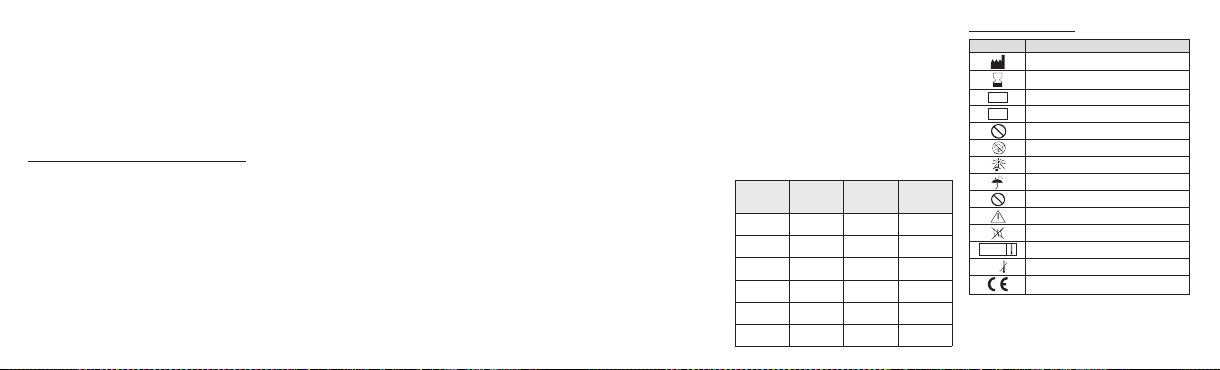
• Preste especial atenção a sinais de embolização mal
STERILIZE
2
2
STERILE
direccionada. Durante a injecção monitorize atentamente os
sinais vitais do doente de modo a incluir SaO2 (por ex.,
hipoxia , alte rações no SNC). Consid ere termina r o
procedimento, investigar a possibilidade de existir um shunt
ou aumentar o tamanho dasmicroesferas caso ocorram sinais
de mau direccionamento ou se desenvolvam sintomas no
doente.
• Considere aumentar o tamanho das microesferas caso os
sinais angi ográficos de e mbolização não apareç am
rapidamente evidentes durante a injecção das microesferas.
Advertências quantoà utilização de microesferas pequenas
• D eve prestar-se pa rticular atenç ão sempre que for
considerada a utilização de agentes embólicos cujo diâmetro
seja inferior ao da capacidade de resolução do equipamento
de imagiologia. A presença de anastomoses arterio-venosas,
ramificações de vasos sanguíneos que se afastem da área
alvo ou vaso s emergen tes não evi dentes a ntes da
emboliz ação, podem origi nar uma e mbolização mal
direccionada e complicações graves.
• Microesferas com tamanho inferior a 100 mícrones irão
geralmente migrar de forma distal para vasos de irrigação
anastomóticos e, portanto, têm maior probabilidade de
terminar a circulação ao tecido distal. O maior potencial para
lesões isquémicas resulta da utilização de microesferas de
tamanho mais peq ueno e deve ser d ada atenção à
consequência desta lesão antes da embolização. As possíveis
consequências incluem inchaço, necrose, paralisia, abcesso
e/ou síndrome pós-embolização mais acentuado.
• O inchaço pós-embolização pode resultar em isquemia no
tecido adjacente à área alvo. Deve ter-se cuidado para evitar
a isquemia de tecido intolerante e não definido como alvo, tal
como o tecido nervoso.
INSTRUÇÕES
• Posicion e o cateter no loc al pretendid o e execute
angiografia de base para avaliar a irrigação de sangue da
lesão.
• As Microesferas Embosphere estão disponíveis numa
diversidade de tamanhos. Devido ao potencial de falha da
embolização e à inerente variabilidade dos tamanhos das
esferas, o médico deve ter a certeza de que selecciona
cuidadosamente o tamanho das Microesferas Embosphere de
acordo com o tamanho dos vasos visados, no nível desejado
de oclusão na vasculatura.
• Seleccione cuidadosamente o tamanho das microesferasde
acordo com o tamanho dos vasos identificados e do cateter
utilizado. As Microesfe ras Embosphere são microesferas
flexíveis que suportam compressão temporária de 20 a 30%
de modo a facilitar a passagem através dos microcateteres.
Estudos demonstrar am uma correl ação di recta en tre o
tamanho das microesferas e o tamanho dos vasos obstruídos.
• Inspeccione a embalagem e a seringa antes de utilizar para
se certificar de que se encontram intactas. A superfície
externa da seringa é estéril.
• Desenrosque a tampa da seringa pré-cheia de Microesfera
Embosphere e deite suave mente o mei o de contr aste
directamente para o interior da seringa reser vatório.
• A suspensão ideal é normalmente obtida com uma mistura
de 50% de meio de contraste e 50% de solução de soro
fisiológico. Para obter uma suspensão homogénea das
Microesferas Embosphere, inverta suavemente a seringa de
20 ml várias vezes. Podem ser adicionados meio de contraste
e solução de NaCl a 0,9% na mesma proporção para obter
uma suspensão mais diluída..
• Nãoutilizeaseringa de20 ml pré-cheia parainjectar as
Microesferas Embosphere através do cateter!
• Remova todo o ar da seringa e ligue-a a um conector da
torneira de três vias.
• Aspire a suspensão utilizando uma seringa pequena (1 a 3
ml) ligada a outro conector da torneira de passagem de três
vias. Evite fazer movimentos de vai e vem a fim de reduzir o
risco de introduzir ar no sistema. Verifique que a quantidade e
concentração pretendidas de microesferas são utilizadas.
• Remova todo o ar da seringa.
• Aperte aseringa ao conector do cateter utilizando oconector
Luer-lock macho da torneira.
• Abra a torneira para ligar a seringa de injecção ao cateter.
• Sob controlo fluoroscópico contínuo, realize a lenta infusão
das microesferas na corrente sanguínea. Injecte sempre em
condições de fluxo livre. O refluxo de microesferas pode
induzir isquemia imediata de tecidos ou vasos sãos.
• Prossiga a infusão atéque seja atingida a desvascularização
pretendida. Estudos demonstraram que as Microesferas
Embosphere penetram de forma mais distal na lesão do que
as partículas em PVA de tamanho semelhante. A redução do
abastecimento de sangue arterial à lesão é, portanto, mais
progressiva.
• No final da infusão, remova o cateter, mantendo ao mesmo
tempo uma aspiração suave, de modo a evitar a deslocação
de quaisquer microesferas residuais que ainda se encontrem
no interior do cateter e, de seguida, feche a torneira de três
vias.
• Remova o cateter.
• Elimine quaisquer Microesferas Embosphere restantes e as
seringas utilizadas.
CONSERVAÇÃO E ARMAZENAMENTO
As Microesferas Embosphere devem ser conservadas num
local fresco,seco e escuro na respectiva seringa eembalagem
originais. Utilize até àdata indicada nos rótulos da embalagem
exterior e do blister. Não congele.
Gama de
tamanhos
(µm)
Código de
cores
1 ml 2 ml
40-120 Laranja S110GH S120GH
100-300 Amarelo S210GH S220GH
300-500 Azul S410GH S420GH
500-700 Vermelho S610GH S620GH
700-900 Verde S810GH S820GH
900-1200 Roxo S1010GH S1020GH
Informação da embalagem:
Símbolo
Designação
Fabricante: Nome e morada
Prazo de validade : ano-mês
Código do lote
LOT
Número de catálogo
REF
Não reestirilizar
Não utilizar se a embalagem estiver danificada
Manter afastado da luz solar
Manter seco
Não reutilizar
Atenção – Consultar as Instruções de utilização
Apirogénico
Esterilizado utilizando vapor
Limite de temperatura inferior
0°C
Logótipo de marca CE - Identificação do organismo
Todos os eventos adversos graves ou ameaçando a vida, ou
causando morte, associados à utilização das Microesferas
Embosphere devem ser relatados ao fabricante do dispositivo.
notificado: 0459
14
Page 15

NEDERLANDS
BESCHRIJVING
®
-microsferen zijn biologisch compatibele, hydrofiele,
Embosphere
niet-absorbeerbare,exact gekalibreerde acrylpolymeermicrosferen
die met varkensgelatinegeïmpregneerdzijn enzijn leverbaar ineen
groteverscheidenheid aan groottenen concentraties.
LEVERINGSVORM
Voorgevulde 20ml-spuit met standaard luerlocktip, individueel
verpakt in een doordrukverpakking en afgesloten met een
®
peel-away Tyvek
drie rokken bestaand zuigeronderdeel van elastomeer.
Inhoud: 1 ml of 2 ml microsferen in een steriele, pyrogeenvrije
0,9% NaCl-oplossing.
INDICATIES
Embosphere-microsferen dienen voor het voor therapeutische
of preoperatieve doeleinden occluderen van bloedvaten bij de
volgende ingrepen:
- embolisatie van hypervasculaire tumoren en gezwellen,
waaronder vleesbomen, meningiomen enz.;
- embolisatie van arterioveneuze misvormingen;
- hemostatische embolisatie.
Microsferen van 40–120 µm dienen meer specifiek voor
embolisatie van meningiomen en levertumoren.
CONTRA-INDICATIES
- onvermogen om vaatocclusie-ingrepen te verdragen;
-deksel. Kunststof schroefdop en zuiger. Uit
- een a natomie van de b loedvaten di e een corre cte
katheterplaatsing onmogelijk maakt;
- aanvoerende slagaders die te klein zijn om ruimte te bieden
voor de geselecteerde microsferen;
- aanwezigheid of vermoeden van vaatspasmen;
- aanwezigheid van distale slagaders die de hersenzenuwen
rechtstreeks van bloed voorzien;
- aanwezi gheid van doorgankelijke anas tomosen tussen
extracraniale en intracraniale bloedvaten;
- arterioveneuze shunts met grote flow of met een diameter
die groter is dan de geselecteerde microsferen;
- longembolie;
- ernstige atherosclerose;
- een bekende allergie voor gelatine.
Microsferen van 40–120 µm en 100–300 µm zijn niet
geschikt voor gebruik in de bronchiale bloedsomloop.
MOGELIJKECOMPLICATIES
Vasculaire embolisatie is een ingreep met hoog risico.Op enig
tijdstip tijdens of na de ingreep kunnen er complicaties
optreden, waaronder onder meer:
- beroerte of herseninfarct;
- occlusie van bloedvaten in gezonde gebieden;
- ruptuur en bloeding van bloedvaten;
- neurologische defecten;
- infectie of hematoom op de injectieplaats;
- allergische reactie, huidirritaties;
- voorbijgaande pijn en koorts;
- vaatspasmen;
- overlijden;
- ischemie op een ongewenste locatie, met inbegrip van
ischemische beroerte, ischemisch infarct (met inbegrip van
myocardinfarct), en weefselnecrose.
- blind heid, gehoor verlies, ve rlies van reuk zin en/of
verlamming.
- aanvullende informatie kan in de waarschuwingen worden
gevonden
LET OP
GEBRUIK DEZE VOORGEVULDE SPUIT NIET OM
EMBOSPHERE-MICROSFEREN RECHTSTREEKS TE
INJECTEREN. DIT IS EEN ‘RESERVOIRSPUIT’. RAADPLEEG
HET GEDEELTE METINSTRUCTIES.
Embosphere-microsferen mogen uitsluitend worden gebruikt door
artsen die gespecialiseerden opgeleidzijn invasculaireembolisatie.
De grootte van de microsferen en het aantal microsferen moeten
zorgvuldig worden geselecteerd op grond van de te behandelen
laesie, geheel en al onder de verantwoordelijkheid van de arts.
Alleen de arts kan besluiten wat het meest geschikte tijdstip is om
de injectie vanmicrosferen testoppen.
Niet gebruiken als de doordrukverpakking, peel-away laag,
schroefdop of spuit beschadigd is. Ditis een wegwerpproduct.
Voer geopende spuiten na gebruik af.Alle ingrepen moeten op
aseptische wijze worden uitgevoerd.
Uitsluitend voor gebruik bij één patiënt – Inhoud bij
leveringsteriel
Niet opnieuw gebruiken, opnieuw verwerken of opnieuw
steriliseren. Hergebruik,herverwerking of hersterilisatie kan de
structurele integriteit van het hulpmiddel in gevaar brengen
en/of kan leiden tot falen van het hulpmiddel; dit kan op zijn
beurt leiden tot letsel, ziekte of overlijden van de patiënt.
Hergebruik, herverwerking of hersterilisatie kan ook een risico
van verontreiniging van het hulpmiddel vormen en/of kan
leiden tot infectie of kruisinfectie van de patiënt, waaronder
onder meer de overdracht van besmettelijke ziekten tussen
patiënten. Verontreiniging van het hulpmiddel kan leiden tot
letsel, ziekte of overlijden van de patiënt.
WAARSCHUWINGEN
• Embosphere-microsferen bevatten varkensgelatine en zouden
daaromeen immuunreactieteweeg kunnen brengen bijpatiënten
die overgevoelig voor collageen of gelatine zijn. Een zorgvuldige
afweging dientplaats tevinden voordat ditproduct gebruikt wordt
bij patiënten van wie vermoed wordt dat ze allergisch zijn voor
injecties met gelatinebevattende stabilisatoren.
• Onderzoek heeft uitgewezen dat Embosphere-microsferen
geen aggregaten vormen en daarom dieper in het vaatstelsel
doordringen dan PVA-partikels van vergelijkbare grootte. Er
dient te worden gezorgd datEmbosphere-microsferen van een
grotere maat worden gekozen bij het emboliseren van
arterio veneuze misvormi ngen met grote shunts om te
voorkomen dat de microsferen in de bloedsomloop van de
longen of het hart terechtkomen.
• Het is mogelijk dat sommige van deEmbosphere-microsferen
enigszins buiten het bereik liggen; daarom moet de arts, na het
arterioveneuze voorkomen in het angiogram in aanmerking te
hebben genomen, zich ervan vergewissen dat hij/zij de grootte
van de Embosphe re-microsferen zorg vuldig selecteert
overeenkomstig de grootte van de te behandelen bloedvaten en
de gewenste mate van occlusiein het vaatstelsel. De grootte van
de Embosphere-microsferen moet zodanig worden geselecteerd
dat ze niet via de slagader in een ader terecht kunnen komen.
• Vanwege de significante complicaties van verkeerd gerichte
embolisatie dient uitzonderlijk behoedzaam te werk worden
gegaan bij ingrepen die extracraniële bloedsomloop van het
hoofd en de hals omvatten en dient de arts de mogelijke
voordelen van het toepassenvan embolisatie af te wegen tegen
de risico’s en mogelijke complicaties van de ingreep. Deze
complicaties zijn onder meer blindheid, gehoorverlies, verlies
van reukzin, verlamming en overlijden.
• De patiënt kan ernstig, door straling geïnduceerd huidletsel
oplopen als gev olg va n lang durige bloot stelling aan
doorlichting, een grote omvang (van de patiënt), schuine
röntgenprojecties en herhaalde blootstelling aan beeldvorming
of röntgenstraling. Raadpleeg het klinische protocol van uw
instelling om te zorgen dat de juiste stralingsdosis wordt
afgegev en voor elk specifi eke type ingreep da t w ordt
uitgevoerd. Artsen moeten patiënten die risico lopen bewaken.
• Het optreden van door straling geïnduceerd letsel van de
patiënt ka n ve rtraagd plaatsv inden. Patië nten moeten
voorgelicht worden over de mogelijke bijwerkingen van straling
en moeten geïnstrueerd worden met wie zij contact moeten
opnemen als zij symptomen vertonen.
• Let speciaal op tekenen van verkeerd gerichte embolisatie.
Tijdens de injectie moeten de vitale functies van de patiënt
zorgvuldig worden bewaakt, met inbegrip van SAO2 (bijv.
hypoxie, veranderingen in het CZS). Er dient te worden
overwogen de ingreep te beëindigen, te onderzoeken of er
15
Page 16

shunts bestaan of een grotere microsfeergrootte te gebruiken
S
TERILIZE
2
2
STERILE
als er tekenen van verkeerd gerichte embolisatie optreden of
als de patiënt symptomen vertoont.
• Overweeg een grotere maat microsferen te gebruiken als
bewijs van embolisatie niet snel duidelijk is in het angiogram
tijdens de injectie van de microsferen.
Waarschuwingen m.b.t. hetgebruik vankleine microsferen
• Een zorgvuldige afweging dient plaats te vinden wanneer
wordt overwogen embolische middelen te gebruiken met een
kleinere diameter dan kan worden weergegeven met het
resolutievermogen van uw beeldvormingsapparatuur. De
aanwezigheid van arterioveneuze anastomosen, bloedvaten in
vertakkingen die van het te behandelen gebied wegleiden of
bloedvaten die zich voordoen maar die vóór embolisatie niet
duidelijk waren, kunnen verkeerd gerichte embolisatie en
ernstige complicaties tot gevolg hebben.
• Microsferen die kleiner dan 100 micron zijn, migreren in het
algemeen distaal naar anastomotische aanvoerendeslagaders
en de kans is daarom groter dat ze de bloedsomloop naar
distaal weefsel beëindigen. Een grotere kans op ischemisch
letsel vloeit voort uit het gebruik van microsferen met een
kleinere grootte; vóór embolisatie moet rekeningmet gevolgen
van dit letsel worden gehouden. De mogelijke gevolgen zijn
zwelling, necrose, verlam ming, abces en/of een heviger
postembolisatiesyndroom.
• Zwelling na embolisatie kan leiden tot ischemie van weefsel
naast de te behandelen plaats. Er dient behoedzaam te werk
worden gegaan om ischemie-intolerant, niet te behandelen
weefsel zoals zenuwweefsel te vermijden.
INSTRUCTIES
• Positioneer de katheter op de gewenste plaats en maak een
aanvangsangiogramom debloedtoevoervan de laesiete evalueren.
• Embosphere-microsferen zijn verkrijgbaar in een brede waaier
vanmaten.Wegenshet gevaarvan verkeerd gerichteembolisatieen
de intrinsieke variabiliteit in sfeergrootten, dient de arts de grootte
van de Embosphere-microsferen nauwkeurig te kiezen volgens de
grootte van de te behandelen doelvaten en de beoogde
vaatocclusie.
• Selecteer de grootte van de microsferen zorgvuldig
overeenkomstig de grootte van de geïdentificeerde bloedvaten en
de gebruikte katheter. Embosphere-microsferen zijn flexibele
partikels die een tijdelijke compressie van 20 tot 30% kunnen
ondergaan om de doorgang door microkatheters te
vergemakkelijken. Onderzoek heeft een directe correlatie
aangetoond tussende groottevan demicrosferenen degroottevan
de geoccludeerde bloedvaten.
•Inspecteer de verpakking en de spuit om te controleren of ze
intact zijn. De buitenkant vande spuitis steriel.
• Schroef de dop van de voorgevulde Embospheremicrosferenspuit los enzuig voorzichtigcontrastmiddelrechtstreeks
in dereservoirspuit op.
• Deidealesuspensiewordt gewoonlijktot stand gebracht met een
mengsel van 50% contrastmiddel en 50% fysiologische
zoutoplossing. Om een homogene suspensie van Embospheremicrosferen tot stand te brengen, keert u de 20ml-spuit diverse
malenom. Contrastmiddel en een 0,9%NaCl-oplossing kunnen in
dezelfde verhouding worden toegevoegd om een meer verdunde
suspensietot standte brengen.
• Gebruik de voorgevulde 20ml-spuit niet om
Embosphere-microsferen via dekatheter teinjecteren!
• Verwijder alle lucht uit de spuit en sluit de spuit aan op één
aanzetstuk van de driewegafsluitkraan.
• Zuig de suspensie op met een kleine spuit (1 tot 3 ml) die
aangesl oten is op e en ander a anzetstuk van de
driewegafsluitkraan. Maak geen bewegingen heen en weer
om het risico te verkleinen dat er lucht in het systeem kan
binnendringen. Controleer of de gewenste hoeveelheid en
concentratie van de microsferen zijn gebruikt.
• Verwijder alle lucht uit de spuit.
• Schroef de spuit op het aanzetstuk van de katheter; gebruik
daarbij de mannelijke luerlockconnector van de afsluitkraan.
• Open de afsluitkraan om de injectiespuit op de katheter aan
te sluiten.
• Infun deer de micr osferen onder continue doorlichting
langzaam in de bloedstroom. Injecteer altijd tijdens vrije
stroom. Reflux van microsferen kan onmiddellijk ischemie van
gezonde weefsels of bloedvaten teweegbrengen.
• Blijf infunderen totdat de gewenste devascularisatie tot
stand is g ebracht. Onderzo ek heef t uitge wezen da t
Embosphere-microsferen meer distaal in de laesie penetreren
dan PVA-partikels van vergelijkbare grootte. Reductie van de
arteriële bloedtoevoer naar de laesie is daarom progressiever.
• Na afloop van de infusie verwijdert u de katheter terwijl u
een voorzichtige aspiratie in stand houdt om te voorkomen dat
resterende microsferen in de katheter van hunplaats raken en
sluit u vervolgens de driewegafsluitkraan.
• Verwijder de katheter.
• Voer resterende Embosphere-microsferen en de gebruikte
spuiten af.
BEWARING EN OPSLAG
Embosphere-microsferen moeten in hun originele spuit en
verpakking op een koele, droge, donkere plaats worden
bewaard. Gebruik de microsferen vóór de op de etiketten op
de buitenverpakking en de doordrukverpakking aangegeven
uiterste gebruiksdatum. Niet invriezen.
Bereik aan
Kleurencode 1 ml 2 ml
grootten (µm)
40-120 Oranje S110GH S120GH
100-300 Geel S210GH S220GH
300-500 Blauw S410GH S420GH
500-700 Rood S610GH S620GH
700-900 Groen S810GH S820GH
900-1200 Paars S1010GH S1020GH
Informatie op de verpakking:
Symbool
Betekenis
Fabrikant: naam en adres
Uiterste gebruiksdatum: jaar-maand
Batchcode
LOT
Catalogusnummer
REF
Niet opnieuw steriliseren
Niet gebruiken als de verpakking beschadigd is
Niet aan zonlicht blootstellen
Droog houden
Niet opnieuw gebruiken
Opgelet – Raadpleeg de gebruiksaanwijzing
Pyrogeenvrij
Gesteriliseerd d.m.v. stoom
Ondergrens temperatuur
0°C
CE markeringslogo – Identificatie aangemelde instantie:
0459
Alle e rnstige of levensbedrei gende bijwe rkingen of
sterfgevallen in verband met het gebruik van Embospheremicrosferen moeten gemeld worden aan de fabrikant van het
hulpmiddel.
16
Page 17

DANSK
BESKRIVELSE
®
Embosphere
mikrosfærer er biokompatible, hydrofile, ikkeabsorberbare,præcist kalibrerede mikrosfæreraf akrylpolymer, der
er imprægneretmed gelatine afporcin oprindelse.De fås iet bredt
udvalg afstørrelser ogkoncentrationer.
LEVERING
20 mlpræfyldtsprøjtemed en standardspids med Luer-lås, der er
individuelt pakket i en blisterbakke og lukket med et aftrækkeligt
®
Tyvek
låg. Skruelåg og stempel af plast. Stempelled i elastomer
med tre skørte.
Indhold: 1 ml eller 2 ml mikrosfærer i en steril, pyrogenfri 0,9 %
NaCl-opløsning.
INDIKATIONER
Embosphere mikrosfærer er beregnet til at okkludere blodkar til
behandlingsmæssige eller præoperative formål i følgende
procedurer:
- Embolisering af hypervaskulære tumorer og processer, inklusive
fibromer iuterus,meningiomer,osv.
- Emboliseringaf arteriovenøse misdannelser.
- Hæmostatisk embolisering.
40-120 µm mikrosfærerer merespecifikt beregnet til embolisering
af meningiomerog levertumorer.
KONTRAINDIKATIONER
- Patienter,der ikke kantåle okkluderendevaskulæreprocedurer.
- Karanatomi,der udelukkerkorrektkateterplacering.
- Forsynende arterier, der er for små til at modtage de valgte
mikrosfærer.
- Tilstedeværelse af eller mistanke om vasospasme.
- Tilstedeværelseaf distalearterier,derdirekteforsynerkranienerver.
- Tilstedeværelse af åbne ekstra-til-intrakranielle anastomoser.
- Arteriovenøse shunts medhøjt flow eller med en diameter, derer
større endde valgte mikrosfærer.
- Lungeemboli.
- Svær aterosklerose.
- Patienter medkendt allergiover forgelatine.
40-120 µm og 100-300 µm mikrosfærer frarådes til brug i
kredsløbet i bronkierne.
POTENTIELLE KOMPLIKATIONER
Vaskulæremboliseringer enhøjrisikoprocedure.Komplikationerkan
opstånår som helst under eller efter proceduren og kan inkludere,
men er ikkebegrænset til:
- Apopleksi eller cerebralt infarkt
- Okklusion afkar i raske områder
- Karruptur og-blødning
- Neurologiskedefekter
- Infektion eller hæmatompå injektionsstedet
- Allergisk reaktion, hudirritationer
- Forbigående smerteog feber
- Vasospasme
- Død
- Iskæmi på et uønsket sted, inklusive iskæmisk slagtilfælde,
iskæmisk infarkt (inklusive myokardieinfarkt) og vævsnekrose
- Blindhed, høretab, tab af lugtesans og/eller paralyse
- Yderligere oplysningerfindes under afsnittetAdvarsler
FORSIGTIG
BRUG IKKE DENNEPRÆFYLDTE SPRØJTE TIL DIREKTE AT
INJICERE EMBOSPHERE MIKROSFÆRER. DET ER EN
”RESERVOIR”-SPRØJTE. SE VENLIGST AFSNITTET MED
INSTRUKTIONER.
Embosphere mikrosfærer må kun anvendes af læger, der er
specialiseret og uddannet i vaskulære emboliseringsprocedurer.
Størrelsen og antallet af mikrosfærer skal omhyggeligt udvælges i
henhold til denlæsion,der skal behandles,og denneudvælgelse er
udelukkende lægens ansvar. Kun lægen kan afgøre det mest
hensigtsmæssige tidspunkt at standse injektionen afmikrosfærer.
Må ikke anvendes, hvis blisterbakken, den aftrækkelige film,
skruelåget eller sprøjten er beskadiget. Dette produkt er til
engangsbrug. Bortskaf åbnede sprøjter efter brug.Alle procedurer
skaludføres ioverensstemmelsemed aseptisk teknik.
Kun tilbrug på enenkelt patient- Indholdetleveres sterilt
Må ikke genbruges, efterbehandles eller resteriliseres. Genbrug,
efterbehandling eller resterilisering kan kompromittere udstyrets
strukturelle integritet og/eller medføre svigt af udstyret, hvilket igen
kan resultere i patientskade, sygdom eller død. Genbrug,
efterbehandling eller resterilisering kan også skabe risiko for
kontaminering af udstyret og/eller medføre infektion eller
krydsinfektion af patienten, inklusive, men ikke begrænset til
overførelse af smitsomme sygdomme fra patient til patient.
Kontaminering afudstyretkan medføre patientskade,sygdom eller
død.
ADVARSLER
• Embosphere mikrosfærer indeholder gelatine afporcinoprindelse
og kan derfor forårsage en immunreaktion hos patienter, der er
overfølsomme over for kollagen eller gelatine.Anvendelse af dette
produkt skal omhyggeligt overvejes, inden produktet bruges til
patienter, hvor der er mistanke om, at de er allergiske over for
injektioner indeholdendestabilisatorer afgelatine.
• Studier har vist, at Embosphere mikrosfærer ikke danner
aggregater, og derfor trænger dybere ind i vaskulaturen
sammenlignet med PVA-partikler af lignende størrelse. Der skal
udvises forsigtighed og vælges Embosphere mikrosfærer i større
størrelser ved embolisering af arteriovenøse misdannelser med
store shunts for at undgå, at sfærerne passerer ind i lunge- eller
hjertekredsløbet.
• Nogleaf Embospheremikrosfærerne kan være ensmuleuden for
området, så lægen skal sikre sig, at størrelsen på Embosphere
mikrosfærerne vælges i henhold til målkarrenes størrelse på det
ønskede okklusionsniveaui vaskulaturen og efterovervejelse afdet
arteriovenøse angiografiske billede. Embosphere mikrosfærers
størrelsebør vælges forat forhindre passage fraarterietil vene.
• På grund af de signifikante komplikationer ved fejl-embolisering,
skal der udvises ekstrem forsigtighed ved alle procedurer, som
involverer detekstrakranialekredsløbomkringhovedetog halsen,og
lægen bør omhyggeligt overveje, om de potentielle fordele ved
emboliseringopvejerprocedurensrisiciog potentielle komplikationer.
Disse komplikationer kan inkludere blindhed, høretab, tab af
lugtesans, paralyse og død.
• Patientenkan fåalvorlig hudskadefra røntgenpå grund af delange
perioder medrøntgengennemlysning, stor patientdiameter, vinklede
røntgenprojektioner og mange billedoptagelser eller radiogrammer.
Der henvises tilhospitalets kliniske protokolfor at sikre, atder gives
den korrekte strålingsdosis for hver specifik proceduretype, der
udføres. Læger bør monitorere patienter, der kan være i
risikogruppen.
• Fremkomst af røntgenfremkaldt skade på patienten kan være
forsinket. Patienter bør rådgives om de potentielle bivirkninger ved
røntgen og informeres om, hvem de skal kontakte, hvis de får
symptomer.
• Vær meget opmærksom på tegn på forkert rettet embolisering.
Patientens puls, åndedræt, temperatur og blodtryk, inklusive SaO2
(f.eks.hypoksi,CNS-ændringer) skal omhyggeligtmonitoreresunder
injektionen. Overvej at afslutte proceduren, undersøge om der er
eventuel shunting,eller øgemikrosfærestørrelse,hvis derer tegn på
forkert målrettet behandling,eller patienten udvikler symptomer.
• Overvej at bruge større mikrosfærer, hvis der ikke hurtigt
fremkommer angiografiskevidens påemboliseringunderinjektion af
mikrosfærerne.
17
Page 18

Advarsler om brug afsmåmikrosfærer
STERILIZE
2
2
STERILE
• Brug af emboliske midler, der er mindre i diameter end
billedbehandlingsudstyrets billedopløsningskapacitet, skal ske efter
nøje overvejelser. Tilstedeværelse af arteriovenøse anastomoser,
grenkar, der fører vækfra målområdeteller begyndende kar,der er
ikke er tydelige inden embolisering, kan føre til forkert rettet
embolisering ogalvorlige komplikationer.
• Mikrosfærer, der er mindre end 100 mikrometer, vil generelt
migrere distalt tilanastomotiske forsynendekar,og harderforstørre
sandsynlighed for at afbryde kredsløbet til distalt væv. Brug af
mikrosfærer i mindre størrelse resulterer i større mulighed for
iskæmiskskade,og konsekvensenaf denne skadeskal tagesunder
overvejelse inden embolisering. De potentielle konsekvenser
inkluderer hævelse, nekrose, paralyse, absces og/eller stærkere
post-emboliseringssyndrom.
• Hævelse efter embolisering kan resultere i iskæmi af væv i
umiddelbar nærhedaf målområdet.Der skaludvisesforsigtighedfor
at undgå iskæmi-intolerant væv, der ikke er i målområdet, f.eks.
nervevæv.
INSTRUKTIONER
• Placér katetret på det ønskedested, og udfør baseline-angiografi
for atevaluere blodforsyningen i læsionen.
• Embosphere mikrosfærer er tilgængelige i flere
størrelsesintervaller. På grund af den potentielle risiko for
fejlembolisering og den iboende variation i kuglestørrelserne skal
lægen være omhyggelig med at vælge størrelsen af Embosphere
mikrosfærer ihenhold til størrelsenpå devalgtekar veddet ønskede
okklusionsniveau i vaskulaturen.
• Udvælg omhyggeligt mikrosfærernes størrelse i henhold til
størrelsen på de identificerede kar og det anvendte kateter.
Embosphere mikrosfærerer fleksiblepartikler, der støttermidlertidig
kompression med 20-30 % for at lette passage gennem
mikrokatetre. Studier har vist en direkte korrelation mellem
mikrosfærernesstørrelse ogstørrelsen påde okkluderedekar.
• Inspicér emballagen ogsprøjten inden brug for atsikre, atde er
intakte.Sprøjtens udvendige overflade er steril.
• Skru låget af Embosphere mikrosfære præfyldt sprøjte, og træk
forsigtigt kontrastmidlet direkteind i reservoir-sprøjten.
• Den ideelle suspension opnås sædvanligvis med en blanding af
50 % kontrastmiddel og 50 % saltvandsopløsning. Vend forsigtigt
20 ml sprøjten op og ned flere gange for at opnå en ensartet
suspension af Embosphere mikrosfærer.Kontrastmiddel og 0,9 %
NaCl-opløsning kan tilsættes i de samme forhold for at opnå en
merefortyndet suspension.
• Brug ikke den 20 ml præfyldte sprøjte til at injicere
Embosphere mikrosfærer gennemkatetret!
• Tøm alluftud af sprøjten,og sætden påen af trevejs-stophanens
muffer.
• Træksuspensionenop med enlille sprøjte (1-3 ml),der er satpå
en anden af trevejs-stophanens muffer. Undgå frem- og
tilbagegåendebevægelser forat reducererisikoenfor at indføreluft
i systemet. Kontrollér, at der bruges den ønskede mængde og
koncentrationaf mikrosfærerne.
• Tøm al luft udaf sprøjten.
• Skru sprøjten på katetermuffen ved hjælp af stophanens hankonnektormed Luer-lås.
• Åbnstophanen forat forbinde injektionssprøjten medkatetret.
• Infundér langsomt mikrosfærerne ind i blodstrømmen under
kontinuerlig gennemlysningskontrol.Injicér altid under forhold med
fritflow.Tilbageløb afmikrosfærerkan fremkaldeøjeblikkeligiskæmi
af sunde væv eller kar.
• Fortsæt infusionen, indtil den ønskede devaskularisering er
opnået. Studier har vist,at Embosphere mikrosfærertrænger mere
distalt ind i læsionen end PVA-partikler af lignende størrelse.
Reduktionaf den arterielleblodforsyningtil læsionener derformere
progressiv.
• Ved afslutningen af infusionen fjernes katetret, samtidig med at
forsigtig aspiration opretholdes for at undgå at forstyrre eventuelle
resterende mikrosfærer, der stadigbefindersig indeni katetret.Luk
dernæst trevejs-stophanen.
• Fjern katetret.
• Bortskaf alle resterende Embosphere mikrosfærer og de brugte
sprøjter.
KONSERVERING OG OPBEVARING
Embosphere mikrosfærer skal opbevares pået køligt,tørt ogmørkt
stedi den oprindelige sprøjteog emballage.Anvendes inden datoen
anført på etiketten på den ydreæske og blisterpakningen.Må ikke
nedfryses.
Størrelsesområde (µm) Farvekode 1 ml 2 ml
40-120 Orange S110GH S120GH
100-300 Gul S210GH S220GH
300-500 Blå S410GH S420GH
500-700 Rød S610GH S620GH
700-900 Grøn S810GH S820GH
900-1200 Lilla S1010GH S1020GH
Information på emballagen:
Symbol
Betegnelse
Producent: Navn og adresse
Anvendes inden: År-måned
Batchkode
LOT
Katalognummer
REF
Må ikke resteriliseres
Må ikke anvendes, hvis emballagen er beskadiget
Holdes væk fra sollys
Holdes tør
Må ikke genbruges
Forsigtig - Se brugsanvisningen
Ikke-pyrogen
Steriliseret med damp
Nedre temperaturgrænse
0°C
CE-mærke logo- Identifikation af anmeldt organ:0459
Alle alvorlige eller livstruende utilsigtede hændelser eller dødsfald
associeret medbrug af Embosphere mikrosfærer skalindberettestil
producenten afudstyret.
18
Page 19

SVENSKA
BESKRIVNING
®
-mikrosfärer är biokompatibla, hydrofila, icke-
Embosphere
absorberbara, exakt kalibrerade mikrosfärer av akrylpolymer
impregnerade med gelatin från gris och finns tillgängliga i ett stort
antalstorlekar ochkoncentrationer.
LEVERANSSÄTT
20 ml förfylld spruta med en vanlig Luerlock-spets, individuellt
förpackadpå blisterbricka förseglad med ettavtagbart Tyvek
Skruvlockoch kolvav plast.Kolvledmed tre elastomerkanter.
Innehåll:1 ml eller 2ml mikrosfäreri steril, pyrogenfri,0,9 % NaCllösning.
INDIKATIONER
Embosphere-mikrosfärer är avsedda att ockludera blodkärl, för
terapeutiska eller preoperativa syften,vid följandeprocedurer:
- Embolisering av hypervaskulära tumörer och processer, inklusive
uterina fibroider, meningiom mm.
- Emboliseringav arteriovenösa missbildningar.
- Hemostatisk embolisering.
40-120 µm mikrosfärer är mer specifikt avsedda för embolisering
av meningiom ochlevertumörer.
®
-lock.
KONTRAINDIKATIONER
- Patienter somej kan genomgåkärlocklusionsprocedurer.
- Kärlanatomi som hindrarkorrektkateterplacering.
- Artärer i kärlförsörjningen som är för små för att ta emot utvalda
mikrosfärer.
- Förekomst aveller misstanke på vasospasm.
- Förekomst avdistala artärer somdirekt försörjer kranialnerver.
- Förekomst avöppetstående extra-till-intrakraniella anastomoser.
- Arteriovenösa högflödesshuntar eller med en diameter större än
de utvaldamikrosfärerna.
- Lungembolism.
- Svår arterioskleros.
- Patienter medkänd allergi mot gelatin.
40-120 µm 100-300 µm mikrosfärer rekommenderas ej för
användning i bronkialcirkulationen.
POTENTIELLA KOMPLIKATIONER
Vaskulär embolisering är en högriskprocedur. Komplikationer kan
uppstå när somhelstunder ellerefter proceduren,och kan omfatta,
men är ej begräsade till,följande:
- Stroke ellercerebralinfarkt
- Ocklusion avkärl som försörjer friskvävnad
- Kärlruptur och blödning
- Neurologiskabortfall
- Infektion eller hematomvid injektionsstället
- Allergisk reaktion, kutan irritation
- Övergående smärtaoch feber
- Vasospasm
- Dödsfall
- Ischemi vid en oönskad lokalisering, inklusive ischemiskt
slaganfall, ischemisk infarkt (inklusive hjärtinfarkt), och
vävnadsnekros
- Blindhet, hörselnedsättning, förlust av luktsinne och/eller
förlamning
- Ytterligare information finns i avsnittet Varningar
VAR FÖRSIKTIG
ANVÄND INTE DENNA FÖRFYLLDA SPRUTA FÖR ATT
INJICERA EMBOSPHERE-MIKROSFÄRER DIREKT. DETTA
ÄR EN ”RESERVOAR”-SPRUTA. SE AVSNITTET MED
INSTRUKTIONER.
Embosphere-mikrosfärer ska endast användas av specialistläkare
som har erhållit utbildning i vaskulära emboliseringsprocedurer.
Storleken på och kvantiteten mikrosfärerna måste noggrant väljas
efter lesionen som ska behandlas och är helt och hållet läkarens
ansvar. Endast läkaren kan bestämma lämplig tidpunkt för när
injektionen avmikrosfärer ska avbrytas.
Får ej användas om blisterbrickan, den avdragbara filmen,
skruvlocket eller sprutan är skadade.Detta är en engångsprodukt.
Kassera öppnade sprutor efter användning. Alla procedurer måste
utföras med iakttagande aven aseptisk teknik.
Endastför enpatientbruk –innehållet tillhandahålls sterilt
Får ej återanvändas, rengöras på nytt eller omsteriliseras.
Återanvänding,rengöring ellerresterilisering kan äventyraenhetens
strukturella integritet och/eller leda till felfunktionav enhetenvilket i
sin tur kan medföra patientskada, sjukdom eller dödsfall.
Återanvändning, rengöring eller resterilisering kan också skapa en
risk för kontaminering av enheten och/eller ledatill patientinfektion
eller korsinfektion, inklusive, men ej begränsat till, överföring av
smittsam(a) sjukdom(ar)från enpatienttill enannan.Kontamination
av enhetenkan resulterai personskada, sjukdom eller att patienten
avlider.
VARNINGAR
• Embosphere-mikrosfärer innehåller gelatin från gris, och kan
därför orsaka enimmunreaktionhos patienter som är överkänsliga
för kollagen eller gelatin. Noggrant övervägande ska ha tagits före
användningen avdenna produkt hos patienter sommisstänksvara
allergiska motinjektioner med gelatinstabiliserare.
• Studierhar visat att Embosphere-mikrosfärer inte bildaraggregat,
och som ett resultat av detta, penetrerar djupare in i kärlsystemet
jämfört med PVA-partiklar av samma storlek. Försiktighet måste
iakttas närstora Embosphere-mikrosfärerväljs vid embolisering av
arteriovenösa missbildningar med stora shuntar för att undvika
passage avsfärer ini lung- ellerkoronarcirkulationen.
• Vissa av Embosphere-mikrosfärerna kan ligga något utanför
intervallet, så läkarenbör vara säkerpå att noggrantvälja storleken
på Embosphere-mikrosfärerna efter målkärlens storlek vid den
önskadenivån för ocklusionen ikärlsystemetochefter beaktandeav
det arteriovenösa angiografiska utseendet. Embospheremikrosfärernas storlek bör väljas så att passage från artär till ven
förhindras.
• På grund av signifikanta komplikationer vid felembolisering, bör
yttersta försiktighet iakttas för alla procedurer som involverar den
extrakraniellacirkulationensom omfattar huvudet och nacken, och
läkaren bör noggrant överväga de potentiella fördelarna med
emboliseringen mot riskerna och procedurens potentiella
komplikationer.Dessakomplikationer kaninkludera blindhet,nedsatt
hörsel,förlust avluktsinne,förlamning ochdödsfall.
• Allvarligstrålningsinducerad hudskada kanuppståpå patientenpå
grund av långa perioder med fluoroskopisk exponering, stor
patientdiameter, vinklade röntgenprojektioner samt vid multipla
körningar för bildregistrering och röntgenbilder. Se det kliniska
protokollet på din inrättning för att säkerställa att den korrekta
stråldosen appliceras för varje specifik procedurtyp som utförs.
Läkarna bör övervakapatienter somkan liggai riskzonen.
• Uppkomstav strålinduceradskada påpatientenkan varafördröjd.
Patienterbör informeras ompotentiellastrålbiverkningar ochvem de
ska kontakta omde får symptom.
• Rikta speciell uppmärksamhet på teckenpå felriktadembolisering.
Underinjektionenövervakapatientens vitalteckennoggrantinklusive
SaO2 (t.ex. hypoxi, CNS-förändringar). Överväg att avsluta
proceduren, undersöka om möjlig shuntning eller ökning av
mikrosfärstorlekenom teckenpå felriktademboliseringuppstår eller
patientenutvecklar symptom.
• Överväg att uppgradera mikrosfärstorleken om inte angiografisk
verifikation av emboliseringen snabbt och tydligt framgår under
mikrosfärinjektionen.
Varningar vid användning av små mikrosfärer
• Noggrant övervägande bör göras närhelst användning planeras
med embolimedel som har mindre diameter än
upplösningskapaciteten hos din bildåtergivande utrustning.
Förekomsten av arteriovenösa anastomoser, kärlgrenar som leder
från målområdet eller uppdykande kärl som inte tidigare syntes
tydligt före emboliseringen kanresulterai felriktad emboliseringoch
svårakomplikationer.
19
Page 20

• Mikrosfärer mindre än 100 mikroner kommer generellt att
STERILIZE
2
2
STERILE
migrera distalt till anastomoserande artärer och är därför mer
benägna att stoppa cirkulationen till distal vävnad. Större potentiell
ischemiskskada sker vidanvändningenav mindremikrosfärstorlek
och övervägande måste göras avseende konsekvensen av denna
skada före emboliseringen. De potentiella konsekvenserna
inkluderar svullnad, nekros,förlamning, abscess och/eller starkare
postemboliseringssyndrom.
• Postemboliseringssvullnad kan leda till ischemi av vävnad nära
målområdet. Försiktighet måste iakttas för att undvika
ischemiintolerant,ej målinriktad vävnad som t.ex.nervvävnad.
INSTRUKTIONER
• Placera katetern vid önskat ställe och utför baslinjeangiografi för
att utvärdera blodförsörjningen till lesionen.
• Embosphere-mikrosfärerfinns tillgängligai en rad olika storlekar.
På grund av potentiell felembolisering och den inneboende
variationen i sfär-storleken bör läkaren välja storleken på
Embosphere-mikrosfärer noggrant utifrån storleken på målkärlen
vid den önskadeocklusionsnivån i blodkärlssystemet.
• Välj noggrant ut mikrosfärstorleken efter kärlets storlek och den
kateter som ska användas. Embosphere-mikrosfärer är flexibla
partiklar som främjar tillfällig kompression med 20-30 % för att
underlätta passagegenom mikrokatetrar.Studier harvisat endirekt
korrelation mellan mikrosfärstorlekar och storlekar på de
ockluderade kärlen.
• Inspektera förpackningen och sprutan före användning för att
säkerställa attde är intakta.Sprutans yttre yta ärsteril.
• Skruva avlocket på Embosphere-mikrosfärerförfyllda spruta och
dra upp kontrastmedel varsamtdirekt ini reservoarsprutan.
• Den idealiska lösningenerhållsvanligtvis med enblandning av 50
% kontrastmedel och 50 % koksaltlösning. För att erhålla en
homogen lösningav Embosphere-mikrosfärer,vänd 20 ml-sprutan
upp och ned flera gånger. Kontrastmedel och 0,9 % NaCl-lösning
kan tillsättas i samma proportioner för att erhålla en mer utspädd
lösning.
• Använd ej den förfyllda 20 ml-sprutan för att injicera
Emboshere-mikrosfärer genomkatetern!
• Avlägsna all luft från sprutan och anslut den till en fattning på
trevägskranen.
• Draupp lösningenmed en liten spruta(1 till 3ml) ansluten till en
annanfattningpå trevägskranen.Undvik moment fram och tillbaka
föratt reducerariskenatt introduceraluft isystemet.Kontrolleraden
önskade kvantiteten och koncentrationen för de mikrosfärer som
används.
• Avlägsna allluft från sprutan.
• Skruva fast sprutan på kateterfattningen med användning av en
Luer-lock-anslutning avhantyp på kranen.
• Öppna kranen för attanslutainjektionssprutantill katetern.
• Under kontinuerligfluoroskopisk kontroll, infundera mikrosfärerna
långsamt in i blodcirkulationen. Injicera alltid under fria
flödesförhållanden.Reflux av mikrosfärer kan inducera omedelbar
ischemi avfriska vävnader eller kärl.
• Fortsätt med infusionen tills den önskade avvaskulariseringen
erhållits. Studier har visat att Embosphere-mikrosfärer penetrerar
mer distalt in i lesionen än PVA-partiklar av samma storlek.
Reduktionen avden arteriellablodförsörjningentill lesionen är därför
mer progressiv.
• I slutet av infusionen, avlägsna katetern under samtidig varsam
aspiration för att undvika att några kvarvarande mikrosfärer,
fortfarande inuti katetern, lossnar.Stäng sedantrevägskranen.
• Ta bort katetern.
• Kassera alla kvarvarande Emboshere-mikrosfärer och använda
sprutor.
SKYDD OCHFÖRVARING
Embosphere-mikrosfärermåste förvaras påen sval,torr,mörk plats
i sin ursprungliga spruta och förpackning. Ska användas före
utgångsdatumet som angespå etiketterna på den yttre lådan och
blisterförpackningen.Får ej nedfrysas.
Storleksintervall
Färgkod 1 ml 2 ml
(µm)
40-120 Orange S110GH S120GH
100-300 Gul S210GH S220GH
300-500 Blå S410GH S420GH
500-700 Röd S610GH S620GH
700-900 Grön S810GH S820GH
900-1200 Lila S1010GH S1020GH
Information om förpackningen:
Symbol
Beteckning
Tillverkare: Namn & adress
Utgångsdatum: år-månad
Batchkod
LOT
Katalognummer
REF
Får ej resteriliseras
Får ej användas om förpackningen är skadad
Skyddas från solljus
Förvara torrt
Får ej återanvändas
Varning – se bruksanvisningen
Icke-pyrogen
Steriliserad med ånga
Nedre temperaturgräns
0°C
CE-märketslogotyp - Anmäld organidentifikation: 0459
Alla allvarliga eller livshotande biverkningar eller dödsfall
associerade med användningen av Embosphere-mikrosfärer ska
rapporteras till tillverkarenav enheten.
20
Page 21

SUOMI
KUVAUS
®
Embosphere
-mikrohiukkaset ovat bioyhteensopivia, hydrofiilisiä,
absorboitumattomia, tarkkaan kalibroituja akryylipolymeerista
valmistettuja mikrohiukkasia, jotka on kyllästetty siasta peräisin
olevalla gelatiinilla. Niistä on saatavana useita eri kokoja ja
pitoisuuksia.
TOIMITUSTAPA
20 ml:n esitäytetty ruisku, jossa on luer-lock-vakiokärki,
yksittäispakattu muovialustalle ja peitetty repäistävällä Tyvek
kannella. Muovinen kierrekorkki ja mäntä. Elastomeeristä
valmistettu kolmikerroksinenmäntäliitos.
Sisältö: 1 ml tai 2 ml mikrohiukkasia steriilissä,pyrogeenittömässä
0,9 %:n keittosuolaliuoksessa.
KÄYTTÖAIHEET
Embosphere-mikrohiukkaset on tarkoitettuverisuoniensulkemiseen
hoitotarkoituksessa ja ennenleikkausta seuraavissatoimenpiteissä:
- hypervaskulaaristen kasvainten ja prosessien embolisaatio,
mukaan lukien kohdunfibroidit, meningioomat jne.
- arteriovenoosisten epämuodostumien embolisaatio
- hemostaattinen embolisaatio.
40–120 µm:n mikrohiukkaset on tarkoitettu erityisesti
meningioomienja maksakasvaintenembolisaatioon.
KONTRAINDIKAATIOT
- potilaat, jotka eivätsiedä verisuoniokkluusiotoimenpiteitä
- verisuonen rakenne, joka estää katetrin sijoittamisen oikein
- sijoittaminen valtimoihin, jotka ovat liian pieniä valituille
mikrohiukkasille
- vasospasmienesiintyminentai epäily
- distaalivaltimot,jotka toimittavatverta suoraan kraniaalihermoihin
- avoimien ekstra-intrakraniaalisten esiintyvien anastomoosien
esiintyminen
- suurivirtauksiset arteriovenoosiset suntit tai läpimitaltaan valittuja
mikrohiukkasiasuuremmat suntit
- keuhkoembolia
®
-
- vakava ateroskleroosi
- potilaat, joillaon tunnettu gelatiiniallergia.
40–120 µm:n ja 100–300 µm:n mikrohiukkasia ei suositella
käytettäväksikeuhkojen verenkierrossa.
MAHDOLLISET KOMPLIKAATIOT
Verisuonen embolisaatio on riskitoimenpide. Komplikaatioita voi
esiintyämillointahansa toimenpiteen aikanatai senjälkeenja niihin
voi kuuluamuun muassa seuraavat:
- halvaustai aivoinfarkti
- terveiden alueidenverisuonien okkluusio
- verisuonen repeytyminen ja verenvuoto
- hermovauriot
- tulehdustai hematoomainjektiokohdassa
- allerginen reaktio, ihoärsytys
- väliaikainenkipu ja kuume
- vasospasmi
- kuolema
- ei-toivotun kohdan iskemia, esimerkiksi iskeeminen aivohalvaus,
iskeeminen lihasinfarkti (sydäninfarkti mukaan luettuna) ja
kudoskuolio
- sokeus, kuulonkatoaminen, hajuaistin katoaminen tai
halvaantuminen.
- Lisätietoja on kohdassaVaroitukset.
HUOMIO
TÄTÄESITÄYTETTYÄ RUISKUAEI SAA KÄYTTÄÄ SUORAAN
EMBOSPHERE-MIKROHIUKKASTEN RUISKUTTAMISEEN.
TÄMÄ ON ”SÄILIÖ”RUISKU. KATSO OHJEKAPPALETTA.
Embosphere-mikrohiukkasia saavat käyttää vain erikoislääkärit,
jotka ovat saaneet koulutuksen verisuonen
embolisaatiotoimenpiteitä varten. Mikrohiukkasten koko ja
lukumäärä on valittava huolellisesti hoidettavan leesion mukaan
lääkärin vastuulla. Ainoastaan lääkäri voi päättää parhaan
mahdollisen ajan mikrohiukkasten ruiskuttamisen lopettamiseksi.
Ei saa käyttää,jos muovialusta,poisrepäistäväkalvo,kierrekorkkitai
ruisku onvaurioitunut.Tämäon kertakäyttöinentuote.Hävitä avatut
ruiskut käytön jälkeen. Kaikkitoimenpiteet onsuoritettava aseptista
menetelmää käyttäen.
Ainoastaan potilaskohtaiseen käyttöön - sisältö
toimitetaan steriilinä
Ei saa käyttää, prosessoida tai steriloida uudelleen. Laitteen
uudelleenkäyttö, -käsittely tai -sterilointi voi vaurioittaa laitetta tai
johtaa tuotteen toimintahäiriöön, mikä puolestaan voi aiheuttaa
potilaan loukkaantumisen, sairastumisen tai kuoleman. Laitteen
uudelleenkäyttö,-prosessointitai -sterilointivoi aikaansaadalaitteen
kontaminaatiovaaran ja/tai aiheuttaa potilaan infektion tai ristiinfektion, muun muassa tarttuvien tautien siirtymisen potilaalta
toiselle. Välineen kontaminoituminen voi aiheuttaa potilaalle
vamman taisairauden taijohtaa kuolemaan.
VAROITUKSET
• Embosphere-mikrohiukkaset sisältävät siasta peräisin olevaa
gelatiinia ja sen vuoksi se voi aiheuttaa immuunireaktion potilaille,
jotka ovat yliherkkiä kollageenille tai gelatiinille. Ennen tämän
tuotteen käyttöä on harkittava huolellisesti sen käyttöä potilaille,
joiden epäilläänolevan allergisia gelatiinistabilointiaineita sisältäville
injektioille.
• Tutkimuksissa on osoitettu, että Embosphere-mikrohiukkaset
eivätmuodostaaggregaattejaja sen vuoksitunkeutuvatsyvemmälle
verisuoniin kuin samankokoiset PVA-hiukkaset. On huolehdittava
suurempien Embosphere-mikrohiukkasten valitsemisesta
suljettaessa arteriovenoosisia epämuodostumia suurilla sunteilla,
jotta hiukkaset eivät pääse keuhkojen tai sepelvaltimoiden
verenkiertoon.
• Jotkin Embosphere-mikrohiukkaset voivat olla hieman
käyttöalueen ulkopuolella, joten lääkärin pitää varmasti valita
Embosphere-mikrohiukkasten koko huolellisesti kohdeverisuonien
koon mukaan halutulla okkluusiotasolla ja anteriovenoosisen
angiografiakuvan perusteella. Embosphere-mikrohiukkasten koko
tulisi valita valtimostalaskimoon pääsyn estämiseksi.
• Virheellisenembolisaationmerkittävien komplikaatioidentakia on
oltava äärettömän huolellinen kaikissa pään ja kaulan alueen
ekstrakraniaaliseen verenkiertoon liittyvissätoimenpiteissäja lääkärin
on harkittava huolellisesti embolisaation edut riskejä ja mahdollisia
toimenpiteen komplikaatioita vastaan. Näitä komplikaatioita ovat
muun muassa sokeus, kuulon katoaminen, hajuaistin katoaminen,
halvaantuminenja kuolema.
• Potilaalla voi esiintyä vakavia säteilyn aiheuttamia ihovaurioita
pitkäaikaisen läpivalaisulle altistumisen, potilaan suuren koon,
kulmassa olevien röntgensäteiden, ja useiden kuvauskertojen tai
röntgenkuvien takia. Katso laitoksen kliinisestä protokollasta, että
oikeaa säteilyannosta käytetään kunkintyyppiselle toimenpiteelle.
Lääkärientulisi tarkkaillamahdollisia riskipotilaita.
• Säteilyn potilaalle aiheuttamat vauriot voivat esiintyä viiveellä.
Potilaille tulisiilmoittaamahdollisistasäteilyn sivuvaikutuksista ja siitä,
keneen hevoivat ottaa yhteyttä,jos oireita esiintyy.
• Kiinnitä erityistä huomiota mahdollisiin väärin kohdistetun
embolisaation merkkeihin. Tarkkaile injektion aikana huolellisesti
potilaan elintoimintoja, mukaan lukien SaO2 (esim. hypoksia,
keskushermoston muutokset). Harkitse toimenpiteen päättämistä,
mahdollisen suntin tutkimista tai mikrohiukkasten koon
suurentamista, jos väärin kohdistamisen merkkejä esiintyy tai
potilaalla näkyy oireita.
• Harkit se su urempien mikrohiukka sten käyttämistä, jos
angiografiassa ei näy nopeasti merkkejä embolisaatiosta
mikrohiukkasten injektion aikana.
Varoituksia pientenmikrohiukkastenkäytöstä
• Huolellista harkintaa vaaditaan aina,kun suunnitellaan sellaisten
emboliahiukkasten käyttöä, jotka ovat läpimitaltaan pienempiä kuin
kuvauslaitteenresoluutiokapasiteetti.Arteriovenoosisetanastomoosit,
21
Page 22

kohdealueeltapois johtavatverisuonihaarattai ennen embolisaatiota
STERILIZE
2
2
STERILE
näkymättömät verisuonet voivat johtaa väärin kohdistettuun
embolisaatioonja vakaviin komplikaatioihin.
• Alle 100 mikronin kokoiset mikrohiukkaset siirtyvät yleensä
distaalisesti anastomoottisiin syöttimiin nähden ja pysäyttävät sen
vuoksi todennäköisemmin distaalisen kudoksen verenkierron.
Suurempi mahdollinen iskeeminen vaurio aiheutuu pienempien
mikrohiukkasten käytöstä,minkävuoksion harkittavatämän vaurion
seurauksia ennen embolisaatiota. Mahdollisia seurauksia ovat
turvotus, nekroosi, halvaantuminen, absessi tai vakavampi
embolisaationjälkeinensyndrooma.
• Embolisaation jälkeinen turvotus voi aiheuttaa iskemiaa
kohdealueen viereiseen kudokseen. On vältettävä huolellisesti
iskemiaintoleransseja,ei-kohdekudoksia,kuten hermokudoksia.
OHJEET
• Laita katetri haluttuun kohtaan ja suorita vertailuangiografia
leesion verenkierron arvioimiseksi.
• Embosphere-mikrohiukkasia on saatavana erikokoisina.
Virheellisen embolisaation mahdollisuuden ja hiukkaskokojen
sisäisen vaihtelevuuden takia lääkärin on varmasti huolellisesti
valittava Embosphere-mikrohiukkasten koko kohdesuonten koon
mukaisesti verisuonten halutulla okkluusiotasolla.
• Valitse mikrohiukkasten koko huolellisesti tunnistettujen
verisuonien ja käytetyn katetrin koon perusteella. Embospheremikrohiukkasetovat joustaviahiukkasia, jotka kestävät 20–30 %:n
väliaikaistakompressiota, jottane voidaansiirtäämikrokatetrienläpi.
Tutkimuksissa on todettu suora yhteys mikrohiukkasten koon ja
suljettujen verisuontenkoon välillä.
• Tarkista pakkausja ruiskuennen käyttöä varmistaaksesi,että ne
ovatehjiä. Ruiskun ulkopinta on steriili.
• Avaa esitäytetyn Embosphere-mikrohiukkasruiskunkorkki ja vedä
varjoainevarovasti säiliöruiskuun.
• Ideaalinen suspensio saavutetaan yleensä sekoittamalla 50 %
varjoainettaja 50% keittosuolaliuosta.Käännä20 ml:nruiskuuseita
kertoja kevyesti ylösalaisin homogeenisen Embospheremikrohiukkassuspension saavuttamiseksi. Varjoaine- ja 0,9 %:n
natriumkloridiliuosta voidaan lisätä samassa suhteessa
laimeamman suspension saavuttamiseksi.
• 20 ml:n esitäytettyä ruiskuaeisaa käyttää Embospheremikrohiukkasten ruiskuttamiseen katetrin läpi!
• Poista kaikki ilma ruiskusta ja liitä se yhteen kolmitiehanan
liitännöistä.
• Vedäsuspensiopieneenruiskuun (1–3 ml), joka on liitetty toiseen
kolmitiehanan liitäntään. Vältä edestakaiset liikkeet pienentääksesi
ilman pääsyriskiä järjestelmään. Tarkista, että käytetään haluttua
mikrohiukkasmäärääja -pitoisuutta.
• Poista kaikkiilma ruiskusta.
• Ruuvaa ruisku katetrin liitäntään sulkuhanan luer-lockurosliittimellä.
• Avaa sulkuhana kytkeäksesi ruiskun katetriin.
• Syötä mikrohiukkaset hitaasti verenkiertoon jatkuvassa
läpivalaisutarkkailussa. Ruiskuta ne aina vapaan virtauksen
olosuhteissa. Mikrohiukkasten takaisin virtaaminen voi aiheuttaa
välittömästi terveellisten kudosten taiverisuonten iskemiaa.
• Jatka infuusiota, kunnes saavutat halutun devaskularisaation.
Tutkimuksissa on todettu, että Embosphere-mikrohiukkaset
tunkeutuvat distaalisemmin leesioon kuin samankokoiset PVAhiukkaset. Leesioon kulkevan valtimoverenvähentyminenon tämän
vuoksi progressiivisempaa.
• Poista katetri infuusion lopuksi ylläpitäen samalla kevyttä
aspiraatiota edelleen katetrin sisällä olevien mikrohiukkasten
irrottamiseksi. Sulje sittenkolmitiehana.
• Poista katetri.
• Hävitä kaikki jäljelle jääneet Embosphere-mikrohiukkaset ja
käytetytruiskut.
SÄILYTYS JAVARASTOINTI
Embosphere-mikrohiukkasia on säilytettävä viileässä, kuivassa ja
Koko (µm) Värikoodi 1 ml 2 ml
40-120 Oranssi S110GH S120GH
100-300 Keltainen S210GH S220GH
300-500 Sininen S410GH S420GH
500-700 Punainen S610GH S620GH
700-900 Vihreä S810GH S820GH
900-1200 Sinipunainen S1010GH S1020GH
pimeässäpaikassaalkuperäisessä ruiskussaan japakkauksessaan.
Ne on käytettäväulompaanlaatikkoonja muovialustaanmerkittyihin
päivämäärään mennessä.Ei saa pakastaa.
Pakkauksen tiedot:
Symboli
Merkitys
Valmistaja: Nimi ja osoite
Viimeinen käyttöpäivä: vuosi-kuukausi
Eräkoodi
LOT
Luettelonumero
REF
Ei saa steriloida uudelleen
Ei saa käyttää, jos pakkaus on vaurioitunut
Suojattava auringonvalolta
Säilytettävä kuivassa
Ei saa käyttää uudelleen
Huomio - katso käyttöohjeita
Ei-pyrogeeninen
Höyrysteriloitu
Lämpötilan alaraja
0°C
CE-merkinnänlogo - Ilmoitusviranomainen:0459
Kaikki Embosphere-mikrohiukkasten käyttöön liittyvät vakavat tai
hengenvaaralliset haittatapahtumat on ilmoitettava laitteen
valmistajalle.
22
Page 23

NORSK
BESKRIVELSE
®
Embosphere
-mikrosfærer er biokompatible, hydrofile, ikkeabsorberbare, og nøyaktig kalibrerte akrylpolymermikrosfærer,
impregnert med gelatin fra svin og er tilgjengelige i en lang rekke
størrelser og konsentrasjoner.
LEVERINGSFORM
20 ml forhåndsfylt sprøyte med en standard Luer-lock-spiss,
individuelt pakket i et blisterbrett forseglet med et avtrekkbart
®
Tyvek
-lokk. Skrulokk og sprøytestempel av plast. Stempelpakning
av elastomer medtre lagflik.
Innhold:1 ml eller2 ml mikrosfærer i steril, pyrogenfri 0,9 % NaCloppløsning.
INDIKASJONER
Embosphere-mikrosfærer erberegnetpå okkludering av blodkar til
terapeutiske eller preoperative formål ved følgende prosedyrer:
- Embolisering av hypervaskulære tumorer og prosesser, inkludert
fibroma uteri,meningiomer,osv.
- Emboliseringav arteriovenøse malformasjoner.
- Hemostatisk embolisering.
40-120 µm mikrosfærerer mer spesifikt beregnet på embolisering
av meningiomerog levertumorer.
KONTRAINDIKASJONER
- Pasienter somikke tåler vaskulære okklusjonsprosedyrer.
- Vaskulær anatomi som hindrerriktig kateterplassering.
- Forsyningssarterier som er for små til å motta de utvalgte
mikrosfærene.
- Forekomst eller mistanke om karspasme.
- Forekomst avdistale arterier som forsynerhjernenervenedirekte.
- Forekomst avåpne ekstra-til-intrakranielle anastomoser.
- Arteriovenøse shunter med høy strømning eller med en større
diameter enn devalgte mikrosfærene.
- Lungemboli.
- Alvorligaterosklerose.
- Pasienter medkjent allergimot gelatin.
Det er ikke anbefalt å bruke 40-120 µm og 100-300 µm
mikrosfærer i bronkialkretsløpet.
POTENSIELLEKOMPLIKASJONER
Vaskulær embolisering er en høyrisiko prosedyre. Det kan
forekomme komplikasjner når som helst under eller etter
prosedyren, og disse kan omfatte, men er ikke begrenset til
følgende:
- Slag eller hjerneslag
- Karokklusjoni sunne gebeter
- Karruptur oghemoragi
- Nevrologisk svekkelse
- Infeksjon eller hematompå injeksjonsstedet
- Allergisk reaksjon, hudirritasjoner
- Forbigående smerteog feber
- Karspasme
- Død
- Iskemi ved uønsket sted, inkludert iskemisk slag, iskemisk infarkt
(inkludert myokardinfarkt) ogvevsnekrose
- Blindhet, hørselstap,tap av luktesans,og/eller lammelse
- SeavsnittetAdvarsler for merinformasjon
FORSIKTIG
IKKE BRUK DENNE FORHÅNDSFYLTE SPRØYTEN TIL
DIREKTE INJISERRING AV EMBOSPHERE-MIKROSFÆRER.
DEN ER E N ”RESERVOAR”-SPRØYTE. SE
INSTRUKSJONSAVSNITTET.
Embosphere-mikrosfærer må kun brukes av leger med
spesialisering og erfaring i vaskulære emboliseringsprosedyrer.
Mikrosfærenes størrelse og mengde skal velges meget nøye i
henhold til lesjonen som skal behandles og utelukkende under
legens ansvar. Bare legen kanbestemmebeste tidspunkt å stoppe
injiseringen avmikrosfærer.
Skal ikke brukes hvis brettpakningen, den avtrekkbare filmen,
skrulokket ellersprøyten erskadet.Dette eret engangsprodukt.Kast
åpnede sprøyter etter bruk.Alle prosedyrer må utføresi henhold til
aseptisk teknikk.
Beregnet kun til bruk av én enkelt pasient – Innholdet
leveressterilt
Skal ikke gjenbrukes, bearbeides eller resteriliseres. Gjenbruk,
bearbeiding eller resterilisering kan ødelegge anordningens
strukturelle integritet og/eller føre til funksjonssvikt, noe som kan
medføre skader, sykdom eller død for pasienten. Dessuten kan
gjenbruk, bearbeiding eller resterilisering utgjøre en risiko for
kontamineringav anordningenog/eller forårsake pasientinfeksjoner
ellerkryssinfeksjonerinkludert,men ikkebegrenset til,overføring av
en eller flere smittsomme sykdommer fra en pasient til en annen.
Kontamineringav anordningen kanføre tilskader,sykdom ellerdød
for pasienten.
ADVARSLER
• Embosphere-mikrosfærer inneholdergelatinfra svinog kanderfor
forårsake en immunreaksjon hos pasienter som er overfølsomme
forkollagenellergelatin.Anvendelseav dette produktetmå vurderes
nøye før det brukes på pasienter som mistenkeså være allergiske
mot injeksjonersom inneholder gelatinstabilisatorer.
• Studier har vist at Embosphere-mikrosfærer ikke danner
aggregater, og dettegjør atde trengerdypereinn ivaskulaturenenn
PVA-partikler av lignende størrelse. Pass på å velge en større
størrelse av Embosphere-mikrosfærer ved embolisering av
arteriovenøse malformasjoner med store shunter for å unngå
sfærenes inntrengning i lungekretsløpeteller koronarkretsløpet.
• Noen av Embosphere-mikrosfærene kan ligge en smule utenfor
verdiområdet. Etter å ha foretatt angiografisk vurdering av det
arteriovenøse utseendet, skal legen derfor velge Embospheremikrosfærenes størrelse meget nøye i henhold til størrelsen på
målkarene og ønsket okklusjonsnivå i vaskulaturen. Embospheremikrosfærenes størrelse skal velges med tanke på å hindre
passasjenfra arterien til venen.
•Pga. de betydelige komplikasjonene som oppstår ved feil
embolisering,må detutvisesekstremforsiktighetnår detutføresen
prosedyre som involverer det ekstrakraniale blodomløpet som
omslutterhodetog halsen,og legenmå nøyeoverveiede potensielle
fordeleneav åanvendeemboliseringmot risikoene og depotensielle
komplikasjonene prosedyren innebærer.Disse komplikasjonene kan
innbefatte blindhet, hørselstap,tap av luktesans,lammelseog død.
• Det kan oppstå alvorlig strålingsindusert hudskade på pasienten
pga. lange perioder med fluoroskopisk eksponering, stor
pasientdiameter, vinklete røntgenprojeksjoner og gjentatte
bildeopptak ellerrøntgenbilder.Se institusjonens kliniske protokoll for
å sørge for at det brukes riktig strålingsdose for hver spesifikke
prosedyretypesom utføres.Legene skal overvåke pasientersom kan
værei fare.
• Starten påen strålingsindusert skade på pasienten kan forsinkes.
Pasienterbør få rådgivningom mulige bivirkninger fra strålingen og
hvemde skal kontakte hvis symptomene oppstår.
• Vær meget oppmerksom på tegn som tilsier feil målretting av
emboliseringen. Pasientens vitale tegn under injiseringen må nøye
overvåkes, slik at de innbefatter SaO2 (f.eks. hypoksi, endringer i
SNS).Hvis der oppstårtegn på feil målretting eller pasienten utvikler
symptomer,skal det vurderes åavslutte prosedyren,samtkontrollere
om det er oppstått shunting eller økning avmikrosfærestørrelsen.
• Under injiseringen av mikrosfærene vurder bruk av større
mikrosfærer hvis det ikke oppstår hurtig angiografisk bevis på at
emboliseringenpågår.
Advarsler om bruk avsmåmikrosfærer
• Overvei nøye dersom du vurderer å bruke emboliske midler som
er mindre i diameter enn oppløsningskapasiteten til ditt
avbildningsutstyrkan vise.Forekomstav arteriovenøse anastomoser,
karforgreninger som førervekkfra målområdet,ellerkar som ikkevar
synlige før emboliseringen, kan føre til feilaktig målretting av
emboliseringenog alvorligekomplikasjoner.
23
Page 24

• Mikrosfærer som er mindre enn 100mikronermigrerervanligvis
STERILIZE
2
2
STERILE
distalt for anastomotiske forsyningsselskar, og det er derfor større
sannsynlighet for atde vil avsperreblodomløpet til distale vev.Bruk
av en mindre mikrosfærestørrelse kan føre til større mulighet for
iskemisk skade og før emboliseringen utføres, må konsekvensene
av denne skaden vurderes. Mulige konsekvenser omfatter:
opphovning, nekrose, lammelse, abscess og/eller sterkere
postemboliseringssyndrom.
• Postemboliseringshovning kan føre tiliskemii vev somgrensertil
målområdet. Det må passes på å unngå iskemi-intolerante, ikkemålrettede vevslik somnervevev.
ANVISNINGER
• Plasserkateteret vedønsket sted og utfør baselinje-angiografi for
å evaluereblodtilførselen til lesjonen.
• Embosphere-mikrosfærer er tilgjengelig i flere størrelser. Pga.
muligheten for feil embolisering og den iboende variasjonen i
sfærestørrelser, må legen velge Embosphere-mikrosfærenes
størrelse med omhu i henhold til størrelsen til målkarene ved det
ønskede okklusjonsnivåeti vaskulaturen.
• Velgmikrosfærenesstørrelse med omhu ihenhold til størrelsentil
de identifiserte karene og anvendt . Embosphere-mikrosfærer er
fleksiblepartikler som tåler 20til 30 %midlertidigkompresjon for å
lette passasjen gjennom mikrokatetre. Studier har vist en direkte
korrelasjon mellom mikrosfærenes størrelse og størrelsen til de
okkluderte karene.
• Undersøk pakningen og sprøyten før bruk for å sikre at de er
intakte.Sprøytens ytre overflate er steril.
• Skru av lokket på Embosphere-mikrosfærenes forhåndsfylte
sprøyte og trekk ut kontrastmiddelet varsomt og direkte inn i
reservoarsprøyten.
• Den ideelle suspensjonen oppnås vanligvis med en blanding av
50 %kontrastmiddelog 50% saltløsning.For å oppnåen homogen
suspensjon av Embosphere-mikrosfærer, snu den 20 ml sprøyten
opp ned flere ganger. Kontrastmiddelet og 0,9 % NaCl-løsningen
kan tilsettes ilike deler forå oppnå enmer fortynnetsuspensjon.
• Bruk ikke den 20 ml forhåndsfylte sprøyten til å injisere
Embosphere-mikrosfærergjennomkateteret!
• Fjernall luftfra sprøyten og kobleden tilen avmuffenepå treveisstoppekranen.
• Trekk opp suspensjonen ved hjelp av en liten sprøyte (1til 3 ml)
koblet tilen annenmuffe på treveis-stoppekranen. Unngå frem-og
tilbakebevegelser for å redusere risikoen for å slippe luft inn i
systemet.Kontrollerat detbrukes ønsketmengdeog konsentrasjon
av mikrosfærer.
• Fjern allluft frasprøyten.
• Skru sprøyten til muffen på kateteret ved bruk av
hannLuerkoplingen på stoppekranen.
• Åpnestoppekranenfor å koble injeksjonssprøyten tilkateteret.
• Infunder mikrosfærene langsomt inn i blodstrømmen, under
uavbrutt fluoroskopisk kontroll. Injiser alltid under fritt flytende
forhold.Tilbakestrømningav mikrosfærer kan umiddelbart indusere
iskemi i sunne vev eller kar.
• Fortsett infusjon til ønsket devaskulariseringer oppnådd.Studier
har vist at Embosphere-mikrosfærer trenger mer distalt inn i
lesjonen enn PVA-partikler av lignende størrelse. Derfor er
reduksjonen avarterieblodets tilførsel tillesjonenmer progressiv.
• Etterat infusjonen er fullført, fjernes kateteret mens du samtidig
opprettholder varsom aspirasjon for å unngå at noen av de
resterende mikrosfærene som fortsatt sitter i kateteret løsner, og
lukkderettertreveis-stoppekranen.
• Fjern kateteret.
• KastresterendeEmbosphere-mikrosfærer og debruktesprøytene
OPPBEVARING OG LAGRING
Embosphere-mikrosfærer må oppbevares på et kjølig, tørt, mørkt
sted i sin opprinnelige sprøyte og pakning. Brukes innen datoen
angitt på etikettene på den ytre esken og blisterpakken. Skal ikke
fryses.
Størrelsesområde
Fargekode 1 ml 2 ml
(µm)
40-120 Oransje S110GH S120GH
100-300 Gul S210GH S220GH
300-500 Blå S410GH S420GH
500-700 Rød S610GH S620GH
700-900 Grønn S810GH S820GH
900-1200 Purpur S1010GH S1020GH
Informasjon på pakningen:
Symbol
Betegnelse
Produsent: Navn og adresse
Brukes innen dato: år-måned
Partikode
LOT
Katalognummer
REF
Skal ikke resteriliseres
Skal ikke brukes hvis pakningen er skadet
Oppbevares utenfor sollys
Oppbevares tørt
Skal ikke gjenbrukes
Forsiktig – se bruksanvisningen
Pyrogenfri
Dampsterilisert
Øvre temperaturgrense
0°C
CE-merkelogo- Identifikasjonsnummer forteknisk
kontrollorgan:0459
Alle alvorlige eller livstruende ugunstige hendelser eller dødsfall
forbundetmed bruk avEmbosphere-mikrosfærermå rapporteres til
produsenten avanordningen.
24
Page 25

ΕΛΛΗΝΙΚΑ
ΠΕΡΙΓΡΑΦΗ
Τα Μικροσφαιρίδια
βιοσυµβατά, υδρόφιλα, µη αορροφήσιµα
και βαθµονοµηµένα µε ακρίβεια
µικροσφαιρίδια αό ακρυλικό ολυµερές
εµοτισµένα µε ζελατίνη χοίρειας
ροέλευσης και διατίθενται σε µεγάλη γκάµα
µεγεθών και συγκεντρώσεων.
ΤΡΟΠΟΣ ΔΙΑΘΕΣΗΣ
Προληρωµένη σύριγγα20 ml µε στάνταράκρο
ασφάλισης Luer, ξεχωριστά συσκευασµένη σε
δίσκο µλίστερ σφραγισµένο µε
αοκολλούµενο καάκι Tyvek®. Πλαστικό
βιδωτό ώµα και έµβολο. Ελαστοµερής
δακτύλιος ασφαλείας εµβόλου τριών
ειέδων.
Περιεχόµενο: 1 ml ή 2 ml µικροσφαιριδίων σε
στείρο, µη υρετογόνοδιάλυµαNaCl 0,9%.
ΕΝΔΕΙΞΕΙΣ
Τα Μικροσφαιρίδια Embosphereέχουν σχεδιαστεί
για την αόφραξη αιµοφόρων αγγείων, για
θεραευτικούς η ροεγχειρητικούς σκοούς,
στιςακόλουθεςδιαδικασίες:
-Εµβολισµός αγγειοβριθών όγκων και
Embosphere®είναι
αγγειοβριθώνεξεργασιών,συµεριλαµβανοµένων
ινοµυωµάτωντηςµήτρας,µηνιγγικώνόγκων,κ.λ.
- Εµβολισµός αρτηριοφλεβωδώνδυσλασιών.
- Αιµοστατικόςεµβολισµός.
Μικροσφαιρίδιαµεγέθους 40-120µm είναιειδικά
σχεδιασµένα για εµβολισµό µηνιγγικών και
ηατικών όγκων.
ΑΝΤΕΝΔΕΙΞΕΙΣ
- Ασθενείς µη δυνάµενοι να υοστούν
διαδικασίες αγγειακής αόφραξης.
- Αγγειακή ανατοµία ου αρεµοδίζει την
ορθήτοοθέτηση καθετήρα.
- Τροφοφόρες αρτηρίες ολύ µικρές για να
δεχθούν ταειλεγµένα µικροσφαιρίδια.
- Παρουσίαή υοψία αγγειόσασµου.
- Παρουσία τελικών αρτηριών ου
τροφοδοτούν αευθείας κρανιακά νεύρα.
- Παρουσία βατών εξωκρανιακών ρος
ενδοκρανιακώναναστοµώσεων.
- Αρτηριοφλεβώδεις εικοινωνίες υψηλής
ροής ή µε διάµετρο µεγαλύτερη αό τα
ειλεγµένα µικροσφαιρίδια.
- Πνευµονική εµβολή.
- Σοβαρή αθηροσκλήρωση.
- Ασθενείςµε γνωστή αλλεργία στηζελατίνη.
Μικροσφαιρίδια µεγέθους 40-120 µm και 100-300
µm δεν συνιστώνται για χρήση στη βρογχική
κυκλοφορία.
ΔΥΝΗΤΙΚΕΣΕΠΙΠΛΟΚΕΣ
Ο εµβολισµός αγγείων είναι µια διαδικασία
υψηλού κινδύνου. Ειλοκές είναι ιθανόν να
εµφανιστούν οοιαδήοτε στιγµή κατά τη
διάρκειαή µετάτη διαδικασίακαι ενδέχεταινα
εριλαµβάνουν,µεταξύάλλων, τις ακόλουθες:
- Εγκεφαλικό εεισόδιο ή εγκεφαλικό
έµφρακτο
- Αόφραξηαγγείωνυγιών εριοχών
- Ρήξη αγγείων και αιµορραγία
- Νευρολογικά ελλείµµατα
- Λοίµωξηή αιµάτωµαστο σηµείο τηςέγχυσης
- Αλλεργική αντίδραση,δερµατικό ερεθισµό
- Παροδικόόνο και υρετό
- Αγγειόσασµο
- Θάνατο
- Ισχαιµία σε ανειθύµητο σηµείο,
συµεριλαµβανοµένων των εριτώσεων
ισχαιµικού εεισοδίου, ισχαιµικού
εµφράγµατος (συµεριλαµβανοµένου
εµφράγµατος του µυοκαρδίου) και νέκρωσης
ιστού
- Τύφλωση, αώλεια ακοής, αώλεια
αίσθησης οσµής,και/ή αράλυση
- Ειλέον ληροφορίες µορείτε να βρείτε
στηνενότητα "Προειδοοιήσεις"
ΠΡΟΣΟΧΗ
ΜΗ ΧΡΗΣΙΜΟΠΟΙΕΙΤΕ ΑΥΤΗ ΤΗΝ
ΠΡΟΠΛΗΡΩΜΕΝΗ ΣΥΡΙΓΓΑ ΓΙΑ ΑΠΕΥΘΕΙΑΣ
ΕΓΧΥΣΗ ΜΙΚΡΟΣΦΑΙΡΙΔΙΩΝ EMBOSPHERE .
EINAI ΣΥΡΙΓΓΑ “ΑΠΟΘΗΚΕΥΣΗΣ”. ΑΝΑΤΡΕΞΤΕ
ΣΤΗΝ ΠΑΡΑΓΡΑΦΟ ΟΔΗΓΙΩΝ.
Τα Μικροσφαιρίδια Embosphere ρέει να
χρησιµοοιούνται µόνο αό ιατρούς
εξειδικευµένους και εκαιδευµένους σε
διαδικασίες εµβολισµού αγγείων. Η ειλογή
του µεγέθους και της οσότητας των
µικροσφαιριδίων ρέει να γίνεται µε
ροσοχή σύµφωνα µε την υό θεραεία
βλάβη και υό την λήρη ευθύνη του ιατρού.
Μόνο ο ιατρός είναι σε θέση να αοφασίσει
ότε είναι η καταλληλότερη στιγµή για να
διακόψει την έγχυση µικροσφαιριδίων.
Μη χρησιµοοιείτε αν ο δίσκος µλίστερ, η
αοκολλούµενη µεµβράνη, το βιδωτό ώµα ή
η σύριγγα έχει υοστεί ζηµιά. Αυτό το ρο8όν
είναι αναλώσιµο. Αορρίψτε τις ανοικτές
σύριγγες µετά τη χρήση. Όλες οι διαδικασίες
ρέει να εκτελούνται εφαρµόζοντας άσητη
τεχνική.
––
Για χρήση σε έναν µόνο ασθενή
εριεχόµενο αρέχεται στείρο
Να µην εαναχρησιµοοιείται και να µην
υοβάλλεται σε νέα εανεεξεργασία ή
εαναοστείρωση. Η εαναχρησιµοοίηση, η
εανεεξεργασία ή εαναοστείρωση
ενδέχεται να θέσουν σε κίνδυνο τη δοµική
ακεραιότητα της συσκευής και/ήνα οδηγήσουν
σε αστοχία της συσκευής, γεγονός ου µε τη
σειρά του ενδέχεται να ροκαλέσει
τραυµατισµό, ασθένεια ή θάνατο του
ασθενούς. Η εαναχρησιµοοίηση, η
εανεεξεργασία ή εαναοστείρωση
ενδέχεται να δηµιουργήσουν κίνδυνο µόλυνσης
της συσκευής και/ή να ροκαλέσουν λοίµωξη ή
µετάδοση λοίµωξης αό ασθενή σε ασθενή,
συµεριλαµβανοµένης, µεταξύ άλλων, της
µετάδοσης λοιµωδών νόσων αό έναν ασθενή
σε άλλον. Ηµόλυνσητης συσκευής ενδέχεται να
οδηγήσει σε τραυµατισµό, ασθένεια ή θάνατο
του ασθενούς.
ΠΡΟΕΙΔΟΠΟΙΗΣΕΙΣ
• Τα Μικροσφαιρίδια Embosphere εριέχουν
ζελατίνηχοίρειαςροέλευσης καισυνεώςθα
µορούσαν να ροκαλέσουν ανοσολογική
αντίδρασησε ασθενείςµε υερευαισθησίαστο
κολλαγόνο ή τη ζελατίνη. Ααιτείται
ροσεκτική αξιολόγηση ριν αό τη χρήση
Το
αυτού του ρο8όντος σε ασθενείς για τους
οοίους υάρχει υοψία αλλεργίας σε
διαλύµατα έγχυσης ου εριέχουν
25
Page 26

σταθεροοιητέςζελατίνης.
• Μελέτες έχουν δείξει ότι τα Μικροσφαιρίδια
Embosphere δεν σχηµατίζουν συσσωµατώµατα
και ως εκ τούτου διεισδύουν βαθύτερα στο
αγγειακό σύστηµα σε σύγκριση µε αρόµοιου
µεγέθους σωµατίδια ολυβυλινικής αλκοόλης
(PVA). Πρέει να είστε ροσεκτικοί ώστε να
ειλέξετε Μικροσφαιρίδια Embosphere
µεγαλύτερου µεγέθους κατά τον εµβολισµό
αρτηριοφλεβωδών δυσλασιών µε µεγάλες
αγγειακέςεικοινωνίες, ώστενα αοφευχθείη
διέλευση των σφαιριδίων εντός της
νευµονικής ή της στεφανιαίας κυκλοφορίας.
• Ορισµένα Μικροσφαιρίδια Embosphere
ενδέχεται να έχουν µέγεθος ελαφρώς
διαφορετικό αό τοειλεγµένο µέγεθος. Μετά
αό αξιολόγηση της αρτηριοφλεβώδους
αγγειογραφικής εικόνας, ο ιατρός ρέει να
είναισίγουρος ότι έχειειλέξειτο µέγεθοςτων
Μικροσφαιριδίων Embosphere σύµφωνα µε το
µέγεθος των στοχευόµενων αγγείων και το
ειθυµητόείεδοαόφραξης του αγγειακού
συστήµατος. Το µέγεθος των
Μικροσφαιριδίων Embosphere ου θα ειλέξετε
ρέει να είναι τέτοιο ώστε να αοφευχθεί η
διέλευσή τους αό αρτηρία σεφλέβα.
• Λόγω των σοβαρών ειλοκών ου µορεί
να συµβούν αό λανθασµένο εµβολισµό,
ρέει να δίδεται ιδιαίτερη ροσοχή κατά τη
διεξαγωγή διαδικασιών, οι οοίες
εριλαµβάνουνεξωκρανιακή κυκλοφορίαου
καλύτει κεφαλή και λαιµό, και ο ιατρός
ρέει νααξιολογήσει ροσεκτικάτα ιθανά
οφέλη του εµβολισµού έναντι των κινδύνων
και των δυνητικών ειλοκών της
διαδικασίας. Οιειλοκές αυτέςείναιδυνατόν
να εριλαµβάνουν τύφλωση, αώλεια ακοής,
αώλεια αίσθησης οσµής, αράλυση και
θάνατο.
• Υάρχει το ενδεχόµενο ρόκλησης σοβαρής
δερµατικής βλάβης του ασθενούς αό
ακτινοβολία λόγω αρατεταµένης
ακτινοσκοικής έκθεσης, µεγάλης σωµατικής
διαµέτρου του ασθενούς, ακτινοσκοικής
αρατήρησης υό γωνία και εανειληµµένων
εκθέσεων για αεικόνιση ή λήψη
ακτινογραφιών. Ανατρέξτε στο κλινικό
ρωτόκολλο του νοσηλευτικού σας ιδρύµατος
γιανα διασφαλίσετετη χορήγησηκατάλληλης
δόσης ακτινοβολίας για κάθε τύο
διαδικασίας ου εκτελείται. Οι ιατροί ρέει
να αρακολουθούν ασθενείς ου µορεί να
διατρέχουν κίνδυνο.
• Η εµφάνιση βλάβης στον ασθενή αό
ακτινοβολία µορεί να καθυστερήσει. Οι
ασθενείς ρέει να ενηµερωθούν για
δυνητικές αρενέργειες αό ακτινοβολία
όως και µε οιον ρέεινα εικοινωνήσουν
αν αρουσιάσουνσυµτώµατα.
• Προσέχετε ιδιαίτερα για ενδείξεις
λανθασµένου εµβολισµού. Κατά τη διάρκεια
της έγχυσης αρακολουθείτε ροσεκτικά τα
ζωτικά σηµεία του ασθενούς
συµεριλαµβανοµένου του κορεσµού
αιµοσφαιρίνης αρτηριακού αίµατος(SAO2) (.χ.
υοξία, αλλαγές του κεντρικού νευρικού
συστήµατος). Εξετάστε το ενδεχόµενο
τερµατισµού της διαδικασίας, διερεύνησης
ιθανής αρτηριοφλεβώδους εικοινωνίας ή
αύξησης του µεγέθους των µικροσφαιριδίων,
αν υάρχουν ενδείξεις λανθασµένου
εµβολισµού ήεµφάνισησυµτωµάτων.
• Εξετάστε το ενδεχόµενο χρήσης
µικροσφαιριδίων µεγαλύτερου µεγέθους αν
δεν εµφανιστεί αγγειογραφική ένδειξη
εµβολισµού σύντοµα κατά τη διάρκεια της
έγχυσης των µικροσφαιριδίων.
Προειδοοιήσεις ου αφορούν τη χρήση
µικροσφαιριδίων
• Ααιτείται ιδιαίτερη ροσοχή όοτε
ροτίθεστε να χρησιµοοιήσετε αράγοντες
εµβολισµού µε διάµετρο µικρότερη αό τη
διακριτική ικανότητα του συστήµατος
αεικόνισης ου χρησιµοοιείτε. Η αρουσία
αρτηριοφλεβωδών αναστοµώσεων,
αγγειακών κλάδων ου αοµακρύνονται αό
την εριοχή-στόχο ήαναδυόµενων αγγείωνµη
εµφανών ριν αό τον εµβολισµό είναι
δυνατόν να οδηγήσει σε λανθασµένο
εµβολισµόκαι σοβαρέςειλοκές.
• Μικροσφαιρίδια µικρότερα αό 100 µm
συνήθως µεταναστεύουν εριφερικά των
αναστοµωτικών τροφοφόρων αγγείων και
εοµένως είναι ιθανότερο ναδιακόψουντην
κυκλοφορία ρος εριφερικούς ιστούς.
Μεγαλύτερη ιθανότητα ισχαιµικής βλάβης
µορεί να ροκύψει αό τη χρήση
µικροσφαιριδίων µικρότερου µεγέθους και
ρέει να δοθεί ροσοχή στις συνέειες
αυτήςτης βλάβης ριν αότον εµβολισµό. Οι
δυνητικές ειλοκές εριλαµβάνουν οίδηµα,
νέκρωση, αράλυση, αόστηµα και/ή
σοβαρότερο σύνδροµο µετά αό εµβολισµό.
• Οίδηµα µετά αό εµβολισµό ενδέχεται να
ροκαλέσει ισχαιµία σειστούςαρακείµενους
στην στοχευόµενη εριοχή. Πρέει να δίδεται
ροσοχή ώστε να αοφεύγονται µη
στοχευόµενοι ιστοί µε µικρή ανοχή στην
ισχαιµία, όωςο νευρικόςιστός.
ΟΔΗΓΙΕΣ
• Τοοθετήστε τον καθετήρα στην ειθυµητή
θέσηκαι διενεργήστεαγγειογραφία αναφοράς
για να εκτιµήσετε την αιµατική αροχή της
βλάβης.
• Τα Μικροσφαιρίδια Embosphere διατίθενται σε
διάφορα µεγέθη. Λόγω του ενδεχοµένου
λανθασµένου εµβολισµού και των διαφόρων
µεγεθών στα οοία είναι διαθέσιµα τα
σφαιρίδια, ο ιατρός ρέει να φροντίσει να
ειλέξει ροσεκτικά το µέγεθος των
Μικροσφαιριδίων Embosphere, σύµφωνα µε το
µέγεθος των αγγείων-στόχων, στα ειθυµητά
είεδα αόφραξης του αγγειακού
συστήµατος.
• Ειλέξτε ροσεκτικά το µέγεθος των
µικροσφαιριδίων σύµφωνα µε το µέγεθος των
αγγείων ου ρόκειταινα εµβολιστούν και του
καθετήρα ου χρησιµοοιείται. Τα
Μικροσφαιρίδια Embosphere είναι εύκαµτα
σωµατίδια τα οοία µορούν να υοστούν
ροσωρινήσυµίεσηκατά 20 έως30%, ώστε να
διευκολύνεται η διέλευσή τους µέσω
µικροκαθετήρων. Μελέτες έχουν δείξει άµεση
συσχέτιση µεταξύ του µεγέθους των
µικροσφαιριδίων και του µεγέθους των
αοφραγµένων αγγείων.
• Ειθεωρήστε τη συσκευασία και τη σύριγγα
ριναό τηχρήση για ναβεβαιωθείτεότι είναι
άθικτα. Η εξωτερική ειφάνεια της σύριγγας
είναιστείρα.
• Ξεβιδώστε το ώµα της ροληρωµένης
σύριγγας µικροσφαιριδίων Embosphere και
αναρροφήστε µε ήιες κινήσεις σκιαγραφικό
µέσοµέσα σ’ αυτήτη σύριγγααοθήκευσης.
26
Page 27

• Το ιδανικό εναιώρηµα ειτυγχάνεται
STERILIZE
2
STERILIZE
2
STERILIZE
2
2
2
STERILIZE
2
2
STERILIZE
2
2
STERILE
συνήθως µε ανάµιξη 50% σκιαγραφικού µέσου
και 50% διαλύµατος φυσιολογικούορού. Για να
ειτευχθεί οµοιογενές εναιώρηµα
Μικροσφαιριδίων Embosphere, αναστρέψτε ήια
τη σύριγγα των 20 ml αρκετές φορές. Για να
άρετε αραιότερο εναιώρηµα, µορείτε να
ροσθέσετε σκιαγραφικό µέσο και διάλυµα
NaCl 0,9% σε ίσες αναλογίες.
• Μη χρησιµοοιείτε την ροληρωµένη
σύριγγα των
Μικροσφαιριδίων
καθετήρα
• Αφαιρέστε όλο τον αέρα αό τη σύριγγακαι
συνδέστε την σε µία θύρα της τρίοδης
στρόφιγγας.
• Αναρροφήστε το εναιώρηµα
χρησιµοοιώντας µια µικρή σύριγγα (1 έως 3
ml) συνδεδεµένη σε µια άλλη θύρα της τρίοδης
στρόφιγγας. Αοφεύγετε να µετακινείτε τη
σύριγγα εµρός-ίσω, για να µειωθεί ο
κίνδυνος εισαγωγής αέρα στο σύστηµα.
Ελέγξτε και βεβαιωθείτε ότι χρησιµοοιούνται
η σωστή οσότητα και συγκέντρωση
µικροσφαιριδίων.
• Αφαιρέστε όλο τοναέρα αό τησύριγγα.
• Βιδώστε τη σύριγγα στον οµφαλό του
καθετήρα, χρησιµοοιώντας τον αρσενικό
σύνδεσµο ασφάλισης Luer της στρόφιγγας.
2200 mmll
για έγχυση
EEmmbboosspphheerree
!
!
µέσω του
• Ανοίξτε τη στρόφιγγα για να συνδέσετε τη
σύριγγα έγχυσηςµε τονκαθετήρα.
• Υό συνεχή ακτινοσκοικό έλεγχο, εγχύστε
αργά µικροσφαιρίδια εντός της αιµατικής
κυκλοφορίας. Η έγχυση ρέει να γίνεται
άντοτε µε ελεύθερη ροή. Παλινδρόµηση
µικροσφαιριδίων είναι δυνατόν να
ροκαλέσει άµεση ισχαιµία υγιών ιστών ή
αγγείων.
• Συνεχίστε την έγχυση έως ότου ειτευχθεί η
ειθυµητή ααγγείωση. Μελέτες έχουν δείξει
ότι τα Μικροσφαιρίδια Embosphere διεισδύουν
βαθύτερα στη βλάβη σε σύγκριση µε
αρόµοιου µεγέθους σωµατίδια
ολυβυλινικής αλκοόλης (PVA). Η µείωση της
αρτηριακής αροχής ρος τη βλάβη γίνεται
εοµένως ιο ροοδευτικά.
• Στο τέλος της έγχυσης, αφαιρέστε τον
καθετήρα διατηρώντας ταυτόχρονα ήια
αναρρόφηση ώστε να αοφευχθεί η
µετακίνηση υολειόµενων µικροσφαιριδίων
ουβρίσκονταιακόµηµέσα στον καθετήρακαι
στη συνέχεια κλείστετην τρίοδη στρόφιγγα.
• Αφαιρέστε τον καθετήρα.
• Αορρίψτε τα υολειόµενα
Μικροσφαιρίδια Embosphere και τις
χρησιµοοιηµένες σύριγγες.
ΔΙΑΤΗΡΗΣΗΚΑΙ ΦΥΛΑΞΗ
Τα Μικροσφαιρίδια Embosphere ρέει να
φυλάσσονται σεδροσερό, ξηρό, σκοτεινό χώρο
στην αρχική τους σύριγγα και στην αρχική
τους συσκευασία. Η ηµεροµηνία λήξης
αναγράφεται στις ετικέτες του εξωτερικού
κουτιού και στη συσκευασία µλίστερ. Μην
ψύχετε.
Εύρος
µεγεθών (µm)
40-120
100-300
300-500
500-700
700-900
900-1200
Χρωµατικός
κωδικός
Πορτοκαλί
Κίτρινο
Κόκκινο
Πράσινο
Μλε
Μοβ
1 ml 2 ml
S110GH S120GH
S210GH S220GH
S410GH S420GH
S610GH S620GH
S810GH S820GH
S1010GH S1020GH
Πληροφορίες
συσκευασία
ου
:
:
Σύµβολο
Ερµηνεία
Κατασκευαστής: Όνοµα & Διεύθυνση
Ηµεροµηνία λήξης: έτος-µήνας
LOT
Κωδικός αρτίδας
REF
Αριθµός καταλόγου
Μην εαναοστειρώνετε
Μη χρησιµοοιείτε εάνη συσκευασία έχει
υοστεί ζηµιά
Διατηρείτε µακριά αότο ηλιακό φως
Διατηρείτε στεγνό
Μην εαναχρησιµοοιείτε
Προσοχή - Ανατρέξτε στις οδηγίεςχρήσης
Μη υρετογόνο
Αοστειρωµένο µε ατµό
Κατώτερο όριο θερµοκρασίας
0°C
Λογότυο σήµανσης CE - Αριθµός
αναγνώρισηςκοινοοιηµένου οργανισµού:
0459
αναγράφονται
στη
Όλες οι σοβαρές ανειθύµητες ενέργειες, οι
ανειθύµητες ενέργειεςου αειλούν τηζωή ή
οι θάνατοι ου σχετίζονται µε τη χρήση
Μικροσφαιριδίων Embosphere ρέει να
αναφέρονται στον κατασκευαστή της
συσκευής.
27
Page 28

TÜRKÇE
TANIM
®
Mikroküreler, domuz jelatini ile döllenmiş, biyo-
Embosphere
uyumlu, hidrofilik, abzorbabl olmayan, hassas bir şekilde
kalibre edilmiş akrilik polimer mikroküreler olup çeşitli boyut
ve konsantrasyonları mevcuttur.
ŞAĞLANMA ŞEKLİ
Soyulabilir Tyvek
ambalajlanmış, standart Luer kilit uçlu 20 mlʼlik önceden
doldurulmuş şırınga. Plastik vidalı kapak ve piston. Elastomer
üç kenarlı piston eklemi.
İçeriği: Pirojensiz, steril, %0.9 NaCl solüsyonu içinde 1 ml
veya 2 ml mikroküre.
ENDİKASYONLAR
Embosphere Mikroküreler aşağıdaki prosedürlerde tedavi ya
da cerrahi öncesi amaçlarıyla kan damarlarında oklüzyon
sağlamak için tasarlanmıştır:
- Uterus fibroidleri, menenjiomlar vb. dahil olmak üzere
hipervasküler tümör ve proseslerin embolizasyonu.
- Arteriovenöz malformasyonların embolizasyonu.
- Hemostatik embolizasyon.
40-120 μmʼlik mikroküreler özellikle menenjiomların ve
karaciğer tümörlerinin embolizasyonu için tasarlanmıştır.
®
kapakla kapalı blister tepsi içinde tek tek
KONTRENDİKASYONLAR
- Vasküler oklüzyon prosedürlerine tolerasyon gösteremeyen
hastalar.
- Doğru kateter yerleşimini engelleyen vasküler anatomi.
- Besleyen arterlerin seçilen mikroküreleri kabul edemeyecek
kadar küçük olması.
- Vazospazm bulunması veya şüphesi.
- Kranial sinirlere doğrudan tedarik eden distal arterler
bulunması.
- Ekstrakranialden intrakraniale patent anastomozlar
bulunması.
- Yüksek akışlı veya seçilen mikrokürelerden daha büyük
çaplı arteriovenöz şantlar.
- Pulmoner embolizm.
- Ağır ateroskleroz.
- Jelatine karşı bilinen alerjisi olan hastalar.
40-120 μmʼlik ve 100-300 μmʼlik mikrokürelerin bronşial
dolaşımda kullanılması önerilmez.
OLASI KOMPLİKASYONLAR
Vasküler embolizasyon yüksek riskli bir prosedürdür.
Prosedür sırasında veya sonrasında her an oluşabilecek
komplikasyonlar arasında aşağıdakiler sayılabilir, ancak
bunlarla sınırlı değildir:
- İnme veya beyin enfarktüsü
- Sağlıklı bölgelerdeki damarların oklüzyonu
- Vasküler rüptür ve hemoraji
- Nörolojik defisitler
- Enjeksiyon yerinde enfeksiyon veya hematom
- Alerjik reaksiyon, kütanöz tahrişler
- Geçici ağrı ve ateş
- Vazospazm
- Ölüm
- İskemik inme, iskemik enfarktüs (miyokard enfarktüsü dahil)
ve doku nekrozu dahil istenmeyen bir konumda iskemi
- Körlük, işitme kaybı, koku alma duyusunun kaybı ve/veya
paraliz
- Uyarılar kısmında ek bilgi bulunmaktadır
DİKKAT
BU ÖNCEDEN DOLDURULMUŞ ŞIRINGAYI,
EMBOSPHERE MİKROKÜRELERİ DOĞRUDAN
ENJEKTE ETMEK İÇİN KULLANMAYIN. BU BİR
“REZERVUAR” ŞIRINGADIR. LÜTFEN TALİMAT
PARAGRAFINA BAŞVURUN.
Embosphere Mikroküreler sadece vasküler embolizasyon
prosedürleri hakkında eğitim görmüş uzman hekimler
tarafından kullanılmalıdır. Mikrokürelerin boyut ve miktarı,
tedavi edilecek lezyona göre, tamamen hekim sorumluluğu
altında dikkatle seçilmelidir. Mikrokürelerin enjeksiyonunu
durdurmak için en uygun zamana sadece hekim karar
verebilir.
Blister tepsisi, soyulabilir tabaka, vidalı kapak veya şırınga
hasarlı görünüyorsa kullanmayın. Bu, tek kullanımlık bir
üründür. Açılmış şırıngaları kullanımdan sonra atın. Bütün
prosedürler bir aseptik teknik uyarınca yapılmalıdır.
Sadece tek bir hastada kullanılmak içindir - İçeriği steril
olarak sağlanmıştır
Tekrar kullanmayın, tekrar işlemden geçirmeyin ve tekrar
sterilize etmeyin. Tekrar kullanmak, tekrar işlemden geçirmek
veya tekrar sterilize etmek cihazın yapısal bütünlüğünü
bozabilir ve/veya cihazın başarısızlığına yol açabilir ve bu da
hastanın yaralanması, hastalanması veya ölümüyle
sonuçlanabilir. Tekrar kullanmak, tekrar işlemden geçirmek
veya tekrar sterilize etmek ayrıca cihaz için bir
kontaminasyon riski oluşturabilir ve/veya bir hastadan
diğerine bulaşıcı hastalık geçmesi dahil ancak bununla sınırlı
olmayarak hasta enfeksiyonuna veya çapraz enfeksiyona
neden olabilir. Cihazın kontaminasyonu hastanın
yaralanmasına, hastalanmasına veya ölümüne yol açabilir.
UYARILAR
• Embosphere Mikroküreler domuz kökenli jelatin içerir ve bu
nedenle kollajen veya jelatine aşırı duyarlı hastalarda bir
immün reaksiyonuna neden olabilir. Bu ürün jelatin
stabilizatörleri içeren enjeksiyonlara karşı alerjisi olduğundan
şüphelenilen hastalarda kullanılmadan önce dikkatle
düşünülmelidir.
• Çalışmalar Embosphere Mikrokürelerin kümeler halinde
birikmediğini ve bunun sonucunda benzer boyutlu PVA
(polivinil alkol) partiküllerine kıyasla vaskülatürün daha
derinlerine girdiğini göstermiştir. Büyük şantlar içeren
arteriovenöz malformasyonları embolize ederken kürelerin
pulmoner veya koroner dolaşıma geçmesini önlemek için
daha büyük boyutlu Embosphere Mikroküreler seçmeye
dikkat edilmelidir.
• Embosphere Mikrokürelerin bazıları belirtilen aralığın biraz
dışında olabileceğinden Embosphere Mikrokürelerin boyutu,
vaskülatür içinde istenen oklüzyon düzeyindeki hedef
damarların boyutuna göre ve arteriovenöz anjiyografi
görüntüsü göz önüne alındıktan sonra hekim tarafından
dikkatle seçilmelidir. Embosphere Mikrokürelerin boyutu
arterden vene geçişi önlemek üzere seçilmelidir.
• Yanlış embolizasyona ilişkin önemli komplikasyonlar
nedeniyle, baş ve boyun ekstrakraniyal dolaşımıyla ilgili
herhangi bir prosedürde son derece dikkatli olunmalı ve
embolizasyon kullanımının olası yararları, prosedürün
risklerine ve olası komplikasyonlarına karşı hekim tarafından
dikkatle tartılmalıdır. Bu komplikasyonlar arasında körlük,
işitme kaybı, koku alma duyusunun kaybı, paraliz ve ölüm
sayılabilir.
• Floroskopiye uzun süre maruz kalma, büyük hasta çapı, açılı
röntgen projeksiyonları ve birden fazla görüntü kayıt veya
radyografi çalışması nedeniyle hastanın cildinde radyasyonun
yol açtığı ciddi yaralanmalar meydana gelebilir. Uygulanan her
bir spesifik prosedür tipine ilişkin doğru radyasyon dozunun
kullanıldığından emin olmak için tesisinizin klinik protokolüne
başvurun. Hekimler risk altında olabilecek hastaları
denetlemelidir.
• Hastada radyasyonun neden olduğu yaralanmanın
başlaması gecikmeli olabilir. Hastalara, radyasyonun olası
yan etkileri ve belirti gördüklerinde kiminle temas kuracakları
konusunda rehberlik sağlanmalıdır.
• Yanlış hedeflenmiş embolizasyon işaretlerine özenle dikkat
edin. Enjeksiyon sırasında hastanın hayati belirtilerini SaO2
dahil olmak üzere (örn. hipoksi, merkezî sinir sistemi
28
Page 29

değişiklikleri) dikkatle izleyin. Herhangi bir yanlış hedefleme
STERILIZE
2
S
TERILIZE
2
S
TERILIZE
2
2
2
STERILIZE
2
2
STERILIZE
2
2
STERILE
işareti oluşur ya da hasta semptomları gelişirse prosedüre
son vermeyi, olası bir şantlamayı araştırmayı veya mikroküre
boyutunu artırmayı düşünün.
• Mikrokürelerin enjeksiyonu sırasında embolizasyonun
anjiyografik kanıtı hemen görülmezse mikrokürelerin
boyutunu artırmayı düşünün.
mikroküre kullanımı hakkında uyarılar
Küçük
• Görüntüleme ekipmanınızın çözünürlük kapasitesinden
daha küçük çaplı embolik ajan kullanımı tasarlandığında
dikkatle düşünülmelidir. Embolizasyondan önce belirgin
olmayan arteriovenöz anastomozların, hedef alandan
uzaklaşan damar dallarının veya sonra ortaya çıkan
damarların mevcut olması embolizasyonun yanlış
hedeflenmesine ve ağır komplikasyonlara yol açabilir.
• 100 mikrondan küçük mikroküreler genellikle anastomotik
besleyicilere distal olarak hareket ederler ve bu nedenle
distal dokuya dolaşımı durdurmaları daha olasıdır. Daha
küçük boyutlu mikroküre kullanımı daha büyük bir iskemik
yaralanma olasılığı ile sonuçlanır ve bu tür bir yaralanmanın
sonuçları embolizasyondan önce göz önüne alınmalıdır.
Olası sonuçlar arasında şişme, nekroz, paraliz, apse ve/veya
daha kuvvetli embolizasyon sonrası sendromu sayılabilir.
• Embolizasyon sonrası şişme, hedef alanın yanındaki
dokuda iskemi ile sonuçlanabilir. Sinirsel doku gibi iskemiye
toleranssız ve hedeflenmeyen dokudan kaçınmak için özen
gösterilmelidir.
TALİMAT
• Kateteri istenen alana konumlandırın ve lezyonun kan
tedarikini değerlendirmek için başlangıç anjiyografisi yapın.
• Embosphere Mikroküreler çeşitli büyüklüklerde
sağlanmaktadır. Yanlış embolizasyon potansiyeli ve küre
büyüklüklerindeki doğal farklılık nedeniyle doktor
Embosphere Mikrokürelerin büyüklüğünü mutlaka damar
sisteminde istenen oklüzyon düzeyindeki hedef damarların
büyüklüğüne göre dikkatle seçmelidir.
• Belirlenen damarların ve kullanılan kateterin boyutuna göre
mikrokürelerin boyutunu dikkatle seçin. Embosphere
Mikroküreler, mikrokateterler içinden geçişi kolaylaştırmak
üzere %20-30 oranında geçici kompresyona olanak tanıyan
esnek partiküllerdir. Çalışmalar, mikrokürelerin boyutu ve
oklüzyonu yapılan damarların boyutu arasında bir direkt
korelasyon göstermiştir.
• Kullanımdan önce ambalaj ve şırınganın hasarsız
olduklarından emin olmak için inceleyin. Şırınganın dış
yüzeyi sterildir.
• Önceden doldurulmuş Embosphere Mikroküre şırıngasının
kapağını çevirerek açın ve kontrast maddeyi direkt olarak
rezervuar şırıngaya yavaşça çekin.
• İdeal süspansiyon genellikle %50 kontrast madde ve %50
salin solüsyon ile elde edilir. Embosphere Mikrokürelerin
homojen bir süspansiyonunu elde etmek için 20 mlʼlik
şırıngayı birkaç kere baş aşağı çevirin. Daha seyreltik bir
süspansiyon elde etmek için aynı oranlarda kontrast madde
ve %0.9 NaCl solüsyonu eklenebilir.
Embosphere Mikroküreleri kateter içinden enjekte etmek
•
için önceden doldurulmuş 20-mlʼlik şırıngayı kullanmayın!
• Şırıngadaki bütün havayı boşaltın ve şırıngayı üç yönlü
stopkokun bir göbeğine takın.
• Üç yönlü stopkokun başka bir göbeğine takılmış küçük bir
şırınga (1-3 ml) kullanarak süspansiyonu çekin. Sistemin
hava alma riskini azaltmak için ileri-geri hareketlerden
kaçının. İstenen miktar ve konsantrasyonda mikroküre
kullanıldığını kontrol edin.
• Şırıngadaki bütün havayı boşaltın.
• Stopkokun erkek Luer kilit konektörünü kullanarak şırıngayı
kateterin göbeğine vidalayarak takın.
• Enjeksiyon şırıngasını katetere bağlamak için stopkoku
açın.
• Sürekli floroskopi kontrolü altında mikroküreleri yavaşça
kan dolaşımına infüze edin. Daima serbest akış koşullarında
enjekte edin. Mikrokürelerin reflüsü, sağlıklı doku veya
damarlarda derhal iskemiye yol açabilir.
• İstenen devaskülarizasyon elde edilene kadar infüzyona
devam edin. Çalışmalar, Embosphere Mikrokürelerin benzer
boyutlu PVA partiküllerine kıyasla lezyonun daha distaline
girdiğini göstermiştir. Dolayısıyla lezyona arteriel kan
tedarikinin azaltılması daha progresiftir.
• İnfüzyonun sonunda, hala kateterin içinde bulunabilecek
mikrokürelerin yerinden oynamasını önlemek için hafif
aspirasyon uygulayarak kateteri çıkarıp, ardından üç yönlü
stopkoku kapatın.
• Kateteri çıkarın.
• Varsa, kalan Embosphere Mikroküreleri ve kullanılmış
şırıngaları atın.
KORUMA VE SAKLAMA
Embosphere Mikroküreler, orijinal şırınga ve ambalaj içinde
serin, kuru ve karanlık bir yerde saklanmalıdır. Dış kutu ve
blister paketi üzerindeki etiketlerde belirtilen tarihe kadar
kullanın. Dondurmayın.
Boyut Aralığı
(µm)
40-120
100-300
300-500
500-700
700-900
900-1200
Renk Kodu
Turuncu
Sarı
Mavi
Kırmızı
Yeşil
Mor
1 ml 2 ml
S110GH S120GH
S210GH S220GH
S410GH S420GH
S610GH S620GH
S810GH S820GH
S1010GH S1020GH
Ambalaj üzerindeki bilgiler:
Sembol
Açıklama
Üretici Firma: Adı ve Adresi
Son kullanma tarihi: yıl-ay
Parti kodu
LOT
Katalog numarası
REF
Tekrar sterilize etmeyin
Ambalaj hasarlıysa kullanmayın
Güneş ışığından uzak tutun
Kuru tutun
Tekrar kullanmayın
Dikkat - Kullanma Talimatına Başvurun
Apirojen
Buhar kullanılarak sterilize edilmiştir
Alt sıcaklık sınırı
0°C
CE işaret logosu - Onaylanmış kuruluştanımı: 0459
Embosphere Mikrokürelerin kullanımıyla bağlantılı bütün ciddi
veya hayatı tehdit edici advers olaylar ya da ölümler cihazın
üreticisine bildirilmelidir.
29
Page 30

ČEŠTINA
POPIS
Mikrokuličky Embosphere
neabsorbovatelné, přesně kalibrované mikrokuličky z
akrylového polymeru impregnované prasečí želatinou.
Dodávají se ve velkém rozsahu velikostí a koncentrací.
STAV PŘI DODÁNÍ
20ml předem naplněná stříkačka se standardním kónusem
Luer Lock, jednotlivě balená v tvarované vaničce uzavřené
odlupovacím víčkem Tyvek
píst. Elastomerový píst s trojím těsněním.
Obsah: 1 ml nebo 2 ml mikrokuliček ve sterilním, pyrogenů
prostém 0,9 % roztoku NaCl.
INDIKACE
Mikrokuličky Embosphere jsou určeny k okluzi cév pro
terapeutické nebo preoperativní účely při následujících
zákrocích:
- Embolizace hypervaskulárních nádorů a procesů včetně
fibroidních nádorů dělohy, meningiomů atd.
- Embolizace arteriovenózních malformací.
- Hemostatická embolizace.
40-120μm mikrokuličky jsou určeny specificky k embolizaci
meningiomů a hepatických nádorů.
®
jsou biokompatibilní, hydrofilní,
®
. Plastový šroubovací uzávěr a
KONTRAINDIKACE
- Pacienti, kteří nejsou schopni podstoupit zákroky s
vaskulární okluzí.
- Vaskulární anatomie znemožňuje správné umístění
katetru.
- Přívodní tepny jsou příliš malé, aby přijaly zvolené
mikrokuličky.
- Přítomnost vasospasmu nebo podezření na něj.
- Přítomnost distálních tepen přímo zásobujících kraniální
nervy.
- Přítomnost patentních extra-intrakraniálních anastomóz.
- Arteriovenózní zkraty s vysokým průtokem nebo s
průměrem větším než zvolené mikrokuličky.
- Plicní embolie.
- Silná ateroskleróza.
- Pacienti se známou alergií na želatinu.
40-120μm a 100-300μm mikrokuličky se nedoporučují pro
použití v bronchiálním oběhu.
MOŽNÉ KOMPLIKACE
Vaskulární embolizace je vysoce rizikový zákrok.
Komplikace mohou nastat kdykoliv během zákroku nebo
po něm a může k nim mimo jiné patřit následující:
- Mrtvice nebo mozkový infarkt
- Okluze cév ve zdravých oblastech
- Protržení cév a krvácení
- Neurologické deficity
- Infekce nebo hematom v místě injekce
- Alergická reakce, podráždění kůže
- Přechodná bolest a horečka
- Vasospasmus
- Smrt
- Ischémie na nežádoucím místě, včetně ischemické
mozkové mrtvice, ischemického infarktu (včetně infarktu
myokardu) a nekrózy tkáně
- Oslepnutí, ohluchnutí, ztráta čichu nebo ochrnutí
- Dodatečné informace jsou obsaženy v části Varování
UPOZORNĚNÍ
NEPOUŽÍVEJTE TUTO PŘEDEM NAPLNĚNOU
STŘÍKAČKU K PŘÍMÉMU VSTŘIKOVÁNÍ
MIKROKULIČEK EMBOSPHERE. TOTO JE
„REZERVOÁROVÁ“ STŘÍKAČKA. VIZ ODSTAVEC
„NÁVOD“.
Mikrokuličky Embosphere smějí používat pouze
specializovaní lékaři vyškolení v postupech vaskulární
embolizace. Velikost a kvalitu mikrokuliček je třeba pečlivě
zvolit podle léze, která se bude léčit. Volba je výlučná
odpovědnost lékaře. O nejvhodnější době zastavení
vstřikování mikrokuliček může rozhodovat pouze lékař.
Nepoužívejte, pokud je tvarovaná vanička, odlupovací
víčko, šroubovací uzávěr nebo stříkačka poškozená. Toto
je výrobek na jedno použití. Otevřené stříkačky po použití
zlikvidujte. Všechny zákroky se musí provádět aseptickou
technikou.
Pouze pro použití u jednoho pacienta – obsah se
dodává sterilní
Nepoužívejte opakovaně, nerenovujte ani neresterilizujte.
Čištění, renovace nebo resterilizace může narušit
strukturální celistvost prostředku nebo způsobit selhání
prostředku, což může mít za následek poranění,
onemocnění nebo smrt pacienta. Čištění, renovace nebo
opakovaná sterilizace může rovněž způsobit riziko
kontaminace prostředku nebo způsobit infekci pacienta
nebo křížovou infekci, včetně, mimo jiné, přenosu
infekčních onemocnění z jednoho pacienta na druhého.
Kontaminace prostředku může mít za následek poranění,
onemocnění nebo smrt pacienta.
VAROVÁNÍ
• Mikrokuličky Embosphere obsahují želatinu prasečího
původu, a proto mohou způsobit imunitní reakci u pacientů,
kteří jsou hypersenzitivní na kolagen nebo želatinu. Je
třeba pečlivě posoudit situaci před použitím tohoto výrobku
u pacientů, u kterých je podezření na alergii na injekce
obsahující želatinové stabilizátory.
• Studie prokazují, že mikrokuličky Embosphere nevytvářejí
shluky, a proto pronikají do cévního řečiště hlouběji než
polyvinylalkoholové (PVA) částice. Při embolizaci
arteriovenózních malformací s velkými zkraty je třeba
pečlivě zvolit větší velikost mikrokuliček Embosphere, aby
se zabránilo proniknutí kuliček do plicního nebo srdečního
oběhu.
• Velikost některých mikrokuliček Embosphere může být
mírně mimo udaný rozsah. Proto lékař musí zajistit, aby
pečlivě zvolil velikost mikrokuliček Embosphere podle
velikosti cílových cév na požadované úrovni okluze v
cévním řečišti a po uvážení arteriovenózního
angiografického obrazu. Velikost mikrokuliček Embosphere
je třeba zvolit tak, aby se zabránilo jejich průchodu z tepen
do žil.
• Vzhledem k závažným komplikacím neúspěšné
embolizace je nutno vynaložit nejvyšší opatrnost při
jakémkoliv zákroku v extrakraniální oblasti pojímající hlavu
a krk. Lékař musí pečlivě zvážit potenciální výhody použití
embolizace oproti rizikům a potenciálním komplikacím
zákroku. Tyto komplikace mohou zahrnovat oslepnutí,
ohluchnutí, ztrátu čichu, ochrnutí a smrt.
• Pacient může utrpět závažné poranění kůže způsobené
ozářením vzhledem k dlouhé expozici při skiaskopii,
velkému průměru těla pacienta, radiologickému zobrazení
pod úhlem a několikanásobnému radiologickému
snímkování. Řiďte se klinickým protokolem své nemocnice,
aby se zajistilo, že se pro každý konkrétní zákrok použije
správná dávka ozáření. Lékaři musí sledovat pacienty, kteří
mohou být vystaveni riziku.
• Poranění pacienta způsobené ozářením se může začít
projevovat později. Pacienty je třeba poučit o potenciálních
vedlejších účincích ozáření a koho musí kontaktovat, pokud
se u nich projeví příznaky.
• Opatrně sledujte příznaky embolizace na nesprávném
místě. Během vstřikování pozorně sledujte životní funkce
pacienta včetně saturace arteriální krve kyslíkem (SaO2)
30
Page 31

(např. hypoxie, změny centrálního nervového systému).
STERILIZE
2
S
TERILIZE
2
S
TERILIZE
2
2
2
STERILIZE
2
2
STERILIZE
2
2
STERILE
Pokud se objeví jakékoliv známky embolizace na
nesprávném místě nebo pacientské příznaky, uvažujte o
ukončení zákroku, vyšetření na možnost vzniku zkratu
nebo zvýšení velikosti mikrokuliček.
• Uvažujte o zvýšení velikosti mikrokuliček, pokud se
během vstřikování mikrokuliček rychle neobjeví
angiografický důkaz embolizace.
Varování pro používání malých mikrokuliček
• Při uvažování o použití embolizačních částic o průměru
menším než rozlišovací schopnost vašeho snímkovacího
zařízení je třeba postupovat s nejvyšší opatrností.
Přítomnost arteriovenózních anastomóz, cévních větví
vedoucích od cílové oblasti nebo cév, které se objeví
později a nebyly viditelné před embolizací, může mít za
následek embolizaci na nesprávném místě a vážné
komplikace.
• Mikrokuličky menší než 100 mikronů obvykle migrují
distálně k anastomotickým přívodním cévám, a proto je
větší pravděpodobnost, že zastaví krevní oběh do distální
tkáně. Použití menších mikrokuliček má za následek
zvýšení pravděpodobnosti ischemického poranění. Před
embolizací je nutno vzít v úvahu následky tohoto poranění.
Potenciální následky zahrnují otok, nekrózu, ochrnutí,
absces nebo silnější postembolizační syndrom.
• Postembolizační otok může mít za následek ischémii
tkáně přilehlé k cílové oblasti. Je třeba se opatrně vyhnout
tkáni, která není cílem zákroku a není schopna tolerovat
ischémii, jako je nervová tkáň.
NÁVOD
• Umístěte katetr na požadované místo a proveďte vstupní
angiografii pro vyhodnocení zásobení léze krví.
• Mikrokuličky Embosphere jsou k dispozici v různých
velikostech. Vzhledem k potenciálně neúspěšné
embolizaci a inherentní různorodosti ve velikosti kuliček se
lékař musí ujistit, že pečlivě zvolil správnou velikost
mikrokuliček Embosphere pro velikost daných cílových cév
a požadovanou výši okluze oběhové soustavy.
• Pečlivě zvolte velikost mikrokuliček podle velikosti
identifikovaných cév a použitého katetru. Mikrokuličky
Embosphere jsou flexibilní částice schopné dočasného
stlačení o 20 až 30 %, což umožňuje průchod mikrokatetry.
Studie prokázaly přímou korelaci mezi velikostí
mikrokuliček a velikostí okludovaných cév.
• Před použitím zkontrolujte, zda jsou obal a stříkačka
neporušené. Externí povrch stříkačky je sterilní.
• Odšroubujte uzávěr předem naplněné stříkačky
mikrokuliček Embosphere a opatrně natáhněte kontrastní
látku přímo do rezervoáru stříkačky.
• Ideální suspenze se obvykle získá se směsí 50 %
kontrastní látky a 50 % fyziologického roztoku. Pro získání
homogenní suspenze mikrokuliček Embosphere 20ml
stříkačku několikrát jemně převraťte. Pro získání řidší
suspenze lze přidat kontrastní látku a 0,9 % roztok NaCl ve
stejném poměru.
• Nepoužívejte tuto 20ml předem naplněnou stříkačku
pro vstřikování mikrokuliček Embosphere katetrem!
• Odstraňte ze stříkačky všechen vzduch a připojte ji k
jednomu ústí třícestného uzavíracího kohoutu.
• Natáhněte suspenzi malou stříkačkou (1 až 3 ml)
připojenou k dalšímu ústí třícestného uzavíracího kohoutu.
Vyhněte se pohybům tam a zpět, abyste snížili riziko
zavedení vzduchu do systému. Zkontrolujte, že se používá
požadované množství a koncentrace mikrokuliček.
• Odstraňte ze stříkačky všechen vzduch.
• Našroubujte stříkačku na ústí katetru s použitím zástrčky
konektoru Luer Lock na uzavíracím kohoutu.
• Otevřete uzavírací kohout a propojte injekční stříkačku s
katetrem.
• Pod stálým skiaskopickým sledováním proveďte pomalou
infuzi mikrokuliček do krevního řečiště. Injikujte vždy za
podmínek volného toku. Reflux mikrokuliček může
způsobit okamžitou ischémii zdravé tkáně nebo cév.
• Pokračujte v infuzi až do dosažení požadované
devaskularizace. Studie prokazují, že mikrokuličky
Embosphere pronikají do léze distálněji než
polyvinylalkoholové (PVA) částice podobné velikosti.
Redukce zásobování léze arteriální krví je proto
progresivnější.
• Po ukončení infuze vyjměte katetr za udržování jemné
aspirace, aby se zamezilo vypuzení případných
reziduálních mikrokuliček zbylých v katetru, potom
uzavřete třícestný uzavírací kohout.
• Vyjměte katetr.
• Zlikvidujte všechny zbývající mikrokuličky Embosphere a
použité stříkačky.
UDRŽOVÁNÍ A SKLADOVÁNÍ
Mikrokuličky Embosphere je nutno skladovat na chladném,
suchém a tmavém místě v originálních lahvičkách a balení.
Použijte do data vyznačeného na štítcích vnější krabice a
tvarované vaničky. Nezmrazujte.
Rozmezí
velikosti
40-120
100-300
300-500
500-700
700-900
900-1200
Barevné
(µm)
kódování
Oranžová
Červená
Žlutá
Modrá
Zelená
Fialová
1 ml 2 ml
S110GH S120GH
S210GH S220GH
S410GH S420GH
S610GH S620GH
S810GH S820GH
S1010GH S1020GH
Informace na balení:
Symbol Určení
Výrobce: Jméno a adresa
Použijte do: rok-měsíc
LOT
Kód šarže
REF
Katalogové číslo
Neresterilizujte
Nepoužívejte, pokud je obal poškozený
Chraňte před slunečním světlem
Uchovávejte v suchu
Nepoužívejte opakovaně
Upozornění - Viz návod k použití
Nepyrogenní
Sterilizováno párou
Spodní hranice teploty
0°C
Logo označení CE - Identifikace notifikované
Všechny vážné nebo životu nebezpečné nežádoucí příhody
nebo úmrtí ve spojení s používáním mikrokuliček
Embosphere se musí hlásit výrobci zařízení.
osoby: 0459
31
Page 32

POLSKI
OPIS
Mikrosfery Embosphere
nieabsorbowalne, precyzyjnie skalibrowane mikrosfery z
polimeru akrylowego, impregnowane żelatyną wieprzową. Są
dostępne w wielu różnych rozmiarach i stężeniach.
POSTAĆ W CHWILI DOSTARCZENIA
wstępnie napełniona strzykawka o pojemności 20 ml ze
standardową końcówką typu Luer-lock, osobno zapakowana na
tacce typu blister z odrywaną przykrywką Tyvek
zakrętka i tłoczek. Potrójna elastomerowa uszczelka tłoczka.
Zawartość: 1 ml lub 2 ml mikrosfer w jałowym, wolnym od
pirogenów roztworze fizjologicznym 0,9% NaCl.
WSKAZANIA
Mikrosfery Embosphere są przeznaczone do okludowania
naczyń krwionośnych, do celów terapeutycznych lub
przedoperacyjnych, w poniższych zabiegach:
- Embolizacja bogato unaczynionych guzów i zmian
guzowatych, w tym mięśniaków macicy, oponiaków itp.
- Embolizacja malformacji tętniczo-żylnych.
- Embolizacja hemostatyczna.
Mikrosfery o rozmiarach 40-120 μm są przeznaczone
specjalnie do embolizacji oponiaków i guzów wątroby.
PRZECIWWSKAZANIA
- Pacjenci niekwalifikujący się do zabiegów okluzji naczyń.
®
to zgodne biologicznie, hydrofilowe,
®
. Plastikowa
- Anatomia naczyń krwionośnych uniemożliwiająca
poprawne umieszczenie cewnika.
- Rozmiar tętnic zaopatrujących zbyt mały, aby przyjąć
wybrane mikrosfery.
- Występowanie lub podejrzenie skurczu naczyń.
- Występowanie dystalnych tętnic bezpośrednio
zaopatrujących nerwy czaszkowe.
- Występowanie drożnych zespoleń zewnątrzczaszkowowewnątrzczaszkowych.
- Przecieki tętniczo-żylne o dużym przepływie lub o średnicy
przekraczającej rozmiar wybranych mikrosfer.
- Zatorowość płucna.
- Poważna miażdżyca tętnic.
- Pacjenci ze stwierdzoną nadwrażliwością na żelatynę.
Mikrosfery o rozmiarach 40-120 μm oraz 100-300 μm nie są
zalecane do stosowania w obrębie krążenia oskrzelowego.
MOŻLIWE POWIKŁANIA
Embolizacja naczyń jest zabiegiem obciążonym dużym ryzykiem.
W dowolnym momencie podczas zabiegu lub po nim mogą
wystąpić powikłania, które mogą obejmować między innymi:
- Udar lub zawał mózgu
- Okluzja naczyń w zdrowych obszarach
- Rozerwanie naczynia i krwotok
- Ubytki neurologiczne
- Zakażenie lub krwiak w miejscu wkłucia
- Reakcja alergiczna, podrażnienie skóry
- Przemijający ból i gorączka
- Skurcz naczyń
- Zgon
- Niedokrwienie w niepożądanej lokalizacji, w tym udar
niedokrwienny, zawał niedokrwienny (w tym zawał mięśnia
sercowego) i martwica tkanek
- Ślepota, utratasłuchu, utrata węchu i/lub paraliż
- Dodatkowe informacje podano w części Ostrzeżenia
PRZESTROGA
NIE UŻYWAĆ TEJ WSTĘPNIE NAPEŁNIONEJ
STRZYKAWKI DO BEZPOŚREDNIEGO
WSTRZYKIWANIA MIKROSFER EMBOSPHERE.
STRZYKAWKA PEŁNI ROLĘ „ZASOBNIKA”. NALEŻY
SIĘ ZAPOZNAĆ Z CZĘŚCIĄ DOTYCZĄCĄ INSTRUKCJI.
Mikrosfery Embosphere muszą być używane wyłącznie
przez lekarzy specjalistów, przeszkolonych w zakresie
zabiegów embolizacji naczyń. Należy starannie dobrać
rozmiar i ilość mikrosfer w zależności od zmiany poddawanej
leczeniu; dobór następuje na wyłączną odpowiedzialność
lekarza. Jedynie lekarz może zadecydować o
najwłaściwszym czasie przerwania wstrzykiwania mikrosfer.
Nie używać, jeśli tacka typu blister, zrywalna folia, zakrętka
lub strzykawka wyglądają na uszkodzone. Jest to produkt
jednorazowego użytku. Wyrzucić otwarte strzykawki po
użyciu. Wszystkie procedury należy wykonywać z
zachowaniem warunków aseptycznych.
Do użytku wyłącznie u jednego pacjenta – Zawartość
dostarczana w postaci jałowej
Nie należy ponownie używać, poddawać procesom ani
sterylizować. Ponowne wykorzystanie lub poddawanie
procesom bądź ponowna sterylizacja może naruszyć
integralność urządzenia i/lub spowodować utratę jego
funkcjonalności, co może z kolei wywołać obrażenia, chorobę
lub zgon pacjenta. Ponowne wykorzystanie lub poddanie
procesom bądź ponowna sterylizacja mogą również
powodować ryzyko zanieczyszczenia urządzenia i/lub
spowodować zakażenie lub zakażenie krzyżowe u pacjenta,
w tym m.in. transmisję choroby zakaźnej (chorób zakaźnych)
pomiędzy pacjentami. Zanieczyszczenie urządzenia może
doprowadzić do obrażeń ciała, choroby lub zgonu pacjenta.
OSTRZEŻENIA
• Mikrosfery Embosphere zawierają żelatynę pochodzenia
wieprzowego. Z tego względu mogą one wywołać reakcję
immunologiczną u pacjentów z nadwrażliwością na kolagen
lub żelatynę. Należy uprzednio starannie rozważyć użycie
tego produktu u pacjentów, u których podejrzewana jest
alergia na wstrzyknięcia zawierające stabilizatory żelatynowe.
• Badania wykazują, że Mikrosfery Embosphere nie tworzą
skupisk i w konsekwencji przenikają głębiej do układu
naczyniowego niż cząstki PVA o podobnej wielkości. Do
embolizacji malformacji tętniczo-żylnych z dużymi
przeciekami należy więc wybierać Mikrosfery Embosphere o
większych rozmiarach. Pomoże to zapobiegać
przedostawaniu się mikrosfer do układu krążenia płucnego
lub wieńcowego.
• Niektóre Mikrosfery Embosphere mogą mieć rozmiar
nieznacznie poza podanym zakresem. Z tego względu lekarz
musi starannie dobrać rozmiar Mikrosfer Embosphere w
zależności od rozmiaru naczyń docelowych oraz żądanego
poziomu okluzji w układzie naczyniowym, biorąc pod uwagę
angiograficzną ocenę żył i tętnic. Rozmiar Mikrosfer
Embosphere powinien być dobrany tak, aby nie dopuścić do
ich przedostania się z tętnicy do żyły.
• Ze względu na znaczące powikłania związane z
nieprawidłową embolizacją, należy zachować wyjątkową
ostrożność w przypadku wszelkich zabiegów obejmujących
krążenie pozaczaszkowe w obrębie głowy i szyi, a lekarz
powinien starannie rozważyć potencjalne korzyści
zastosowania embolizacji oraz ryzyko i potencjalne powikłania
związane z zabiegiem. Powikłania te mogą obejmować
ślepotę, utratę słuchu, utratę węchu, paraliż oraz zgon.
• U pacjentów mogą wystąpić poważne obrażenia skóry
wywołane promieniowaniem, spowodowane przez długie
okresy naświetlania fluoroskopowego, dużą średnicę ciała
pacjenta, projekcje rentgenowskie wykonywane pod różnymi
kątami oraz wykonywanie rejestracji wielu obrazów lub
radiogramów. W celu zapewnienia, że dla każdego
konkretnego typu przeprowadzanego zabiegu stosowana jest
właściwa dawka promieniowania, należy zastosować się do
obowiązującego w placówce protokołu klinicznego. Lekarze
powinni monitorować stan pacjentów, w przypadku których
może wystąpić zagrożenie.
• Obrażenia pacjenta wywołane promieniowaniem mogą
wystąpić z opóźnieniem. Należy poinformować pacjentów o
możliwych skutkach ubocznych promieniowania oraz o tym,
do kogo powinni się zwrócić w razie wystąpienia objawów.
• Należy zwrócić szczególną uwagę na objawy embolizacji
innego miejsca niż zaplanowane. Podczas wstrzykiwania
należy starannie kontrolować parametry życiowe pacjenta, w
tym SaO2 (np. wykrywanie hipoksji, zmian w obrębie
ośrodkowego układu nerwowego). Jeżeli pojawią się
jakiekolwiek oznaki embolizacji niewłaściwego obszaru lub u
32
Page 33

pacjenta wystąpią jej objawy, należy rozważyć zakończenie
STERILIZE
2
STERILIZE
2
S
TERILIZE
2
2
2
STERILIZE
2
2
STERILIZE
2
2
STERILE
procedury, przeprowadzenie badania pod kątem obecności
przecieku lub zwiększenie rozmiaru mikrosfer.
• Jeżeli podczas wstrzykiwania mikrosfer embolizacja nie
zostanie szybko potwierdzona angiograficznie, należy
rozważyć zwiększenie ich rozmiaru.
Ostrzeżenia dotyczące użycia małych mikrosfer
• Należy starannie rozważyć przypadki, w których planowane
jest użycie czynników embolizujących których średnica jest
mniejsza niż zdolność rozdzielcza używanego sprzętu do
obrazowania. Występowanie zespoleń tętniczo-żylnych,
odgałęzień naczyń odchodzących od obszaru docelowego,
albo naczyń, których obecność nie była uwidoczniona przed
embolizacją, może prowadzić do embolizacji niewłaściwego
obszaru i do poważnych powikłań.
• Mikrosfery mniejsze niż 100 mikronów na ogół będą
podlegały migracji w kierunku dystalnym, do naczyń
zasilających zespolenie, zatem jest bardziej
prawdopodobne, że zamkną krążenie w tkance położonej
dystalnie. Użycie mikrosfer o mniejszym rozmiarze wiąże się
z większym prawdopodobieństwem uszkodzeń
niedokrwiennych, dlatego przed embolizacją należy
rozważyć skutki takich uszkodzeń. Potencjalne skutki to
między innymi: opuchlizna, martwica, paraliż, ropień i/lub
silniejszy zespół poembolizacyjny.
• Opuchlizna poembolizacyjna może doprowadzić do
niedokrwienia tkanki sąsiadującej z obszarem docelowym.
Należy zachować ostrożność, aby nie nastąpiło to w
tkankach nietolerujących niedokrwienia, nie będących celem
embolizacji, takich jak tkanka nerwowa.
INSTRUKCJE
• Ustawić cewnik w żądanym miejscu i przeprowadzić
podstawową angiografię w celu oceny zaopatrzenia zmiany
w krew.
• Mikrosfery Embosphere są dostępne w wielu rozmiarach.
Ze względu na możliwość nieprawidłowej embolizacji oraz
nieuniknione różnice rozmiarów sfer, lekarz powinien
starannie dobrać rozmiar Mikrosfer Embosphere w
zależności od rozmiaru naczyń docelowych oraz żądanego
poziomu okluzji w układzie naczyniowym.
• Starannie dobrać rozmiar mikrosfer w zależności od
rozmiaru zidentyfikowanych naczyń oraz używanego
cewnika. Mikrosfery Embosphere są giętkimi cząstkami,
które wytrzymują czasową kompresję o 20 do 30%, co
ułatwia ich przechodzenie przez mikrocewniki. Badania
wykazują bezpośrednią korelację pomiędzy rozmiarem
mikrosfer i rozmiarem zamykanych naczyń.
• Przed użyciem sprawdzić, czy opakowanie i strzykawka są
nienaruszone. Zewnętrzna powierzchnia strzykawki jest
jałowa.
• Odkręcić zakrętkę wstępnie napełnionej strzykawki z
Mikrosferami Embosphere i delikatnie wciągnąć środek
cieniujący bezpośrednio do strzykawki pełniącej rolę
zasobnika.
• Idealną zawiesinę uzyskuje się zazwyczaj stosując
mieszaninę 50% środka cieniującego i 50% roztworu soli
fizjologicznej. Delikatnie odwrócić kilka razy strzykawkę o
pojemności 20 ml w celu uzyskania jednorodnej zawiesiny
Mikrosfer Embosphere. Aby uzyskać bardziej rozcieńczoną
zawiesinę, można dodać w tej samej proporcji środek
cieniujący oraz roztwór fizjologiczny 0,9% NaCl.
•
Nie używać wstępnie napełnionej strzykawki o pojemności
20 ml do wstrzykiwania Mikrosfer Embosphere przez cewnik!
• Usunąć ze strzykawki całe powietrze i podłączyć ją do
jednej z końcówek trójnika z zaworem odcinającym.
• Zassać zawiesinę za pomocą małej strzykawki (od 1 do 3
ml), podłączonej do drugiej końcówki trójnika. Unikać ruchów
do przodu i do tyłu, aby zmniejszyć ryzyko wprowadzenia
powietrza do układu. Sprawdzić, czy używana jest żądana
ilość i stężenie mikrosfer.
• Usunąć całe powietrze ze strzykawki.
• Przykręcić strzykawkę do końcówki cewnika, używając
męskiego złącza typu Luer-lock trójnika.
• Otworzyć zawór odcinający, aby połączyć strzykawkę
iniekcyjną z cewnikiem.
• Pod ciągłą kontrolą fluoroskopową powoli wstrzyknąć
mikrosfery do krwiobiegu. Wstrzykiwanie należy zawsze
prowadzić w warunkach swobodnego przepływu. Refluks
mikrosfer może doprowadzić do natychmiastowego
niedokrwienia zdrowych tkanek lub naczyń.
• Kontynuować infuzję do uzyskania pożądanej
dewaskularyzacji. Badania wykazują, że Mikrosfery
Embosphere przenikają głębiej do zmiany niż cząstki PVA o
podobnym rozmiarze. Zatem redukcja dopływu krwi tętniczej
do zmiany następuje na głębszym poziomie.
• Po zakończeniu infuzji wyjąć cewnik, pozostawiając
delikatne zasysanie, w celu uniknięcia uwolnienia mikrosfer
pozostałych jeszcze w cewniku, a następnie zamknąć trójnik
z zaworem odcinającym.
• Wyjąć cewnik.
• Wyrzucić niezużyte Mikrosfery Embosphere i zużyte
strzykawki.
KONSERWACJA I PRZECHOWYWANIE
Mikrosfery Embosphere należy przechowywać w oryginalnej
strzykawce i opakowaniu w chłodnym, suchym i ciemnym
miejscu. Zużyć przed datą podaną na etykiecie na opakowaniu
zewnętrznym oraz na opakowaniu typu blister. Nie zamrażać.
Zakres
rozmiarów
40-120
100-300
300-500
500-700
700-900
900-1200
Oznaczenie kolorem
(µm)
Pomarańczowy
Żółty
Niebieski
Czerwony
Zielony
Fioletowy
1 ml 2 ml
S110GH S120GH
S210GH S220GH
S410GH S420GH
S610GH S620GH
S810GH S820GH
S1010GH S1020GH
Informacje na opakowaniu
Symbol Opis
Producent: Nazwa i adres
Data ważności: rok-miesiąc
LOT
Kod partii
REF
Numer katalogowy
Nie sterylizować ponownie
Nie używać, jeżeli opakowanie jest uszkodzone
Chronić przed światłem słonecznym
Chronić przed wilgocią
Nie używać ponownie
Przestroga – Zapoznać się z Instrukcją użycia
Niepirogenny
Sterylizowany metodą parową
Dolne ograniczenie temperatury
0°C
Oznaczenie CE - Identyfikator jednostki
notyfikowanej: 0459
Wszystkie poważne lub zagrażające życiu zdarzenia
niepożądane lub zgony, związane z użyciem Mikrosfer
Embosphere, należy zgłaszać do producenta wyrobu.
33
Page 34

ROMÂNĂ
DESCRIERE
Microsferele Embosphere
biocompatibile, hidrofilice, neabsorbabile şi precis calibrate,
impregnate cu gelatină porcină şi disponibile într-o largă
gamă de dimensiuni şi concentraţii.
MODUL DE FURNIZARE
Seringă de 20 ml preumplută cu un vârf Luerlock standard,
ambalată individual în tavă preformată şi sigilată cu un capac
decolabil din Tyvek
Conector de piston cu trei pliuri din elastomer.
Conţinut: 1 ml sau 2 ml de microsfere în soluţie sterilă,
apirogenă de clorură de sodiu 0,9%.
INDICAŢII
Microsferele Embosphere sunt concepute să ocluzioneze
vasele sanguine, în scopuri terapeutice sau preoperative, în
următoarele proceduri:
- Embolizarea tumorilor şi proceselor hipervasculare, inclusiv
fibroame uterine, meningioame etc.
- Embolizarea malformaţiilor arteriovenoase.
- Embolizarea hemostatică.
Microsferele de 40-120 μmsunt concepute mai specific pentru
embolizarea meningioamelor şi tumorilor hepatice.
®
sunt microsfere din polimer acrilic,
®
. Dopînşurubabil şi piston din plastic.
CONTRAINDICAŢII
- Pacienţii care nu pot tolera procedurile de ocluzie vasculară.
- Anatomie vasculară care împiedică plasarea corectă a
cateterului.
- Artere de alimentare prea mici pentru a accepta microsferele
selectate.
- Prezenţă sau suspiciune de vasospasm.
- Prezenţa arterelor distale care alimentează direct nervii
cranieni.
- Prezenţa anastomozelor deschise extra-intracraniene.
- Şunturi arteriovenoase cu debit ridicat sau cu diametru mai
mare decât microsferele selectate.
- Embolism pulmonar.
- Ateroscleroză severă.
- Pacienţii cu alergie cunoscută la gelatină.
Microsferele de 40-120 μm şi cele de 100-300 μm nu sunt
recomandate pentru utilizare în circulaţia pulmonară.
COMPLICAŢII POTENŢIALE
Embolizarea vasculară este o procedură cu risc înalt.
Complicaţii pot interveni oricând în timpul sau după procedură
şi acestea pot include dar nu se limitează la următoarele:
- Accident vascular cerebral sau infarct cerebral
- Ocluzionarea vaselor în regiunile sănătoase
- Ruptură vasculară şi hemoragie
- Deficituri neurologice
- Infecţie sau hematom la locul injecţiei
- Reacţie alergică, iritaţii cutanate
- Durere şi febră tranzitorie
- Vasospasm
- Deces
- Ischemie localizată în mod nedorit, inclusiv atacul ischemic,
infarctul ischemic (inclusiv infarctul miocardic) şi necroza
ţesuturilor
- Orbire, pierdereaauzului, pierderea mirosului, şi/sau
paralizie
- Informaţiile suplimentare se găsesc în secţiunea Avertizări.
PRECAUŢIE
A NU SE FOLOSI ACEASTĂ SERINGĂ PREUMPLUTĂ
PENTRU A INJECTA DIRECT MICROSFERELE
EMBOSPHERE. ACEASTA ESTE O SERINGĂ
„REZERVOR”. VĂ RUGĂM SĂ CONSULTAŢI
PARAGRAFUL CU INSTRUCŢIUNI.
Microsferele Embosphere trebuie folosite numai de către
medici specialişti instruiţi în procedurile de embolizare
vasculară. Dimensiunea şi cantitatea microsferelor trebuie
selectată cu grijă în funcţie de leziunea tratată şi complet sub
responsabilitatea medicului. Numai medicul poate decide
când este cel mai adecvat să se oprească injecţia cu
microsfere.
A nu se folosi dacă tava preformată, pelicula decolabilă, dopul
înşurubabil sau seringa sunt deteriorate. Acesta este un
produs de unică folosinţă. Eliminaţi seringile deschise, după
folosire. Toate procedurile trebuie efectuate conform unei
tehnici aseptice.
Exclusiv de unică folosinţă – Conţinut furnizat steril
A nu se reutiliza, reprocesa sau resteriliza. Reutilizarea,
reprocesarea sau resterilizarea poate compromite integritatea
structurală a dispozitivului şi/sau poate duce la nefuncţionarea
dispozitivului, ceea ce la rândul său poate rezulta în rănirea,
îmbolnăvirea sau decesul pacientului. Reutilizarea,
reprocesarea sau resterilizarea poate de asemenea să
creeze riscul contaminării dispozitivului şi/sau cauza infecţia
sau infecţia încrucişată a pacientului, inclusiv dar nelimitată la
transmiterea bolii(bolilor) infecţioase de la un pacient la altul.
Contaminarea dispozitivului poate duce la rănire, îmbolnăvire
sau decesul pacientului.
AVERTIZĂRI
• Microsferele Embosphere conţin gelatină de origină porcină,
aşadar, pot produce o reacţie imună la pacienţii cu
hipersensibilitate la colagen sau gelatină. Trebuie acordată o
atenţie specială înainte de folosirea acestui produs la pacienţii
suspecţi de a fi alergici la injecţii care conţin stabilizatori de
gelatină.
• Studiile au indicat că Microsferele Embosphere nu formează
agregate şi, de aceea, penetrează mai adânc în vasculatură
comparativ cu particulele din polivinil alcool de mărime
similară. Trebuie avută grijă să se selecteze Microsfere
Embosphere de dimensiuni mai mari atunci când se
embolizează malformaţii arteriovenoase cu şunturi mari,
pentru a se evita trecerea sferelor în circulaţia pulmonară sau
coronară.
• Unele Microsfere Embosphere pot depăşi uşor gama
dimensiunilor, de aceea medicul trebuie să se asigure că alege
cu grijă mărimea Microsferelor Embosphere în funcţie de
mărimea vaselor vizate la nivelul dorit de ocluzie în vasculatură
şi după ce a luat în considerare aspectul angiografic
arteriovenos. Mărimea Microsferelor Embosphere trebuie
selectată pentru a se preveni trecerea de la arteră la venă.
• Datorită complicaţiilor semnificative ale embolizării greşite,
trebuie avută extrem de multă grijă pentru orice proceduri care
implică circulaţia extracranială din zona capului şi a gâtului şi
medicul va trebui să evalueze cu grijă potenţialele beneficii ale
utilizării embolizării comparativ cu riscurile şi potenţialele
complicaţii ale procedurii. Aceste complicaţii pot include
orbirea, pierderea auzului, pierderea mirosului, paralizie şi
deces.
• Poate apare o leziune cutanată gravă a pacientului, cauzată
de radiaţie, datorită perioadelor lungi de expunere
fluoroscopică, diametrului mare al pacientului, proiecţiilor
unghiulare ale radiografiei şi seriilor multiple de înregistrări
imagistice sau radiografii. Consultaţi protocolul clinic al
centrului dumneavoastră să vă asiguraţi că se aplică doza
corectă de radiaţie pentru fiecare tip specific de procedură
efectuată. Medicii trebuie să monitorizeze pacienţii care pot
prezenta risc.
• Apariţia leziunii produse de radiaţie a pacientului poate fi
întârziată. Pacienţii trebuie să fie informaţi despre potenţialele
efecte secundare produse de radiaţie şi pe cine trebuie să
contacteze dacă apar simptome.
• Acordaţi atenţie deosebită semnelor de embolizare cu ţintă
greşită. În timpul injecţiei monitorizaţi cu grijă semnele vitale
34
Page 35

ale pacientului inclusiv procentajul de hemoglobină
STERILIZE
2
STERILIZE
2
S
TERILIZE
2
2
2
STERILIZE
2
2
STERILIZE
2
2
STERILE
disponibilă saturată cu oxigen (SaO2) (de exemplu, hipoxia,
modificări ale sistemului nervos central). Luaţi în considerare
terminarea procedurii, investigarea existenţei unei posibile
şuntări, sau mărirea dimensiunii microsferelor dacă apar orice
semne de ţintă greşită sau simptome ale pacientului.
• Luaţi în considerare creşterea mărimii microsferelor dacă
evidenţa angiografică de embolizare nu apare repede în
timpul injectării microsferelor.
ări despre folosirea microsferelor mici
Avertiz
• Trebuie avută grijă deosebită când se plănuieşte folosirea
agenţilor embolici cu diametru mai mic decât capacitatea de
rezoluţie a echipamentului dumneavoastră de imagistică.
Prezenţa anastomozelor arteriovenoase, ramificaţiilor
vasculare îndepărtându-se de regiunea vizată sau vaselor
emergente neevidente înainte de embolizare poate duce la
embolizare cu ţintă greşită şi complicaţii grave.
• Microsferele mai mici de 100 microni vor migra, în general,
distal faţă de arterele de alimentare anastomotice şi astfel au
o mai mare probabilitate de a termina circulaţia către ţesutul
distal. Un potenţial mai ridicat de rănire ischemică rezultă din
folosirea microsferelor de mărime mai mică şi consecinţa
acestei răniri trebuie luată în considerare înainte de
embolizare. Posibile consecinţe includ: edem, necroză,
paralizie, abces şi/sau sindrom post embolizare mai intens.
• Edemul de post-embolizare poate duce la ischemia ţesutului
adiacent regiunii vizate. Aveţi grijă să evitaţi ţesutul nevizat,
intolerant la ischemie, cum ar fi ţesutul nervos.
INSTRUCŢIUNI
• Poziţionaţi cateterul la locul dorit şi efectuaţi angiografia
iniţială pentru a evalua alimentarea sangvină a leziunii.
• Microsferele Embosphere sunt disponibile într-o gamă
variată de dimensiuni. Datorită potenţialei embolizări greşite şi
a variabilităţii inerente a dimensiunilor sferelor, medicul trebuie
să se asigure că selectează dimensiunea Microsferelor
Embosphere corect conform dimensiunii vaselor de sânge
vizate la nivelul dorit al ocluziei vasculare.
• Selectaţi cu grijă mărimea microsferelor în funcţie de
mărimea vaselor identificate şi cateterului folosit. Microsferele
Embosphere sunt particule flexibile care permit compresie
temporară de 20 până la 30% pentru a facilita trecerea prin
microcatetere. Studiile au indicat o corelaţie directă între
mărimea microsferelor şi mărimea vaselor ocluzionate.
• Inspectaţi ambalajul şi seringa înainte de folosire pentru a vă
asigura că sunt intacte. Suprafaţa externă a seringii este
sterilă.
• Deşurubaţi dopul seringii preumplute cu Embosphere
Microsfere şi trageţi uşor agent de contrast direct în seringa
rezervor.
• Suspensia ideală se obţine de obicei cu un amestec de 50%
agent de contrast şi 50% soluţie salină. Pentru obţinerea unei
suspensii omogene de Microsfere Embosphere, răsturnaţi
uşor seringa de 20 ml de câteva ori. Agentul de contrast şi
soluţia de clorură de sodiu 0,9% pot fi adăugate în aceleaşi
proporţii pentru a se obţine o suspensie mai diluată.
• Nu folosiţi seringa preumplută de 20 ml pentru a injecta
Microsfere Embosphere prin cateter!
• Îndepărtaţi tot aerul din seringă şi conectaţi-o la un suport al
robinetului de închidere cu trei căi.
• Trageţi suspensia folosind o seringă mică (1 - 3 ml)
conectată la un alt suport al robinetului de închidere cu trei căi.
Evitaţi mişcările înainte şi înapoi pentru a reduce riscul
introducerii aerului în sistem. Verificaţi dacă se foloseşte
cantitatea şi concentraţia de microsfere dorită.
• Scoateţi tot aerul din seringă.
• Înşurubaţi seringa pe suportul cateterului, folosind
conectorul tată Luerlock al robinetului de închidere.
• Deschideţi robinetul pentru a conecta seringa de injecţie la
cateter.
• Sub control fluoroscopic continuu, injectaţi încet microsferele
în fluxul sanguin. Întotdeauna injectaţi în condiţii de curgere
liberă. Refluxul microsferelor poate induce ischemia imediată
a ţesuturilor sau vaselor sănătoase.
•Continuaţi infuzia până când obţineţi devascularizarea
dorită. Studiile au indicat că Microsferele Embosphere
penetrează mai distal în leziune decât particulele din polivinil
alcool de mărime similară. Reducerea alimentării leziunii cu
sânge arterial este astfel progresiv mai adâncă.
• La sfârşitul infuziei, scoateţi cateterul în timp ce menţineţi o
aspiraţie uşoară, pentru a evita dislocarea oricăror microsfere
reziduale aflate încă în interiorul cateterului, apoi închideţi
robinetul de închidere cu trei căi.
• Îndepărtaţi cateterul.
• Eliminaţi orice rest de Microsfere Embosphere şi seringile
folosite.
PĂSTRARE ŞI DEPOZITARE
Microsferele Embosphere trebuie păstrate într-un loc rece,
uscat, întunecat, în seringa şi ambalajul original. Folosiţi până
la data indicată pe etichetele cutiei exterioare şi ambalajului
preformat. A nu se congela.
Gama
dimensiunilor
(µm)
40-120
100-300
300-500
500-700
700-900
900-1200
Cod culoare
Portocaliu
Galben
Albastru
Roşu
Verde
Violet
1 ml 2 ml
S110GH S120GH
S210GH S220GH
S410GH S420GH
S610GH S620GH
S810GH S820GH
S1010GH S1020GH
Informaţii pe ambalaj:
Simbol Semnificaţie
Producător: Numele şi Adresa
A se folosi până la data: anul-luna
LOT
Codul lotului
REF
Număr catalog
A nu se resteriliza
A nu se folosi dacă ambalajul este deteriorat
A se feri de lumină
A se păstra uscat
A nu se refolosi
Precauţie - Consultaţi Instrucţiunile de folosire
Apirogenic
Sterilizat cu abur
Limita inferioară de temperatură
0°C
Sigla marcajului CE - Numărul de identificare a
organismului notificat: 0459
Toate evenimentele adverse serioase sau care pun viaţa în
pericol sau decesele asociate cu folosirea Microsferelor
Embosphere trebuie raportate la producătorul dispozitivului.
35
Page 36

БЪЛГАРСКИ
ОПИСАНИЕ
Микросферите Еmbosphere
хидрофилни, нерезорбируеми, точно калибрирани
микросфери от акрилен полимер, импрегнирани със
свински желатин, и се предлагат в широка гама от
размери и концентрации.
ТЪРГОВСКИ ВИД
Предварително напълнена спринцовка от 20 ml със
стандартен заключващ накрайник Луер, отделно
опакована в блистерна опаковка, която е запечатана с
обелващ се капак Tyvek
и бутало. Еластомерно бутално съединение с три поли.
Съдържание: 1 ml или 2 ml микросфери в стерилен,
непирогенен 0,9% разтвор на NaCl.
ПОКАЗАНИЯ
Микросферите Еmbosphere са предназначени да
запушват кръвоносни съдове с терапевтична или
предоперативна цел, при следните процедури:
- Емболизация на хиперваскуларни тумори и процеси,
включително фиброиди на матката, менингиоми и др.
- Емболизация на артериовенозни малформации.
- Хемостатична емболизация.
Микросферите с размер 40-120 μm са предназначени поспециално за емболизация на менингиоми и
®
са биосъвместими,
®
. Пластмасова капачка на винт
чернодробни тумори.
ПРОТИВОПОКАЗАНИЯ
- Пациенти, които не могат да понасят процедури на
съдова оклузия.
- Съдова анатомия, изключваща възможността за
правилно поставяне на катетъра.
- Прекалено малки захранващи артерии, които не могат
да поемат избраните микросфери.
- Наличен или суспектен вазоспазъм.
- При дистални артерии, които директно
кръвоснабдяват черепно-мозъчните нерви.
- При съществуващи проходими екстра- към
интракраниални анастомози.
- Артериовенозни шънтове с интензивен поток или с
диаметър, по-голям от този на избраните микросфери.
- Белодробна емболия.
- Тежка атеросклероза.
- Пациенти с известна алергия към желатин.
Микросферите с размер 40-120 μm и 100-300 μm не се
препоръчват за използване в бронхиалното
кръвообращение.
ВЪЗМОЖНИ УСЛОЖНЕНИЯ
Съдовата емболизация е високорискова процедура.
Усложнения може да се появят по всяко време по време
на или след процедурата, и може да включват, но не се
ограничават само до:
- Инсулт или мозъчно инфарциране
- Оклузии на съдове в здрави области
- Разкъсване на съд и кръвоизлив
- Неврологични дефицити
- Инфекция или хематом в мястото на инжектиране
- Алергична реакция, кожни дразнения
- Преходна болка и повишена температура
- Вазоспазъм
- Смърт
- Исхемия с нежелана локализация, включително
исхемичен инсулт, исхемичен инфаркт (включително
инфаркт на миокарда) и тъканна некроза
- Ослепяване, загуба на слуха, загуба на обонянието
и/или парализа
- Допълнителна информация е дадена в точка
„Предупреждения“
ВНИМАНИЕ
НЕ ИЗПОЛЗВАЙТЕ ТАЗИ ПРЕДВАРИТЕЛНО
НАПЪЛНЕНА СПРИНЦОВКА ЗА ДИРЕКТНО
ИНЖЕКТИРАНЕ НА МИКРОСФЕРИТЕ EMBOSPHERE.
ТОВА Е “РЕЗЕРВОАРНА” СПРИНЦОВКА. МОЛЯ,
ПРОЧЕТЕТЕ ПАРАГРАФА С ИНСТРУКЦИИТЕ.
Микросферите Еmbosphere трябва да бъдат използвани
само от лекар, специално обучен да извършва
процедурите на съдова емболизация. Размерът и
количеството на микросферите трябва да се избира
внимателно, в съответствие с третираната лезия, като
лекарят носи цялата отговорност за избора. Само
лекарят може да определи най-подходящият момент за
спиране на инжектирането на микросфери.
Да не се използват, ако блистерната опаковка,
обелващият се филм/капак, капачката на винт или
спринцовката са повредени. Това е продукт за
еднократна употреба. След употреба изхвърлете
отворените спринцовки. Всички процедури трябва да
бъдат направени с асептична техника.
Само за еднократна употреба - съдържанието се
доставя стерилно
Да не се използват, обработват или стерилизират
повторно. Повторното използване, обработване или
стерилизиране може да наруши структурната цялост на
изделието и да доведе до неговата повреда, което от
своя страна може да доведе до нараняване, заболяване
или смърт на пациента. Повторното използване,
обработване или стерилизиране може да създаде и риск
от замърсяване на изделието и/или да причини
инфекция на пациента или кръстосана инфекция,
включително, но без да се ограничава само до
предаването на инфекциозно заболяване/ния от един
пациент на друг. Замърсяването на изделието може да
доведе до нараняване, заболяване или смърт на
пациента.
ПРЕДУПРЕЖДЕНИЯ
• Микросферите Еmbosphere съдържат свински желатин
и по тази причина биха могли да причинят имунна
реакция при пациенти, които са свръхчувствителни към
колаген или желатин. Необходима е внимателна
преценка преди използването на този продукт при
пациенти, за които се подозира, че са алергични към
инжекции, съдържащи желатинови стабилизатори.
• Проучванията показват, че микросферите Embosphere
не формират агрегати и в резултат на това проникват подълбоко във васкулатурата в сравнение с частици от
поливинилалкохол (РVА) със същия размер. Внимателно
трябва да се изберат микросфери Embosphere с поголеми размери, когато се емболизират артерио-венозни
малформации с големи шънтове, за да се избегне
преминаване на сферите в белодробното или
коронарното кръвообращение.
• Някои от микросферите Embosphere може да бъдат
донякъде извън диапазона, така че след преценка на
артерио-венозното и ангиографско изобразяване
лекарят трябва да се увери, че внимателно е подбрал
микросферите Embosphere според размера на целевите
съдове на желаното ниво на оклузия във васкулатурата.
Размерът на микросферите Embosphere трябва да се
избере така, че да се предотвратява преминаването на
кръв от артерия към вена.
• Поради значителните усложнения на неправилната
емболизация, особено внимание е необходимо при
всякакви процедури засягащи екстракраниалното
кръвообращение на главата и шията, а лекарят трябва
внимателно да прецени потенциалните ползи от
36
Page 37

използване на емболизация спрямо рисковете и
потенциалните усложнения на процедурата. Тези
усложнения могат да включват ослепяване, загуба на
слуха, загуба на обонянието и/или парализа и смърт.
• Пациентът може да получи тежко увреждане,
индуцирано от рентгеновите лъчи при продължителни
периоди на флуороскопско визуализиране, голям
диаметър на пациента, рентгенографски проекции под
ъгъл и множество рентгенографски снимки или
рентгенографии. Вижте рентгенографския протокол на
вашето лечебно заведение, за да се гарантира
прилагането на подходяща доза радиация за всяка
извършена процедура от определен вид. Лекарите
трябва да наблюдават пациенти, които може да бъдат
рискови.
• Началото на индуцираното от рентгеновите лъчи
увреждане може да бъде забавено. На пациентите
трябва да бъдат обяснени потенциалните нежелани
реакции от рентгеновото облъчване и да им бъде
указано на кого трябва да се обадят, ако забележат
поява на симптоми.
• Особено внимателни трябва да бъдете за признаци на
емболизация извън целта. При инфекция внимателно
наблюдавайте жизнените показатели на пациента,
включително и насищането с кислород (SaO2) (напр.
хипоксия, промени в ЦНС). Преценете дали се налага
прекратяване на процедурата, като проверите за
възможно шънтиране, или увеличите размера на
микросферите, ако при пациента се появят признаци на
пропускане на целта или симптоми.
• Преценете дали да бъде увеличен размерът на
микросферите, ако скоро след началото на
инжектирането на микросферите няма ангиографски
данни за емболизация.
Предупреждения относно използването на малки
микросфери
• Внимателно трябва да се планира използването на
емболизиращи продукти, които са с по-малък диаметър
от разделителната способност на вашето
визуализиращо оборудване. Наличието на артериовенозни анастомози, разклонения на съдове,
отвеждащи встрани от прицелната зона, или появата на
съдове, които не са били видими преди емболизацията,
може да доведе до емболизация извън целта и тежки
усложнения.
• Микросфери, по-малки от 100 микрона, обикновено
мигрират дистално от анастомозните фидъри и поради
това при тях има по-голяма вероятност да прекъснат
кръвообращението в дисталните тъкани. Потенциална
по-голямо исхемично увреждане се получава от
използването на микросфери с по-малък размер и
трябва да се направи преценка за последствията от
това увреждане преди извършване на емболизация.
Потенциалните последствия включват: оток, некроза,
парализа, абсцес и/или по-изразен постемболизационен
синдром.
• Постемболизационният оток може да доведе до
исхемия на тъканите в съседство с целевата зона.
Трябва да се внимава, за да се избегне нецелева тъкан,
непонасяща исхемия, като нервната тъкан.
ИНСТРУКЦИИ
• Позиционирайте катетъра в желаното място и
направете изходна ангиография, за да оцените
кръвоснабдяването на лезията.
• Микросферите Embosphere се предлагат в цяла гама от
размери. Поради възможността за погрешна
емболизация и присъщата за сферите вариабилност в
размера, лекарят трябва внимателно да избира размера
на микросферите Embosphere според размера на
прицелните съдове на желаното ниво на оклузия в
съдовата система.
• Внимателно изберете размера на микросферите
според размера на идентифицираните съдове и
използвания катетър. Микросферите Embosphere са
гъвкави микросфери, които поддържат временна
компресия от 20 до 30%, за да улеснят преминаването
през микрокатетрите. Проучванията показват, че
съществува пряка връзка между размера на
микросферите и размера на запушените съдове.
• Проверете опаковката и спринцовката преди употреба,
за да се уверите, че не са повредени. Външната
повърхност на спринцовката е стерилна.
• Развийте капачката на предварително напълнената
спринцовка Embosphere и внимателно изтеглете
контрастното вещество директно в резервоарната
спринцовка.
• Идеалната суспензия обикновено се получава със смес
от 50% контрастно вещество и 50% физиологичен
разтвор. За да получите хомогенна суспензия на
микросферите Embosphere, внимателно обърнете
спринцовката от 20 ml няколко пъти. Контрастното
вещество и 0,9% разтвор на NaCl могат да бъдат
добавени в същите пропорции, за да се получи поразредена суспензия.
• Не използвайте предварително напълнената
спринцовка от 20 ml за инжектиране на
микросферите Embosphere през катетъра!
• Отстранете всичкия въздух от спринцовката и я
свържете към един от изходите на трипътното кранче.
• Изтеглете суспензията, използвайки малка спринцовка
(1 до 3 ml), свързана към другия отвор на трипътното
кранче. Избягвайте движения напред и назад, за да
намалите риска от вкарване на въздух в системата.
Проверете дали се използва желаното количество и
концентрация на микросфери.
• Отстранете всичкия въздух от спринцовката.
• Завийте спринцовката върху втулката на катетъра,
използвайки мъжкия конектор тип Луер на кранчето.
• Отворете кранчето, за да свържете инжекционната
спринцовка с катетъра.
• Под непрекъснат флуороскопски контрол, бавно
инфузирайте микросферите в кръвообращението.
Винаги инжектирайте в условия на безпрепятствен
кръвоток. Рефлукс на микросфери може да причини
незабавна исхемия на здрава тъкан или съдове.
• Продължете инфузията, докато се постигне желаната
деваскуларизация. Проучванията показват, че
микросферите Embosphere проникват по-дистално в
лезията отколкото частиците от поливинил алкохол
(РVА) с подобен размер. По тази причина редуцирането
на артериалното кръвоснабдяване на лезията прониква
по-надълбоко във васкулатурата.
• В края на инфузията свалете катетъра, като
поддържате лека аспирация, за да избегнете нежелано
поставяне на останалите в катетъра микросфери, след
което затворете трипътното кранче.
• Извадете катетъра.
• Изхвърлете останалите микросфери Embosphere и
използваните спринцовки.
37
Page 38

ЗАПАЗВАНЕ И СЪХРАНЕНИЕ
STERILIZE
2
STERILIZE
2
STERILIZE
2
2
2
STERILIZE
2
2
STERILIZE
2
2
STERILE
Микросферите Embosphere трябва да се съхраняват на
студено, сухо, тъмно място в тяхната оригинална
спринцовка и опаковка. Да се използват преди датата,
отбелязана върху външната опаковка и опаковката на
блистера. Да не се замразяват.
Размери
(µm)
Цветен код
40-120
оранжев
100-300
300-500
500-700
700-900
900-1200
пурпурен
жълт
син
червен
зелен
S110GH S120GH
S210GH S220GH
S410GH S420GH
S610GH S620GH
S810GH S820GH
S1010GH S1020GH
1 ml 2 ml
Информация върху опаковката:
Символ Означение
Производител: Име и адрес
Годен до: година-месец
LOT
Партиден код
REF
Каталожен №
Да не се стерилизира повторно
Да не се използва, ако опаковката е повредена
Да се пази от слънчева светлина
Да се пази на сухо
Да не се използва повторно
Внимание - Прочетете инструкциите за употреба
Непирогенен
Стерилизирано с пара
Долна температурна граница
0°C
Графично изображение на “СЕ” маркировкаИдентификация на оторизираното тяло: 0459
Всички сериозни или животозастрашаващи нежелани
събития или смъртни случаи, свързани с използването
на микросферите Embosphere, трябва да се съобщават
на производителя на изделието.
38
Page 39

MAGYAR
LEÍRÁS
Az Embosphere® Mikrogömbök biokompatibilis, hidrofil, nem
felszívódó, pontosan kalibrált, sertés zselatinnal impregnált
poliakril-mikrogömbök, amelyek számos különböző méretben
és koncentrációban rendelkezésre állnak.
KISZERELÉS
20 ml-es előre megtöltött fecskendő standard Luer-záras
véggel, egyenként csomagolva, lehántható Tyvek
lezárt blisztertálcában. Műanyag csavaros kupak és dugattyú.
Három tömítőgyűrűs elasztomer dugattyúilleszték.
Tartalom: 1 ml vagy 2 ml mikrogömb steril, pirogénmentes
0,9%-os NaCl oldatban.
JAVALLATOK
Az Embosphere Mikrogömbök rendeltetése a vérerek
okklúziója terápiás és preoperatív célokból, a következő
eljárásokban:
- Hypervascularis tumorok és folyamatok, többek között
méhmióma, meningióma stb. embolizációja.
- Arteriovenózus malformációk embolizációja.
- Hemosztatikus embolizáció.
40-120 μm-es mikrogömböket specifikusan a meningiómák
és májtumorok embolizációjára tervezték.
®
fedéllel
ELLENJAVALLATOK
- A vaszkuláris okklúziós eljárásokat elviselni képtelen
betegek.
- A helyes katéter-behelyezést kizáró vaszkuláris anatómia.
- A kezelendő helyet tápláló artériák túl szűkek a kiválasztott
mikrogömbök befogadásához.
- Vasospazmus fennállása vagy gyanúja.
- Cranialis idegeket közvetlenül tápláló disztális artériák
jelenléte.
- Átjárható extra-intracranialis anasztomózisok jelenléte.
- Nagy átfolyású, vagy a kiválasztott mikrogömböknél
nagyobb átmérőjű arteriovenózus söntök.
- Tüdőembólia.
- Súlyos atherosclerosis.
- Ismert zselatinallergia.
40-120 μm-es és 100-300 μm-es mikrogömbök használata a
bronchialis keringésben nem ajánlott.
LEHETSÉGES KOMPLIKÁCIÓK
A vascularis embolizáció magas kockázatú eljárás. Az eljárás
során vagy azt követően bármikor előfordulhatnak
komplikációk, többek között, de nem kizárólag a következők:
- Stroke vagy agyi infarctus
- Érelzáródás egészséges területeken
- Érruptúra és haemorrhagia
- Neurológiai tünetek
- Fertőzés vagy haematoma az injekció helyén
- Allergiás reakció, bőrirritáció
- Átmeneti fájdalom és láz
- Érspazmus
- Halál
- Ischaemia nemkívánatos helyen, többek között ischaemiás
stroke, ischaemiás infarktus (többek között infarctus
myocardii) és szövetnekrózis
- Vakság, hallásvesztés, szaglásvesztés és/vagy paralysis
- További információ található a FIGYELMEZTETÉSEK c.
részben.
FIGYELEM!
TILOS EZT AZ ELŐRE MEGTÖLTÖTT FECSKENDŐT AZ
EMBOSPHERE MIKROGÖMBÖK KÖZVETLEN
BEFECSKENDEZÉSÉRE HASZNÁLNI! EZ EGY
„TARTÁLYKÉNT” SZOLGÁLÓ FECSKENDŐ. OLVASSA
EL AZ „UTASÍTÁSOK” CÍMŰ BEKEZDÉST.
Az Embosphere Mikrogömböket csak a vaszkuláris
embolizációs eljárások végzésére kiképzett szakorvosok
használhatják. A mikrogömbök méretét és mennyiségét nagy
gondossággal, a kezelendő léziónak megfelelően kell
kiválasztani, és ezért teljes mértékben az orvos felelős. Csak
az orvos döntheti el, mikor a legmegfelelőbb leállítani a
mikrogömbök befecskendezését.
Ha a csomagolótálca, a lehántható fólia, a csavaros kupak
vagy a fecskendő megsérült, tilos felhasználni! Ez eldobható
termék. A kinyitott fecskendőket használat után helyezze a
hulladékba. Minden eljárást aszeptikus technikával kell
végezni.
Kizárólag egy beteghez való felhasználására – A csomag
tartalma steril kiszerelésű
Újra felhasználni, újra feldolgozni vagy újra sterilizálni tilos! Az
újrafelhasználás, az újrafeldolgozás és az újrasterilizálás
veszélyeztetheti az eszköz szerkezeti integritását és/vagy az
eszköz meghibásodásához vezethet, ami a beteg sérülését,
betegségét vagy halálát okozhatja. Az újrafelhasználás, az
újrafeldolgozás és az újrasterilizálás az eszköz
szennyeződésének veszélyét is megteremheti és/vagy a
beteg fertőzését vagy keresztfertőzését okozhatja, beleértve,
de nem kizárólag a fertőző betegség(ek) átvitelét egyik
betegről a másikra. Az eszköz szennyeződése a beteg
sérüléséhez, betegségéhez vagy halálához vezethet.
FIGYELMEZTETÉSEK
• Az Embosphere Mikrogömbök sertésből származó zselatint
tartalmaznak, ezért immunreakciót okozhatnak olyan
betegeknél, akik túlérzékenyek a kollagénre vagy a zselatinra.
Alaposan meg kell fontolni azt, hogy használható-e ez a
termék olyan betegeknél, akikről gyanítható, hogy allergiásak
a zselatineredetű stabilizátorokat tartalmazó injekciókra.
• A vizsgálatok azt mutatták, hogy az Embosphere
Mikrogömbök nem képeznek aggregátumokat, és ennek
eredményeképp mélyebbre behatolnak az érrendszerbe, mint
a hasonló méretű PVA részecskék. Nagy söntöket tartalmazó
arteriovenózus malformációk embolizációjakor vigyáznia kell
arra, hogy nagyobb méretű Embosphere Mikrogömböket
válasszon, nehogy a gömbök bejussanak a kisvérkörbe vagy
a koszorúérrendszerbe.
• Az Embosphere Mikrogömbök egy része a megadott
tartománytól kissé eltérő méretű lehet, ezért rendkívül fontos,
hogy az orvos az Embosphere Mikrogömbök méretét a
célerek méretének és az érrendszerben elérni kívánt elzárási
mértéknek megfelelően, az arteriovenózus angiográfiás
megjelenés mérlegelését követően válassza ki. Az
Embosphere Mikrogömbök méretét úgy kell kiválasztani, hogy
ne juthassanak át az artériából a vénába.
• A rossz helyen végzett embolizáció okozta jelentős
komplikációk miatt rendkívüli óvatossággal kell eljárni minden,
a fejet és a nyakat magában foglaló, extracranialis keringést
érintő eljárásnál, és az orvosnak gondosan mérlegelnie kell
egyrészt az embolizáció alkalmazásának lehetséges hasznát,
másrészt az eljárás kockázatát és lehetséges komplikációit. E
komplikációk között előfordulhat vakság, hallásvesztés,
szaglásvesztés. paralysis és halál.
• A hosszú ideig tartó fluoroszkópos expozíciók, a beteg nagy
átmérője, a röntgenfelvételeknél alkalmazott ferde projekciók
és az ismételt képrögzítés vagy felvételkészítés miatt a beteg
súlyos sugárzás-indukált bőrsérülést szenvedhet. Olvassa el
az Ön intézményének klinikai protokollját annak biztosítására,
hogy minden specifikus eljárástípus végzésekor a megfelelő
sugárdózis kerüljön alkalmazásra. Az orvosoknak
monitorozniuk kell a kockázatnak kitett betegeket.
• A beteg sugárzás-indukált sérülése késve jelentkezhet. A
betegeket tájékoztatni kell a sugárzás lehetséges
mellékhatásairól és arról, hogy kihez forduljanak, ha tüneteket
tapasztalnak.
• Gondosan figyelje, hogy nem jelentkeznek-e célt tévesztett
embolizáció jelei. Injekció közben gondosan monitorozza a
beteg életjeleit, beleértve a SaO2-t is (pl. hypoxia, központi
39
Page 40

idegrendszeri változások stb.) Ha a célt tévesztett
STERILIZE
2
STERILIZE
2
STERILIZE
2
2
2
STERILIZE
2
2
STERILIZE
2
2
STERILE
embolizáció bármilyen jele mutatkozik, vagy a betegnél
tünetek jelentkeznek, fontolja meg az eljárás befejezését,
sönt esetleges jelenlétének kivizsgálását, vagy a
mikrogömbméret növelését.
• Ha a mikrogömbök befecskendezése során az orvos nem
nyer rövid időn belül angiográfiás bizonyítékot az embolizáció
megtörténtére, fontolja meg a mikrogömbök méretének
emelését.
Kis mikrogömbök használatával kapcsolatos figyelmeztetések
• Alaposan meg kell gondolni olyan embolizáló ágensek
használatát, amelyek átmérője kisebb a rendelkezésre álló
képalkotó berendezés feloldóképességénél. Az
arteriovenózus anasztomózisok, a célterületről elvezető
oldalerek vagy az embolizáció előtt nem látható kilépő erek
jelenléte célt tévesztett embolizációhoz és súlyos
komplikációkhoz vezethet.
• A 100 mikronnál kisebb mikrogömbök általában túljutnak az
anasztomózist tápláló ereken, és ezért nagyobb
valószínűséggel zárják el a disztális szövetekbe irányuló
keringést. A kisebb méretű mikrogömbök használata nagyobb
lehetséges ischaemiás károsodást okozhat, ezért
embolizáció előtt meg kell fontolni e károsodás
következményeit. A lehetséges következmények többek
között: duzzadás, necrosis, paralysis, abscessus és/vagy
erősebb posztembolizációs szindróma.
• A posztembolizációs duzzadás ischaemiát okozhat a
célterülettel szomszédos szövetben. Gondosan el kell kerülni
az ischaemia iránt intoleráns nem-célszövetet, például az
idegszövetet.
UTASÍTÁSOK
• A katétert pozicionálja a kívánt helyre és végezzen
referencia-angiográfiát a lézió vérellátásának értékelésére.
• Az Embosphere Mikrogömbök széles mérettartományban
kaphatók. A rossz helyen végzett embolizáció lehetősége és
a gömbméretek inherens változatossága miatt az orvosnak
gondosan kell eljárnia az Embosphere Mikrogömbök
méretének kiválasztásakor, a vaszkulatúrában lévő okklúzió
kívánt szintjénél található célvéredények mérete szerint.
• Gondosan, az azonosított erek és a használt katéter
méretének megfelelően válassza ki a mikrogömb-méretet. Az
Embosphere Mikrogömbök rugalmas részecskék, amelyek a
mikrokatétereken való áthaladás érdekében 20-30%-os
átmeneti kompressziót képesek elviselni. A vizsgálatok
közvetlen korrelációt állapítottak meg a mikrogömbök mérete
és az elzárt erek mérete között.
• Használat előtt vizsgálja meg a csomagolást és a
fecskendőt, hogy sértetlenek-e. A fecskendő külső felülete
steril.
• Csavarja le az Embosphere Mikrogömbök előre megtöltött
fecskendő kupakját, és óvatosan szívja fel a kontrasztanyagot
közvetlenül a tartályként szolgáló fecskendőbe.
• Az ideális szuszpenzió általában kontrasztanyag és
fiziológiás sóoldat 1:1 arányú keverékével állítható elő.
Többször óvatosan fordítsa át a 20 ml-es fecskendőt, hogy
homogén Embosphere Mikrogömb szuszpenziót kapjon.
Hígabb szuszpenzió készítéséhez egyenlő arányban
kontrasztanyag és 0,9% NaCl oldat adható.
Tilos a 20 ml-es előre megtöltött fecskendővel a katéteren
•
át befecskendezni az Embosphere Mikrogömböket!
• Szorítsa ki az összes levegőt a fecskendőből, és
csatlakoztassa a háromjáratú elzárócsap egyik kónuszához.
• Szívja fel a szuszpenziót egy kisméretű (1-3 ml-es)
fecskendőbe, amely a háromjáratú elzárócsap egy másik
kónuszához van csatlakoztatva. Kerülje az előre-hátra
mozgatást, nehogy levegő jusson a rendszerbe. Ellenőrizze,
hogy a mikrogömbök a kívánt mennyiségben és
koncentrációban vannak-e jelen.
• Szorítsa ki az összes levegőt a fecskendőből.
• Az elzárócsap dugós Luer-záras csatlakozójának
segítségével csatlakoztassa a fecskendőt a katéter
kónuszához.
• Az elzárócsap kinyitásával hozza létre a kapcsolatot az
injekciós fecskendő és a katéter között.
• Folyamatos fluoroszkópos ellenőrzés mellett lassan
infundálja a mikrogömböket a véráramba. Mindig szabad
folyási körülmények között végezze a beinjektálást. A
mikrogömbök visszaáramlása azonnali ischaemiát okozhat
az egészséges szövetekben vagy erekben.
• Folytassa az infúziót mindaddig, amíg a kívánt
devaszkularizációt el nem érte. A vizsgálatok azt mutatták,
hogy az Embosphere Mikrogömbök disztálisabban
behatolnak a lézióba, mint a hasonló méretű PVA részecskék.
A lézió artériás vérellátásának csökkenése ezért mélyebbre
eljut.
• Az infúzió befejeztével távolítsa el a katétert, közben enyhe
szívást tartva fel benne, nehogy a katéterben esetleg
visszamaradt mikrogömbök kiszabaduljanak, majd zárja el a
háromjáratú elzárócsapot.
• Távolítsa el a katétert.
• Helyezze a hulladékba az Embosphere Mikrogömbök
maradékát és a használt fecskendőket.
KONZERVÁLÁS ÉS TÁROLÁS
Az Embosphere Mikrogömbök eredeti fecskendőjükben és
csomagolásukban, hűvös, száraz, sötét helyen tárolandók. A
külső dobozon és a bliszter csomagolásán lévő címkéken
feltüntetett lejárati idő előtt felhasználandó. Fagyasztani tilos.
Mérettartomány
(µm)
40-120
100-300
300-500
500-700
700-900
900-1200
Színkód
Narancsszínű
Sárga
Piros
Zöld
Kék
Lila
1 ml 2 ml
S110GH S120GH
S210GH S220GH
S410GH S420GH
S610GH S620GH
S810GH S820GH
S1010GH S1020GH
A csomagoláson található információ:
Jel Megnevezés
Gyártó: Név és cím
Lejárati idő: év-hónap
LOT
Gyártási tétel száma
REF
Katalógusszám
Újra sterilizálni tilos!
Ha a csomagolás sérült, tilos felhasználni!
Napfénytől védve tárolandó
Szárazon tartandó
Újra felhasználni tilos!
Figyelem – lásd a használati utasítást
Nem lázkeltő
Gőzzel sterilizálva
Alsó hőmérséklethatár
0°C
CE jelölés - A kijelölt szervezet azonosító kódja:
Az Embosphere Mikrogömbök használatával kapcsolatos
minden súlyos vagy életveszélyes nem kívánatos eseményt
vagy halálesetet jelenteni kell az eszköz gyártójának.
0459
40
Page 41

LATVIEŠU
APRAKSTS
®
Embosphere
Microspheres ir biosavietojamas, hidrofīlas,
neabsorbējamas, precīzi kalibrētas akrilpolimēra mikrosfēras,
kas impregnētas ar cūkas želatīnu un kurām ir pieejams plašs
izmēru un koncentrāciju diapazons.
PIEGĀDES VEIDS
20 ml pilnšļirce ar standarta Luer-lock galu, atsevišķi iepakota
formētā paplātē, kas noslēgta ar noplēšanu Tyvek
Plastmasas skrūvējamais vāciņš un virzulis. Elastomēra
trīskāršais virzuļa savienojums.
Saturs: 1 ml vai 2 ml mikrosfēru sterilā, apirogēnā 0,9% NaCl
šķīdumā.
INDIKĀCIJAS
Embosphere Microspheres ir paredzētas asinsvadu oklūzijai
terapeitiskiem vai preoperatīviem mērķiem šādu procedūru
gadījumā:
- hipervaskulāru audzēju un procesu, to skaitā dzemdes
miomu, meningiomu u.c., embolizācijai;
- arteriovenozu malformāciju embolizācijai;
- hemostatiskai embolizācijai.
40-120 μm mikrosfēras ir vairāk paredzētas meningiomu un
aknu audzēju embolizācijai.
®
vāku.
KONTRINDIKĀCIJAS
- Pacienti, kuri nav spējīgi izturēt asinsvadu oklūzijas
procedūras.
- Asinsvada anatomija, kas nepieļauj pareizu katetra
ievietošanu.
- Pievadošās artērijas ir pārāk mazas, lai tajās varētu ievadīt
izvēlētās mikrosfēras.
- Vazospazma vai aizdomas par to.
- Pacientam ir distālās artērijas, kas tieši apgādā kraniālos
nervus.
- Pacientam ir caurejamas anastomozes, kas savieno
ekstrakraniālos asinsvadus ar intrakraniālajiem asinsvadiem.
- Arteriovenozie šunti ar lielu plūsmas ātrumu vai diametru,
kas lielāks nekā izvēlētajām mikrosfērām.
- Plaušu embolija.
- Smaga ateroskleroze.
- Pacienti ar zināmu alerģiju pret želatīnu.
40-120 μm un 100-300 μm mikrosfēras neiesaka izmantot
bronhu asinsvadu sistēmā.
IESPĒJAMĀS KOMPLIKĀCIJAS
Asinsvadu embolizācija ir augsta riska procedūra.
Komplikācijas var veidoties jebkurā procedūras brīdī vai pēc
procedūras, un tās ietver, bet neaprobežojas ar, turpmāk
minēto:
- insults vai cerebrāls infarkts;
- asinsvadu oklūzija slimības neskartos rajonos;
- asinsvada plīsums un asiņošana;
- neiroloģisks deficīts;
- infekcija vai asinsizplūdums injekcijas vietā;
- alerģiska reakcija, ādas kairinājums;
- pārejošas sāpes un drudzis;
- vazospazma;
- nāve;
- išēmija nevēlamā lokalizācijā, tostarp išēmisks insults,
išēmisks infarkts (t.sk., miokarda infarkts) un audu nekroze;
- aklums, dzirdes zudums, ožas zudums un/vai paralīze.
- Papildu informācija atrodama sadaļā „Brīdinājumi”.
UZMANĪBU!
NEIZMANTOJIET ŠO PILNŠĻIRCI TIEŠAI EMBOSPHERE
MICROSPHERES INJEKCIJAI. TĀ IR „REZERVUĀRA”
ŠĻIRCE. LŪDZU, SKATIET INSTRUKCIJU PUNKTU.
Embosphere Microspheres drīkst lietot tikai ārsti, kuri
specializējušies un apmācīti asinsvadu embolizācijas
procedūrās. Mikrosfēru lielums un daudzums rūpīgi jāizvēlas
atbilstoši ārstējamajam bojājumam, un par to pilnībā atbild
ārsts. Lēmumu par vispiemērotāko laiku mikrosfēru injekcijas
pārtraukšanai var pieņemt tikai ārsts.
Nelietojiet, ja ir bojāta formētā paplāte, noplēšamā plēve,
skrūvējamais vāciņš vai šļirce. Šis ir vienreizējas lietošanas
izstrādājums. Pēc lietošanas izmetiet atvērtās šļirces. Visas
procedūras jāveic, ievērojot aseptisku tehniku.
Lietošanai tikai vienam pacientam – piegādes brīdī
saturs ir sterils
Nelietojiet atkārtoti, neapstrādājiet atkārtoti un nesterilizējiet
atkārtoti. Atkārtota lietošana, atkārtota apstrāde vai atkārtota
sterilizācija var ietekmēt ierīces struktūras viengabalainību
un/vai izraisīt ierīces darbības traucējumus, kas, savukārt,
var izraisīt pacienta traumu, slimību vai nāvi. Atkārtota
lietošana, atkārtota apstrāde vai atkārtota sterilizācija var radīt
arī ierīces kontaminācijas risku vai pacienta inficēšanos,
savstarpēju inficēšanos, tai skaitā, bet ne tikai, infekcijas
slimības/-u pārnešanu no viena pacienta otram. Ierīces
kontaminācija var izraisīt pacienta traumu, slimību vai nāvi.
BRĪDINĀJUMI
• Embosphere Microspheres satur cūkas izcelsmes želatīnu
un tādēļ var izraisīt imūnsistēmas reakciju pacientiem, kuriem
ir paaugstināta jutība pret kolagēnu vai želatīnu. Šī
izstrādājuma lietošana rūpīgi jāapsver pacientiem, kuriem
pastāv aizdomas par alerģisku reakciju uz injicējamiem
preparātiem, kas satur želatīna stabilizatorus.
• Pētījumos konstatēts, ka Embosphere Microspheres
neveido kompleksus un tādējādi iekļūst dziļāk asinsvadu
sistēmā salīdzinājumā ar līdzīga izmēra PVA daļiņām. Jābūt
piesardzīgiem un, embolizējot arteriovenozas malformācijas
ar lieliem šuntiem, jāizvēlas lielāka izmēra Embosphere
Microspheres, lai nepieļautu sfēru nokļūšanu plaušu vai
koronāro asinsvadu sistēmā.
• Dažu Embosphere Microspheres izmērs var nedaudz
neatbilst diapazonam, tādēļ pēc angiogrāfijā redzamās
arteriovenozās sistēmas izvērtēšanas ārstam jābūt
pārliecinātam par rūpīgu Embosphere Microspheres izmēra
izvēli atbilstoši mērķa asinsvadu izmēram vēlamajā oklūzijas
vietā asinsvadu sistēmā. Jāizvēlas tāds Embosphere
Microspheres izmērs, lai nepieļautu to nokļūšanu no artērijas
vēnā.
• Tā kā nepareizas embolizācijas gadījumā iespējamas
būtiskas komplikācijas, īpaša piesardzība jāievēro, veicot
jebkādas procedūras, kas skar ekstrakraniālo asinsriti, kas
aptver galvu un kaklu, un ārstam rūpīgi jāizvērtē embolizācijas
izmantošanas iespējamie ieguvumi salīdzinājumā ar
iespējamām procedūras komplikācijām. Komplikācijas var
ietvert aklumu, dzirdes zudumu, ožas zudumu, paralīzi un
nāvi.
• Nopietni starojuma izraisīti ādas bojājumi pacientam var
rasties, ja pastāv ilgstoša fluoroskopiska ekspozīcija, liels
pacienta diametrs, tiek uzņemtas slīpās rentgena projekcijas
vai arī sērijas ar vairākiem attēliem vai rentgenogrammām. Lai
nodrošinātu, ka katrai specifiskajai procedūrai tiek izmantota
pareiza starojuma deva, skatiet iestādes klīnisko protokolu.
Ārstiem jāuzrauga iespējamie riska grupas pacienti.
• Starojuma izraisīts bojājums pacientam var attīstīties
novēloti. Pacienti jāinformē par iespējamām starojuma
blakusparādībām, kā arī viņiem jāpastāsta, ar ko jāsazinās
simptomu parādīšanās gadījumā.
• Īpašu uzmanību pievērsiet neprecīzas embolizācijas
pazīmēm. Injekcijas laikā rūpīgi uzraugiet pacienta dzīvībai
svarīgos rādītājus, kuriem jāietver SaO2 (piem., hipoksija,
izmaiņas CNS). Ja parādās jebkādas neprecīzas
41
Page 42

embolizācijas pazīmes vai pacienta simptomi, apsveriet
STERILIZE
2
STERILIZE
2
STERILIZE
2
2
2
STERILIZE
2
2
STERILIZE
2
2
STERILE
procedūras pārtraukšanu, iespējama šunta meklēšanu vai
mikrosfēru izmēra palielināšanu.
• Apsveriet mikrosfēru izmēra palielināšanu, ja mikrosfēru
injekcijas laikā ātri neparādās angiogrāfisks embolizācijas
apstiprinājums.
Brīdinājumi par mazu mikrosfēru izmantošanu
• Vienmēr rūpīgi jāapsver tādu embolizācijas līdzekļu
izmantošana, kuru diametrs ir mazāks nekā attēlveidošanas
aprīkojuma izšķirtspēja. Arteriovenozo anastomožu esamība,
asinsvadu zari, kas virzās prom no mērķa rajona vai „jaunu”
asinsvadu, kurus neredzēja pirms embolizācijas, negaidīta
parādīšanās var izraisīt neprecīzu embolizāciju un smagas
komplikācijas.
• Mikrosfēras, kas mazākas nekā 100 mikroni, parasti
pārvietojas distāli aiz pievadošām anastomozēm un tādēļ,
ticamāk, pārtrauks distālo audu asinscirkulāciju. Lielāki
iespējamie išēmiskie bojājumi rodas, izmantojot mazāka
izmēra mikrosfēras, un pirms embolizācijas jāapsver šāda
bojājuma sekas. Iespējamās sekas ietver tūsku nekrozi,
paralīzi, abscesu un/vai smagāku pēcembolizācijas
sindromu.
• Pēcembolizācijas tūska var radīt mērķa zonai pieguļošo
audu išēmiju. Jārīkojas piesardzīgi, lai netiktu skarti išēmiju
slikti panesoši audi, kas nav mērķaudi, piemēram, nervaudi.
NORĀDĪJUMI
• Pozicionējiet katetru vēlamajā vietā un veiciet sākuma
stāvokļa angiogrāfiju, lai novērtētu asins piegādi bojājumam.
• Ir pieejams dažādu Embosphere Microspheres klāsts.
Iespējamas nepareizas embolizācijas un raksturīgas sfēru
lieluma mainības dēļ ārstam jābūt drošam par pareizu
Embosphere Microspheres izvēli atkarībā no mērķa
asinsvadu izmēra nepieciešamajā asinsvadu oklūzijas
līmenī.
• Rūpīgi izvēlieties mikrosfēru izmēru atbilstoši identificēto
asinsvadu izmēram un izmantotajam katetram. Embosphere
Microspheres ir elastīgas daļiņas, kuras iespējams īslaicīgi
saspiest par 20 līdz 30%, lai atvieglotu pārvietošanu cauri
mikrokatetriem. Pētījumos ir konstatēta tieša saistība starp
mikrosfēru izmēru un okludēto asinsvadu izmēru.
• Pirms lietošanas pārbaudiet iepakojumu un šļirci, lai
pārliecinātos, ka tie nav bojāti. Šļirces ārējā virsma ir sterila.
• Atskrūvējiet Embosphere Microspheres pilnšļirces vāciņu un
uzmanīgi ievelciet kontrastvielu tieši rezervuāra šļircē.
• Ideālu suspensiju parasti iegūst, izveidojot maisījumu ar
50% kontrastvielas un 50% NaCl šķīduma. Lai iegūtu
homogēnu Embosphere Microspheres suspensiju, uzmanīgi
apgrieziet 20 ml šļirci vairākas reizes. Lai iegūtu lielāku
suspensijas atšķaidījumu, var pievienot kontrastvielu un 0,9%
NaCl vienādās proporcijās.
• Neizmantojiet 20 ml pilnšļirci Embosphere
Microspheres injekcijai caur katetru!
• Izspiediet no šļirces visu gaisu un pievienojiet to vienai
trīszaru noslēgkrāna galviņai.
• Ievelciet suspensiju, izmantojot mazu šļirci (no 1 līdz 3 ml),
kas pievienota citai trīszaru noslēgkrāna galviņai. Izvairieties
pārvietot virzuli uz priekšu un atpakaļ, lai samazinātu gaisa
iekļūšanas risku sistēmā. Pārbaudiet, vai tiek izmantots
vēlamais mikrosfēru daudzums un koncentrācija.
• Izspiediet no šļirces visu gaisu.
• Uzskrūvējiet šļirci uz katetra galviņas, izmantojot
noslēgkrāna spraudņa tipa Luer-lock savienotāju.
• Atveriet noslēgkrānu, lai savienotu injekcijas šļirci ar katetru.
• Pastāvīgā fluoroskopiskā kontrolē lēnām ievadiet
mikrosfēras asinsrites sistēmā. Injekcijas laikā plūsmai
vienmēr jābūt brīvai. Mikrosfēru atvilnis var izraisīt tūlītēju
veselo audu vai asinsvadu išēmiju.
• Turpiniet infūziju, līdz ir sasniegta vēlamā devaskularizācija.
Pētījumos ir konstatēts, ka Embosphere Microspheres iekļūst
distālāk bojājumā nekā līdzīga izmēra PVA daļiņas. Tādējādi
arteriālās asins plūsmas piegādes samazinājums bojājumam
ir izteiktāks.
• Infūzijas beigās izņemiet katetru, vienlaicīgi veicot vieglu
aspirāciju, lai nepieļautu katetra iekšpusē esošo mikrosfēru
atlieku izkustēšanos, pēc tam noslēdziet trīszaru
noslēgkrānu.
• Izņemiet katetru.
• Izmetiet atlikušās Embosphere Microspheres un izmantotās
šļirces.
KONSERVĀCIJA UN UZGLABĀŠANA
Embosphere Microspheres jāuzglabā vēsā, sausā, tumšā
vietā oriģinālajā šļircē un iepakojumā. Izlietojiet līdz datumam,
kas norādīts uz ārējās kārbas un blistera iepakojuma
marķējuma. Nesasaldēt.
Izmēru
diapazons
40-120
100-300
300-500
500-700
700-900
900-1200
Krāsas kods
(µm)
Oranžs
Dzeltens
Sarkans
Zaļš
Violets
Zils
1 ml 2 ml
S110GH S120GH
S210GH S220GH
S410GH S420GH
S610GH S620GH
S810GH S820GH
S1010GH S1020GH
Informācija uz iepakojuma:
Simbols Nozīme
Ražotājs: Nosaukums un adrese
Izmantot līdz datumam: gads-mēnesis
LOT
Sērijas kods
REF
Kataloga numurs
Nesterilizēt atkārtoti
Nelietot, ja iepakojums ir bojāts
Sargāt no saules gaismas
Nesamitrināt
Nelietot atkārtoti
Uzmanību! Skatīt lietošanas instrukciju
Apirogēns
Sterilizēts, izmantojot tvaiku
Zemākā temperatūras robeža
0°C
CE marķējuma logotips - Uzraugošās iestādes
Par visiem nopietniem vai dzīvībai bīstamiem nevēlamiem
notikumiem vai nāves gadījumiem, kas saistīti ar Embosphere
Microspheres lietošanu, jāziņo ierīces ražotājam.
identifikācija: 0459
42
Page 43

LIETUVIŠKAI
APRAŠYMAS
®
„Embosphere
“ mikrosferos yra biologiškai suderinamos,
hidrofilinės, neabsorbuojamosios, tiksliai sukalibruotos akrilo
polimero mikrosferos, impregnuotos kiaulių kilmės želatina; jų
galima įsigyti įvairių dydžių ir koncentracijų.
KAIP TIEKIAMA
20 ml užpildytas švirkštas su standartiniu fiksuojamosios
Luerio jungties antgaliu, atskirai supakuotas lizdiniame dėkle,
užsandarintame „Tyvek
užsukamasis dangtelis ir stūmoklis. Elastomerinė trisluoksnė
stūmoklio sandūra.
Pakuotės sudėtis: 1 ml arba 2 ml mikrosferų steriliame,
nepirogeniškame 0,9% NaCl fiziologiniame tirpale.
INDIKACIJOS
„Embosphere“ mikrosferos yra skirtos kraujagyslėms užkimšti
gydymo arba priešoperaciniais tikslais, atliekant tokias
procedūras:
- Hipervaskulinių navikų ir darinių, įskaitant gimdos fibroidus,
meningiomas ir kt., embolizacija.
- Arterioveninių malformacijų embolizacija.
- Hemostazinė embolizacija.
40–120 μm mikrosferos yra specifiškiau pritaikytos
meningiomų ir kepenų navikų embolizacijai.
®
“ nuplėšiamąja plėvele. Plastikinis
KONTRAINDIKACIJOS
- Pacientai, negalintys toleruoti kraujagyslių okliuzijos
procedūrų.
- Anatominiai kraujagyslių ypatumai, trukdantys tinkamai
įstatyti kateterį.
- Maitinančiosios arterijos per mažos, kad tilptų pasirinktos
mikrosferos.
- Esamas arba įtariamas vazospazmas.
- Distalinių arterijų, tiesiogiai maitinančių galvos nervus,
buvimas.
- Esama atvira ekstrakranijinė-intrakranijinė anastomozė.
- Didžiasraučiai arba didesnio skersmens nei pasirinktos
mikrosferos arterioveniniai nuosrūviai.
- Plaučių embolija.
- Sunki aterosklerozė.
- Pacientai, kuriems nustatyta alergija želatinai.
40–120 μm ir 100–300 μm mikrosferų nerekomenduojama
naudoti bronchų kraujagyslėse.
GALIMOS KOMPLIKACIJOS
Kraujagyslių embolizacija yra labai rizikinga procedūra.
Atliekant procedūrą ar po jos bet kada gali pasireikšti
komplikacijų, tarp kurių gali būti tokios:
- Insultas arba smegenų infarktas
- Sveiko baseino kraujagyslių okliuzija
- Kraujagyslių plyšimas ir hemoragija
- Neurologinis deficitas
- Infekcija arba hematoma injekcijos vietoje
- Alerginė reakcija, odos sudirginimas
- Laikinas skausmas ir karščiavimas
- Vazospazmas
- Mirtis
- Išemija netinkamoje vietoje, įskaitant išeminį insultą, išeminį
infarktą (įskaitant miokardo infarktą) ir audinio nekrozę
- Aklumas, apkurtimas, uoslės praradimas ir (arba) paralyžius
- Papildomą informaciją žr. skyriuje „Įspėjimai“
DĖMESIO
NEGALIMA „EMBOSPHERE“ MIKROSFERŲ TIESIOGIAI
ŠVIRKŠTI ŠIUO UŽPILDYTU ŠVIRKŠTU. TAI
ŠVIRKŠTAS-„REZERVUARAS“. SKAITYKITE
NURODYMŲ SKYRIŲ.
„Embosphere“ mikrosferas gali naudoti tik gydytojai
specialistai, išmanantys kraujagyslių embolizacijos procedūrų
taikymo metodiką. Gydytojas, prisiimdamas visą atsakomybę,
privalo pagal gydomą pažeidimą atidžiai parinkti mikrosferų
dydį ir kiekį. Tik gydytojas gali nuspręsti, kada yra pats
tinkamiausias laikas nutraukti mikrosferų injekciją.
Negalima naudoti, jei yra pažeistas lizdinis dėklas,
nuplėšiamoji plėvelė, užsukamasis dangtelis arba švirkštas.
Tai yra vienkartinis gaminys. Naudotus atidarytus švirkštus
išmeskite. Visas procedūras būtina atlikti laikantis metodinių
aseptikos reikalavimų.
Skirta naudoti tik vienam pacientui – pakuotės turinys
yra sterilus
Negalima pakartotinai naudoti, apdoroti ar sterilizuoti.
Pakartotinai naudojant, apdorojant ar sterilizuojant galima
pažeisti įtaiso struktūrinį vientisumą arba sąlygoti įtaiso
gedimą, galintį sukelti paciento sužalojimą, ligą ar mirtį.
Pakartotinai naudojant, apdorojant ar sterilizuojant, taip pat
kyla pavojus įtaisą užteršti ir (arba) galima pacientą užkrėsti
infekcija arba sukelti kryžminę infekciją, įskaitant infekcinės (ių) ligos (-ų) užkrato plitimą tarp pacientų ir kt. Įtaiso užteršimo
pasekmės gali būti paciento sužalojimas, liga arba mirtis.
ĮSPĖJIMAI
• „Embosphere“ mikrosferų sudėtyje yra kiaulių kilmės
želatinos, todėl jos gali sukelti imuninę reakciją pacientams,
kuriems pasireiškia padidėjęs jautrumas kolagenui arba
želatinai. Būtina nuodugniai apsvarstyti šio gaminio skyrimą
pacientams, jei įtariama alergija injekciniams tirpalams, kurių
sudėtyje yra želatinos stabilizatorių.
• Tyrimais nustatyta, kad „Embosphere“ mikrosferos
nesijungia į agregatus, todėl giliau prasiskverbia į kraujagyslių
tinklą, lyginant su panašaus dydžio PVA dalelėmis.
Embolizuojant arteriovenines malformacijas su dideliais
nuosrūviais, būtina rinktis didesnes „Embosphere“
mikrosferas, apsisaugant, kad mikrosferų nepatektų į plaučių
arba širdies kraujotaką.
• Kai kurios „Embosphere“ mikrosferos gali šiek tiek neatitikti
nurodyto dydžio, todėl gydytojui privalu atidžiai parinkti
„Embosphere“ mikrosferų dydį atsižvelgiant į gydomų
kraujagyslių dydį ir pageidaujamą okliuzijos lygį kraujagyslių
sistemoje, pirmiausia įvertinus angiografinį arterioveninių
darinių vaizdą. Renkantis „Embosphere“ mikrosferų dydį
reikia numatyti, kad jos nepatektų iš arterijos į veną.
• Netinkama embolizacija sukelia reikšmingas komplikacijas,
todėl būtina imtis ypatingų atsargumo priemonių atliekant bet
kokią procedūrą, apimančią ekstrakranijinę kraujotaką,
įskaitant galvą ir kaklą, o gydytojas turi atidžiai apsvarstyti
galimą embolizacijos taikymo naudą ir galimų procedūros
komplikacijų riziką. Tarp šių komplikacijų gali būti aklumas,
apkurtimas, uoslės praradimas, paralyžius ir mirtis.
• Radioaktyvioji apšvita dėl ilgalaikio fluoroskopijos poveikio,
stambiam kūno sudėjimui skirtų nuostatų, kampinių rentgeno
spindulių projekcijų ir daugybinių vaizdų registravimo ar
radiogramų serijų gali sukelti sunkius odos sužalojimus.
Vadovaukitės savo įstaigos klinikinio protokolo taisyklėmis, kad
užtikrintumėte, jog kiekvienai atliekamai specifinio pobūdžio
procedūrai taikoma tinkama radioaktyviosios apšvitos dozė.
Pacientus, kuriems kyla rizika, gydytojas turi stebėti.
• Pirmieji radioaktyviosios apšvitos sukelto sužalojimo
požymiai pacientui gali pasireikšti vėliau. Pacientams reikia
išaiškinti galimą šalutinį radioaktyviosios apšvitos poveikį ir
nurodyti, kur kreiptis pastebėjus simptomų.
• Ypatingą dėmesį reikia kreipti, ar neatsiranda nenumatytoje
vietoje atliktos embolizacijos požymių. Injekcijos metu atidžiai
stebėkite paciento gyvybinių funkcijų rodiklius, tarp jų SaO2
(pvz., dėl hipoksijos, CNS pokyčių). Atsiradus bet kokių
injekcijos nenumatytoje vietoje požymių arba pacientui
pasireiškus simptomų, rekomenduojama nutraukti procedūrą,
ištirti, ar neatsirado nuosrūvio, arba padidinti mikrosferų dydį.
• Patartina padidinti mikrosferas, jei mikrosferų injekcijos metu
greitai neišryškėja embolizacijos angiografinis vaizdas.
43
Page 44

Įspėjimai dėl mažų mikrosferų naudojimo
STERILIZE
2
STERILIZE
2
STERILIZE
2
2
2
STERILIZE
2
2
STERILIZE
2
2
STERILE
• Nuodugnus įvertinimas būtinas kiekvieną kartą ketinant
naudoti embolines medžiagas, kurios yra mažesnio
skersmens nei vaizdinių tyrimų įrangos skiriamoji geba. Prieš
embolizaciją nepastebėjus arterioveninių anastomozių arba iš
gydomosios srities ar iš gretimų kraujagyslių atsišakojančių
šalutinių kraujagyslių, embolizaciją galima atlikti nenumatytoje
vietoje, sukeliant sunkias komplikacijas.
• Smulkesnės nei 100 mikronų mikrosferos paprastai migruoja
distaliai nuo anastomozinių maitinanančiųjų kraujagyslių,
todėl kelia didesnę grėsmę atkirsti kraujotaką į distalinius
audinius. Naudojant mažesnio dydžio mikrosferas, kyla
didesnio išeminio sužalojimo galimybė, todėl į galimas tokio
sužalojimo pasekmes būtina atsižvelgti prieš embolizaciją.
Tarp pasekmių gali būti tinimas, nekrozė, paralyžius,
abscesas ir (arba) stipresnis poembolizacinis sindromas.
• Poembolizacinis tinimas gali sukelti gretimų gydomosios
srities audinių išemiją. Būtina imtis atsargumo priemonių
siekiant apsaugoti išemijos netoleruojančius gydyti
nenumatytus audinius, pvz., nervinius audinius.
NURODYMAI
• Nustatę kateterį pageidaujamoje vietoje, atlikite pradinį
angiografinį tyrimą pažeidimo kraujotakai įvertinti.
•„Embosphere“ mikrosferos tiekiamos įvairių dydžių. Dėl
netinkamos embolizacijos tikimybės ir būdingųjų sferų dydžio
skirtumų gydytojas turi ypač kruopščiai parinkti „Embosphere“
mikrosferų dydį pagal tikslinių gyslų dydį numatytame
kraujagyslių okliuzijos lygyje.
• Atsižvelgdami į nustatytą kraujagyslių dydį ir naudojamą
kateterį, kruopščiai parinkite mikrosferų dydį. „Embosphere“
mikrosferos yra lanksčios dalelės, kurios atlaiko laikiną 20–
30% suspaudimą praėjimui mikrokateteriais palengvinti.
Tyrimais nustatytas tiesioginis ryšys tarp mikrosferų dydžio ir
užkimštų kraujagyslių dydžio.
• Prieš naudodami apžiūrėkite, ar nepažeista pakuotė ir
švirkštas. Išorinis švirkšto paviršius yra sterilus.
• Atsukite „Embosphere“ užpildyto švirkšto dangtelį ir atsargiai
įtraukite kontrastinės medžiagos tiesiai į švirkštą-rezervuarą.
• Optimali suspensija paprastai gaunama sumaišius 50%
kontrastinės medžiagos ir 50% fiziologinio tirpalo. 20 ml
švirkštą švelniai keletą kartų pavartykite, kad susidarytų
vienalytė „Embosphere“ mikrosferų suspensija. Norint
suspensiją labiau praskiesti, galima lygiomis santykinėmis
dalimis pridėti kontrastinės medžiagos ir 0,9% NaCl tirpalo.
• Negalima 20 ml užpildyto švirkšto naudoti
„Embosphere“ mikrosferoms įšvirkšti per kateterį!
• Iš švirkšto pašalinkite visą orą ir jį prijunkite prie trikrypčio
kranelio movinės jungties.
• Suspensiją įsiurbkite į mažos talpos švirkštą (1–3 ml),
prijungtą prie kitos trikrypčio kranelio movinės jungties.
Stenkitės nestumdyti pirmyn ir atgal, kad būtų mažesnė rizika
į sistemą patekti oro. Patikrinkite, ar naudojamas reikiamas
mikrosferų kiekis ir koncentracija.
• Iš švirkšto išstumkite visą orą.
• Per trikrypčio kranelio kištukinę fiksuojamąją Luerio jungtį
užsukite švirkštą ant kateterio movinės jungties.
• Atidarykite kranelį injekciniam švirkštui su kateteriu sujungti.
• Nuolat stebėdami fluoroskopiškai, lėtai sušvirkškite
mikrosferas į kraujotaką. Visada švirkškite laisvosios tėkmės
sąlygomis. Mikrosferų refliuksas gali sukelti staigią sveikų
audinių ir kraujagyslių išemiją.
• Tęskite infuziją, kol pasieksite pageidaujamą
devaskuliarizaciją. Tyrimais nustatyta, kad „Embosphere“
mikrosferos į pažeidimą prasiskverbia distaliau negu
panašaus dydžio PVA dalelės. Taigi didesne progresija
susilpnėja arterinio kraujo priplūdimas į pažeidimą.
• Užbaigę infuziją, ištraukite kateterį kartu palaikydami
nesmarkią aspiraciją, kad neištrūktų jokių kateterio viduje
užsilikusių mikrosferų dalelių, paskui uždarykite trikryptį
kranelį.
• Ištraukite kateterį.
• Išmeskite visas likusias „Embosphere“ mikrosferas ir
naudotus švirkštus.
KONSERVAVIMAS IR LAIKYMAS
„Embosphere“ mikrosferas būtina laikyti vėsioje, sausoje,
tamsioje vietoje gamintojo švirkšte ir pakuotėje. Galima
naudoti iki išorinės dėžutės ir lizdinės pakuotės etiketėse
nurodytos datos. Negalima užšaldyti.
Dydžio ribos
(µm)
40-120
100-300
300-500
500-700
700-900
900-1200
Spalvinis kodavimas
Oranžinė
Geltona
Mėlyna
Raudona
Žalia
Violetinė
1 ml 2 ml
S110GH S120GH
S210GH S220GH
S410GH S420GH
S610GH S620GH
S810GH S820GH
S1010GH S1020GH
Informacija ant pakuotės:
Simbolis Apibrėžtis
Gamintojas: pavadinimas ir adresas
Tinka iki: metai-mėnuo
LOT
Partijos kodas
REF
Katalogo numeris
Pakartotinai nesterilizuoti
Nenaudoti, jeigu pažeista pakuotė
Saugoti nuo saulės šviesos
Laikyti sausai
Nenaudoti pakartotinai
Dėmesio! Žr. naudojimo instrukciją
Nepirogeniška
Sterilizuota garais
Apatinė temperatūros riba
0°C
CE ženklo logotipas – Paskelbtosios įstaigos
kodas: 0459
Apie visus sunkius arba gyvybei pavojingus nepageidaujamus
reiškinius, susijusius su „Embosphere“ mikrosferų naudojimu,
reikia pranešti įtaiso gamintojui.
44
Page 45

SLOVENČINA
POPIS
Mikroguľôčky Embosphere
nevstrebateľné, presne kalibrované mikroguľôčky z
akrylového polyméru napustené bravčovou želatínou a
dodávajú sa v širokej škále veľkostí a koncentrácií.
SPÔSOB DODANIA
Vopred naplnená striekačka s objemom 20 ml so špičkou so
štandardným luerovým uzáverom, jednotlivo balená v
blistrovej tácke, utesnená odlepovacím vrchnákom z
materiálu Tyvek
Elastomérový kĺb trojcípeho piestu.
Obsah: 1 ml alebo 2 ml mikroguľôčky v sterilnom,
nepyrogénnom 0,9 % roztoku NaCl.
INDIKÁCIE
Mikroguľôčky Embosphere sú určené na oklúziu ciev, na
terapeutické alebo predoperačné účely, pri nasledujúcich
zákrokoch:
- embolizácia hypervaskulárnych nádorov a procesov,
vrátane fibroidov v maternici, meningiómov atď.
- embolizácia arteriovenóznych malformácií
- hemostatická embolizácia
40-120 μm mikroguľôčky sú konkrétne určené na embolizáciu
meningiómov a nádorov pečene.
®
sú biokompatibilné, hydrofilné,
®
. Plastový závitový uzáver a piest.
KONTRAINDIKÁCIE
- Pacienti neschopní tolerovať zákroky oklúzie ciev
- Anatómia ciev, brániaca správnemu umiestneniu katétra
- Prívodné tepny príliš malé na prijatie zvolených
mikroguľôčok
- Prítomnosť cievneho kŕča alebo podozrenie naň
- Prítomnosť distálnych artérií priamo zásobujúcich hlavové
nervy
- Prítomnosť priechodných extra až intrakraniálnych
anastomóz
- Arteriovenózne skraty s vysokým prietokom alebo
priemerom väčším než zvolené mikroguľôčky
- Pľúcna embólia
- Silná ateroskleróza
- Pacienti so známou alergiou na želatínu
40-120 μm a 100-300 μm mikroguľôčky sa neodporúčajú na
použitie v prieduškovom obehu.
MOŽNÉ KOMPLIKÁCIE
Embolizácia cievy je vysoko rizikový zákrok. Komplikácie
môžu nastať kedykoľvek počas zákroku alebo po ňom a
môžu zahŕňať, okrem iného, nasledujúce:
- apoplexiu alebo mozgový infarkt
- oklúziu ciev v zdravých oblastiach
- prasknutie a krvácanie cievy
- neurologické nedostatky
- infekciu alebo hematóm v mieste podania injekcie
- alergickú reakciu, podráždenie kože
- prechodnú bolesť a horúčku
- cievny kŕč
- smrť
- ischémiu na nežiaducom mieste vrátane ischemickej
porážky, ischemického infarktu (vrátane infarktu myokardu) a
nekrózy tkaniva
- oslepnutie, stratusluchu, stratu čuchu alebo paralýzu
- Ďalšie informácie sa uvádzajú v časti Varovania.
UPOZORNENIE
NEPOUŽÍVAJTE VOPRED NAPLNENÚ STRIEKAČKU NA
PRIAME VSTREKOVANIE MIKROGUĽÔČOK
EMBOSPHERE. TOTO JE „ZÁSOBNÍKOVÁ“
STRIEKAČKA. POZRITE SI ODSEK S POKYNMI.
Mikroguľôčky Embosphere smú používať len špecializovaní
lekári vyškolení na zákroky embolizácie ciev. Veľkosť a
množstvo mikroguľôčok sa musí opatrne zvoliť podľa lézie,
ktorá sa bude liečiť, výlučne podľa zodpovednosti lekára. Len
lekár môže rozhodnúť, kedy je najvhodnejší čas na
zastavenie injekcie mikroguľôčok.
Nepoužívajte, ak blistrová tácka, odlepovacia fólia, závitový
uzáver alebo striekačka sú poškodené. Toto je jednorazový
produkt. Otvorené striekačky po použití zlikvidujte. Všetky
postupy sa musia vykonávať aseptickou technikou.
Na použitie len u jedného pacienta – obsah sa dodáva sterilný
Nepoužívajte, nespracovávajte ani nesterilizujte opakovane.
Opakované použitie, spracovanie alebo sterilizácia môže
oslabiť štrukturálnu neporušenosť zariadenia alebo viesť k
zlyhaniu zariadenia, čo môže potom spôsobiť zranenie,
ochorenie alebo smrť pacienta. Opakované použitie,
spracovanie alebo sterilizácia môžu tiež vytvoriť riziko
kontaminácie zariadenia alebo spôsobiť infekciu alebo
krížovú infekciu pacienta, vrátane, okrem iného, prenosu
infekčných chorôb z jedného pacienta na druhého.
Kontaminácia zariadenia môže viesť k zraneniu, ochoreniu
alebo smrti pacienta.
VAROVANIA
• Mikroguľôčky Embosphere obsahujú želatínu bravčového
pôvodu, a preto môžu vyvolať imunitnú reakciu u pacientov,
ktorí sú precitlivení na kolagén alebo želatínu. Pozorné
zváženie sa vyžaduje pred použitím tohto produktu u
pacientov, u ktorých existuje podozrenie na alergiu na injekcie
obsahujúce želatínové stabilizátory.
• Štúdie preukázali, že mikroguľôčky Embosphere
nevytvárajú zhluky a z toho dôvodu prenikajú hlbšie do ciev v
porovnaním s polyvinylalkoholovými časticami podobnej
veľkosti. Musí sa dať pozor a pri embolizácii arteriovenóznych
malformácií s veľkými skratmi sa musia zvoliť mikroguľôčky
Embosphere väčšej veľkosti, aby sa zabránilo prieniku
guľôčok do pľúcneho alebo koronárneho obehu.
• Niektoré mikroguľôčky Embosphere môžu byť mierne mimo
rozsahu, preto lekár musí opatrne zvoliť veľkosť mikroguľôčok
Embosphere podľa veľkosti cieľových ciev na požadovanej
úrovni oklúzie v cieve a po zvážení vzhľadu arteriovenóznej
angiografie. Veľkosť mikroguľôčok Embosphere sa musí
zvoliť tak, aby sa zabránilo ich prechodu z tepny do žily.
• Kvôli vážnym komplikáciám pri nesprávnej embolizácii sa
musí dávať extrémny pozor pri všetkých postupoch
zahŕňajúcich extrakraniálny obeh zasahujúcich oblasť hlavy a
krku a lekár musí pozorne zvážiť potenciálne prínosy použitia
embolizácie oproti rizikám a možným komplikáciám zákroku.
Tieto komplikácie môžu zahŕňať oslepnutie, stratu sluchu,
stratu čuchu, paralýzu a smrť.
• Vážne ožarovaním spôsobené poranenia kože môžu
vzniknúť u pacientov kvôli dlhým dobám fluoroskopickej
expozície, veľkému priemeru pacienta, RTG projekcii pod
uhlom a viacerým záznamom snímok alebo rádiografii. Pozrite
si klinický protokol vášho zariadenia na zaistenie aplikácie
správnej dávky ožarovania pre každý špecifický typ
vykonaného zákroku. Lekári musia monitorovať pacientov,
ktorí môžu byť rizikoví.
• Začiatok ožarovaním spôsobeného zranenia pacienta môže
byť oneskorený. Pacienti musia byť poučení o možných
vedľajších účinkoch ožarovania a koho majú kontaktovať v
prípade, že sa u nich prejavia príznaky.
• Pozorne sledujte, či nevidieť známky nesprávne cielenej
embolizácie. Počas podávania injekcie pozorne sledujte
pacientove vitálne funkcie vrátane SaO2 (napríklad hypoxiu,
zmeny centrálneho nervového systému). Ak sa vyvinú
akékoľvek známky nesprávneho cielenia alebo sa u pacienta
prejavia príznaky, zvážte ukončenie zákroku a vyšetrenie
možného skratu alebo zväčšenie veľkosti mikroguľôčok.
• Ak sa angiograficky preukáže, že embolizácia nezačne byť
rýchlo viditeľná počas podávania injekcie mikroguľôčok,
zvážte zväčšenie veľkosti mikroguľôčok.
45
Page 46

Varovania o použití malých mikroguľôčok
STERILIZE
2
STERILIZE
2
STERILIZE
2
2
2
STERILIZE
2
2
STERILIZE
2
2
STERILE
• Pozorné zváženie sa vyžaduje vždy, keď sa uvažuje o
použití embolických látok, ktoré majú menší priemer, než je
schopnosť rozlíšenia vašich zobrazovacích prístrojov.
Prítomnosť arteriovenóznych anastomóz, cievnych vetiev
vedúcich smerom preč od cieľovej oblasti alebo objavenia
ciev, ktoré neboli viditeľné pred embolizáciou, môže viesť k
nesprávne cielenej embolizácii a ťažkým komplikáciám.
• Mikroguľôčky menšie než 100 mikrónov zvyčajne putujú
distálne do anastomotických prívodov, a preto
pravdepodobnejšie zastavia obeh v distálnom tkanive. Väčšia
možnosť ischemického zranenia je spôsobená použitím
menšej veľkosti mikroguľôčok a musí sa zvážiť následok tohto
zranenia ešte pred embolizáciou. Možné následky zahŕňajú
opuch, nekrózu, paralýzu, absces alebo silný
postembolizačný syndróm.
• Postembolizačný opuch môže spôsobiť ischémiu tkaniva
priľahlého k cieľovej oblasti. Musí sa dať pozor a vyhnúť sa
necieľovému tkanivu, ktoré netoleruje ischémiu, ako napríklad
nervovému tkanivu.
POKYNY
• Katéter umiestnite na požadované miesto a vykonajte
základnú angiografiu na vyhodnotenie krvného zásobovania
lézie.
• Mikroguľôčky Embosphere sú k dispozícii v rôznych
veľkostiach. Vzhľadom na možnosť nesprávneho vykonania
embolizácie a prirodzenú variabilitu veľkostí guľôčok musí
lekár pozorne zvoliť veľkosť mikroguľôčok Embosphere podľa
rozmerov cieľových ciev na žiaducej úrovni oklúzie v cievnej
sústave.
• Veľkosť mikroguľôčok pozorne zvoľte podľa veľkosti
identifikovaných ciev a použitého katétra. Mikroguľôčky
Embosphere sú pružné častice, ktoré podporujú dočasnú
kompresiu o 20 až 30 % na pomoc pri priechode cez
mikrokatétre. Štúdie preukázali priamu súvislosť medzi
veľkosťou mikroguľôčok a veľkosťou upchaných ciev.
• Pred použitím skontrolujte obal a striekačku, či sú
neporušené. Vonkajší povrch striekačky je sterilný.
• Uzáver vopred naplnenej striekačky Embosphere odkrúťte a
jemne natiahnite kontrastnú látku priamo do zásobníkovej
striekačky.
• Ideálna suspenzia sa zvyčajne získa zmiešaním 50 %
kontrastnej látky a 50 % fyziologického roztoku. Na získanie
homogénnej suspenzie mikroguľôčok Embosphere
niekoľkokrát jemne preklopte striekačku s objemom 20 ml. Na
získanie zriedenejšieho roztoku možno pridať kontrastnú látku
a 0,9 % roztok NaCl v tom istom pomere.
• Nepoužívajte vopred naplnenú striekačku s objemom 20 ml
na vstrekovanie mikroguľôčok Embosphere cez katéter!
• Zo striekačky odstráňte všetok vzduch a napojte ju na jedno
z hrdiel trojcestného kohútika.
• Suspenziu natiahnite pomocou malej striekačky (1 až 3 ml)
pripojenej k inému hrdlu trojcestného kohútika. Vyhýbajte sa
pohybu dopredu a dozadu, aby sa znížilo riziko vpustenia
vzduchu do systému. Skontrolujte, či sa používa požadované
množstvo a koncentrácia mikroguľôčok.
• Zo striekačky odstráňte všetok vzduch.
• Striekačku zakrúťte na hrdlo katétra pomocou zasúvacieho
konektora s luerovým uzáverom na kohútiku.
• Kohútik otvorte na spojenie injekčnej striekačky s katétrom.
• Za neustálej fluoroskopickej kontroly mikroguľôčky pomaly
vstrekujte do krvného obehu. Vždy vstrekujte za podmienok
voľného toku. Reflux mikroguľôčok môže vyvolať okamžitú
ischémiu zdravého tkaniva alebo ciev.
• Pokračujte vo vstrekovaní dovtedy, kým sa nedosiahne
požadovaná devaskularizácia. Štúdie preukázali, že
mikroguľôčky Embosphere prenikajú do lézie distálnejšie než
polyvinylalkoholové častice podobnej veľkosti. Zníženie
prítoku arteriálnej krvi do lézie je preto progresívnejšie.
• Na konci infúzie katéter vyberte, pričom udržiavajte jemnú
aspiráciu, aby nedošlo k uvoľneniu nejakých zvyškových
mikroguľôčok, ktoré sú ešte stále vo vnútri katétra, a potom
zatvorte trojcestný kohútik.
• Odstráňte katéter.
• Všetky zvyšné mikroguľôčky Embosphere a použité
striekačky zlikvidujte.
KONZERVÁCIA A UCHOVÁVANIE
Mikroguľôčky Embosphere sa musia uchovávať na
chladnom, suchom, tmavom mieste v pôvodných
striekačkách a balení. Použite do dátumu exspirácie
uvedeného na označení vonkajšej škatule a blistrového
balenia. Nezmrazujte.
Rozsah
veľkostí
40-120
100-300
300-500
500-700
700-900
900-1200
Farebný kód
(µm)
oranžová
modrá
červená
zelená
fialová
žltá
1 ml 2 ml
S110GH S120GH
S210GH S220GH
S410GH S420GH
S610GH S620GH
S810GH S820GH
S1010GH S1020GH
Informácie na obale:
Symbol Označenie
Výrobca: Meno a adresa
Dátum exspirácie: rok-mesiac
LOT
Kód dávky
REF
Katalógové číslo
Nesterilizujte opakovane
Nepoužívajte, ak je obal poškodený
Uchovávajte mimo slnečného svetla
Uchovávajte v suchu
Nepoužívajte opakovane
Upozornenie – pozri návod na použitie
Nepyrogénne
Sterilizované parou
Dolný teplotný limit
0°C
Logo označenia CE - identifikácia
Všetky vážne alebo život ohrozujúce nežiaduce udalosti alebo
smrti spojené s použitím mikroguľôčok Embosphere sa musia
hlásiť výrobcovi zariadenia.
notifikovaného orgánu: 0459
46
Page 47

EESTI
KIRJELDUS
Mikrosfäärid Embosphere
seaželatiiniga immutatud bioühilduvad hüdrofiilsed
mitteabsorbeeruvad täpselt kalibreeritud mikrosfäärid, mis on
saadaval laias suuruste ja kontsentratsioonide vahemikus.
TURUSTAMISVIIS
Eeltäidetud 20 ml süstal Luer-tüüpi otsaga, eraldi pakendatud
mahakooritava Tyvek
plastikkork ja kolb. Elastomeerist kolmeosaline kolviliigend.
Pakendi sisu: 1 ml või 2 ml mikrosfääre steriilses
pürogeenivabas 0,9% NaCl lahuses.
NÄIDUSTUSED
Mikrosfäärid Embosphere on ette nähtud veresoonte
okludeerimiseks ravi või operatsioonieelsetel eesmärkidel
järgmistes protseduurides:
- hüpervaskulaarsete kasvajate ja protsesside
emboliseerimine, k.a emaka fibroomid, meningioomid jne;
- arteriovenoossete väärarendite emboliseerimine;
- hemostaatiline emboliseerimine.
40–120 μm mikrosfäärid on peamiselt ette nähtud
meningioomide ja maksakasvajate emboliseerimiseks.
VASTUNÄIDUSTUSED
- Veresoone okludeerimist mittetaluvad patsiendid.
®
on akrüülpolümeerist
®
-kaanega blisteralusel. Ärakeeratav
- Kateetri nõuetekohast paigutust takistav
vaskulaaranatoomia.
- Liiga väikesed toitearterid valitud mikrosfääride
vastuvõtmiseks.
- Vasospasmi olemasolu või selle kahtlus.
- Vahetult kraniaalnärve toitvate distaalsete arterite
olemasolu.
- Avatud ekstra-intrakraniaalsete anastomooside olemasolu.
- Suure vooluga arteriovenoossed šundid, mille läbimõõt
ületab valitud mikrosfääride suurust.
- Kopsuemboolia.
- Raskekujuline ateroskleroos.
- Patsiendid teadaoleva allergiaga želatiini suhtes.
40–120 μm ja 100–300 μm mikrosfäärid ei ole soovitatavad
kasutamiseks bronhiaalses vereringes.
VÕIMALIKUD TÜSISTUSED
Vaskulaarne emboliseerimine on kõrge riskitasemega
protseduur. Tüsistused on võimalikud igal ajal protseduuri
käigus või pärast selle lõpetamist ning nende hulka võivad
muu hulgas kuuluda järgmised:
- rabandus või ajuinfarkt;
- veresoonte oklusioon tervetes piirkondades;
- veresoone rebend ja verejooks;
- neuroloogilised defitsiidid;
- infektsioon või hematoom süstekohas;
- allergiline reaktsioon, nahaärritused;
- mööduv valu ja palavik;
- vasospasm;
- surm;
- isheemia soovimatus kohas, sh isheemiline insult,
isheemiline infarkt (sh müokardi infarkt), koe nekroos;
- nägemiskaotus, kuulmiskaotus, lõhnatunde kaotus ja/või
halvatus.
- Lisateavet leiate lõigust „Hoiatused“.
ETTEVAATUST
ÄRGE KASUTAGE SEDA EELTÄIDETUD SÜSTALT
MIKROSFÄÄRIDE EMBOSPHERE OTSESEKS
SÜSTIMISEKS. SEE ON „MAHUTI“-SÜSTAL. VT JUHISTE
JAOTIST.
Mikrosfääre Embosphere on lubatud kasutada ainult
vaskulaarse emboliseerimise protseduuride alase
ettevalmistusega arstidel. Mikrosfääride suurus ja kogus tuleb
hoolikalt valida vastavalt ravitavale kahjustusele ning selle
eest vastutab täielikult arst. Ainult arst määrab kõige
sobivama aja mikrosfääride süstimise lõpetamiseks.
Mitte kasutada kahjustatud blisteraluse, ärakeeratava korgi
või süstla korral. See on ühekordselt kasutatav toode.
Kõrvaldage avatud süstlad pärast kasutamist. Kõikide
protseduuride teostamisel tuleb järgida aseptilist tehnikat.
Kasutamiseks ainult ühel patsiendil - Sisu on
turustamisel steriilne
Mitte korduvalt kasutada, töödelda ega steriliseerida. Korduv
kasutamine, töötlemine või steriliseerimine võib kahjustada
seadme struktuurset terviklikkust ning põhjustada selle tõrget,
mis võib omakorda põhjustada patsiendi vigastust, haigust või
surma. Korduv kasutamine, töötlemine või steriliseerimine
võib samuti tekitada seadme saastumisohu ning patsiendi
nakkust või ristnakkust, seal hulgas nakkushaigus(t)e
ülekannet ühel patsiendilt teisele. Seadme saastumine võib
põhjustada patsiendi vigastusi, haigestumist või surma.
HOIATUSED
• Mikrosfäärid Embosphere sisaldavad seapäritolu želatiini
ning võivad seetõttu põhjustada immuunreaktsioone
kollageeni või želatiini suhtes ülitundlikel patsientidel. Antud
toote kasutamist tuleb hoolikalt kaaluda
želatiinstabilisaatoreid sisaldavate süstide suhtes allergia
kahtlusega patsientidel.
• Uuringud on näidanud, et Mikrosfäärid Embosphere ei
moodusta agregaate ning tungivad seetõttu sügavamale
soonestikku samas suuruses PVA-osakestega võrreldes.
Suurte šuntidega arteriovenoossete väärarendite
emboliseerimisel tuleb sfääride kopsu- või
koronaarvereringesse sattumise vältimiseks valida suurema
läbimõõduga Mikrosfäärid Embosphere.
• Osa Mikrosfääre Embosphere võivad suurusvahemikust
veidike välja jääda ning seetõttu tuleb arstil hoolikalt valida
Mikrosfääride Embosphere suurus vastavalt sihtveresoonte
suurusele soovitud oklusioonitasemel veresoonkonnas ja
arvestades eelnevalt arteriovenoosse angiograafia tulemusi.
Mikrosfääride Embosphere suuruse valik peab vältima nende
arterist veeni sattumist.
• Tõsiste tüsistuste tõttu väärembolisatsiooni korral tuleb
kõikide protseduuride teostamisel, mis hõlmavad pea
kolbavälist vereringet, toimida äärmiselt ettevaatlikult ning
arstil tuleb hoolikalt kaaluda emboliseerimisest saadavat
võimalikku kasu võrreldes protseduuriga seotud riskide ja
võimalike tüsistustega. Nende tüsistuste hulka kuuluvad
nägemiskadu, kuulmiskadu, lõhnatunde kadu, halvatus ja
surm.
• Pikaajaliste röntgenuuringute, patsiendi suure läbimõõdu,
röntgenkiirguse nurga all projektsioonide ja mitmekordsete
ülesvõtete või radiograafia tõttu võib patsiendil tekkida
kiirgusest tingitud tõsine nahavigastus. Õige kiirgusannuse
tagamiseks vastavalt igale teostatava protseduuri tüübile
järgige oma asutuse raviprotokolli. Arstidel tuleb jälgida
võimalikke ohustatud patsiente.
• Patsiendi kiirgusest tingitud vigastus võib ilmneda viivitusega.
Patsiente tuleb nõustada kiirguse võimalike kõrvaltoimete
suhtes ning teavitada neid, kelle poole pöörduda sümptomite
tekkimisel.
• Jälgige hoolikalt emboliseerimise vale sihtkoha märke.
Süstimise ajal jälgige hoolikalt patsiendi SAO2-ga seotud
elulisi näitajaid (nt hüpoksiat ja muutusi kesknärvisüsteemis).
Mis tahes vale sihtkoha märkide ilmnemisel või patsiendil
sümptomite tekkimisel kaaluge protseduuri katkestamist ja
võimaliku šundi olemasolu uurimist või mikrosfääride
läbimõõdu suurendamist.
• Kui angiograafia ei näita mikrosfääride süstimise ajal kiiret
emboliseerumist, kaaluge suuremate mikrosfääride
kasutamist.
47
Page 48

Hoiatused väikeste mikrosfääride kasutamise suhtes
STERILIZE
2
STERILIZE
2
STERILIZE
2
2
2
STERILIZE
2
2
STERILIZE
2
2
STERILE
• Hoolikalt tuleb kaaluda sellise emboliseerimismaterjali
kasutamist, mille osakeste läbimõõt on väiksem teie
kuvaseadmete eraldusvõimest. Arteriovenoossete
anastomooside, sihtkohast eemale suunduvate harusoonte
või enne emboliseerimist märkamata jäänud veresoonte
ilmnemine võib põhjustada emboliseerimise vale sihtkoha
koos raskete tüsistustega.
• Mikrosfäärid läbimõõduga alla 100 mikroni migreeruvad
üldiselt anastomootiliste toitesoonte suhtes distaalselt ning
võivad seetõttu tõenäolisemalt katkestada distaalse koe
verevarustuse. Väiksemate mikrosfääride kasutamisega
kaasneb suurem võimaliku isheemilise vigastuse oht ning
enne emboliseerimist tuleb kaaluda selle vigastuse tagajärgi.
Võimalike tagajärgede hulka kuuluvad paistetus, nekroos,
halvatus, abstsess ja/või raskem embolisatsioonijärgne
sündroom.
• Embolisatsioonijärgne paistetus võib põhjustada isheemiat
sihtkohaga piirnevates kudedes. Hoolikalt tuleb vältida
sihtkohta mittekuuluvate isheemiat mittetaluvate kudede,
nagu näiteks närvikude, kaasamist.
JUHISED
• Viige kateeter soovitud kohta ning teostage algseisundi
angiograafia kahjustuse verevarustuse hindamiseks.
• Mikrosfäärid Embosphere on saadaval mitmes suuruses.
Väärembolisatsiooni ja mikrosfääride suuruste varieeruvuse
tõttu peab arst Mikrosfääride Embosphere suuruse väga
hoolikalt valima, arvestades sihtsoonte suurust soonestiku
soovitud oklusioonitasandil.
• Valige hoolikalt mikrosfääride suurus vastavalt tuvastatud
veresoonte ja kasutatava kateetri suurusele. Mikrosfäärid
Embosphere on elastsed, mikrokateetrite läbimiseks ajutiselt
20–30% ulatuses kokkusurutavad osakesed. Uuringud on
näidanud otsest korrelatsiooni mikrosfääride ja
okludeeritavate veresoonte suuruse vahel.
• Vaadake pakend ja süstal enne kasutamist üle ning
veenduge, et need ei ole kahjustatud. Süstla välispind on
steriilne.
• Keerake Mikrosfääridega Embosphere eeltäidetud süstla
kork maha ning tõmmake kontrastaine õrnalt otse
mahutisüstlasse.
• Ideaalse suspensiooni annab tavaliselt 50% kontrastaine ja
50% füsioloogilise lahuse segu. Mikrosfääride Embosphere
homogeense suspensiooni saavutamiseks pöörake 20 ml
süstal õrnalt mõned korrad ümber. Suspensiooni
lahjendamiseks võib lisada võrdsetes osades kontrastainet ja
0,9% NaCl lahust.
• Ärge kasutage 20 ml eeltäidetud süstalt Mikrosfääride
Embosphere läbi kateetri süstimiseks!
• Eemaldage süstlast kogu õhk ning ühendage see ühega
kolmepoolse sulgurventiili muhvidest.
• Tõmmake suspensioon väikse (1–3 ml), kolmepoolse
sulgurventiili teise muhviga ühendatud süstla abil üles. Õhu
süsteemi sattumise ohu vähendamiseks vältige edasi ja
tagasi liigutamist. Kontrollige mikrosfääride kogust ja
kontsentratsiooni.
• Eemaldage süstlast kogu õhk.
• Keerake süstal sulgurventiili Luer-liidese abil kateetri
muhvile.
• Avage sulgurventiil injektsioonisüstla kateetriga
ühendamiseks.
• Süstige mikrosfäärid pideva fluoroskoopilise kontrolli all
verre. Süstige alati vaba voolu tingimustes. Mikrosfääride
tagasivool võib põhjustada kohest tervete kudede või
veresoonte isheemiat.
• Jätkake infusiooni koni soovitud devaskularisatsiooni
saavutamiseni. Uuringud on näidanud, et Mikrosfäärid
Embosphere tungivad kahjustusse distaalsemalt kui samas
suuruses PVA-osakesed. Seetõttu on kahjustuse arteriaalse
verevarustuse vähenemine sügavam.
• Infusiooni lõpetamisel eemaldage kateeter, säilitades kerget
aspiratsiooni kateetrisse jäänud mikrosfääride paigal
hoidmiseks ning sulgege seejärel kolmepoolne sulgurventiil.
• Eemaldage kateeter.
• Kõrvaldage kõik järelejäänud Mikrosfäärid Embosphere ja
kasutatud süstlad.
SÄILITAMINE JA HOIUSTAMINE
Mikrosfääre Embosphere tuleb hoida jahedas kuivas pimedas
kohas nende esialgses süstlas ja pakendis. Kasutada kuni
väliskarbi ja blisterpakendi etikettidel näidatud kuupäevani.
Mitte külmutada.
Suurusvahemik
(µm)
40-120
100-300
300-500
500-700
700-900
900-1200
Värvikood
Oranž
Kollane
Sinine
Punane
Roheline
Tumelilla
1 ml 2 ml
S110GH S120GH
S210GH S220GH
S410GH S420GH
S610GH S620GH
S810GH S820GH
S1010GH S1020GH
Pakendil näidatud teave:
Sümbol Tähistus
Tootja: nimi ja aadress
Kõlblik kuni: aasta-kuu
LOT
Partii kood
REF
Katalooginumber
Mitte uuesti steriliseerida
Kahjustatud pakendi korral mitte kasutada
Hoida eemal päikesevalgusest
Hoida kuivana
Mitte korduvalt kasutada
Ettevaatust - lugege kasutusjuhendit
Mittepürogeenne
Steriliseeritud auruga
Temperatuuri alampiir
0°C
Kõikidest Mikrosfääridega Embosphere seotud tõsistest või
eluohtlikest kõrvaltoimetest ja surmajuhtumitest tuleb teatada
seadme tootjale.
EÜ logo – Teavitatud asutuse tähis: 0459
48
Page 49

РУССКИЙ
ОПИСАНИЕ
Микросферы Embosphere® представляют собой
биосовместимые, гидрофильные, нерассасывающиеся,
точно калиброванные микросферы из акрилового
полимера, пропитанные свиным желатином и
выпускаемые в широком диапазоне размеров и
концентраций.
ФОРМА ПОСТАВКИ
Предварительно наполненный шприц вместимостью 20
мл со стандартным наконечником Люэра,
индивидуально упакованный на блистерном лотке,
герметически закрытом отрывающейся крышкой
®
. Пластмассовый навинчивающийся колпачок и
Tyvek
поршень. Уплотнитель поршня с тремя кольцами из
эластомера.
Содержимое: 1 или 2 мл микросфер в стерильном, не
содержащем пирогенов физиологическом растворе с
0,9% NaCl.
ПОКАЗАНИЯ
Микросферы Embosphere предназначены для
окклюзии кровеносных сосудов в терапевтических или
предоперационных целях при следующих процедурах:
- Эмболизации гиперваскулярных опухолей и
процессов, включая маточные фиброиды, менингиомы
и пр.
- Эмболизации артериовенозных аномалий.
- Гемостатической эмболизации.
Микросферы диаметром 40-120 мкм предназначены
главным образом для эмболизации менингиом и
опухолей печени.
ПРОТИВОПОКАЗАНИЯ
- Пациенты, не переносящие процедуры окклюзии
сосудов.
- Пациенты, чье анатомическое строение сосудов не
позволяет правильно установить катетер.
- Диаметр питающих артерий слишком мал для
введения выбранных микросфер.
- Наличие вазоспазма или подозрение на него.
- Наличие дистальных артерий, непосредственно
снабжающих черепные нервы.
- Наличие у пациента анастомозов между
внечерепными и внутричерепными сосудами.
- Артериовенозные шунты с высоким объемом потока
или с диаметром, превышающим диаметр выбранных
микросфер.
- Легочная эмболия.
- Тяжелый атеросклероз.
- Пациенты с известной аллергией на желатин.
Микросферы диаметром 40-120 и 100-300 мкм не
рекомендуется использовать в системе
кровоснабжения бронхов.
ВОЗМОЖНЫЕ ОСЛОЖНЕНИЯ
Процедура эмболизации связана с высоким риском.
Осложнения могут возникнуть в любое время в ходе и
после процедуры и могут включать, в числе прочих,
следующие:
- Инсульт или ишемический инсульт
- Окклюзию сосудов на здоровых участках
- Разрыв сосуда и геморрагию
- Неврологические расстройства
- Инфекцию или гематому в месте инъекции
- Аллергическую реакцию, кожные раздражения
- Перемежающуюся боль и лихорадку
- Вазоспазм
- Смерть
- Ишемия нежелательной локализации, в том числе
ишемический инсульт, ишемический инфаркт (в том
числе инфаркт миокарда) и некроз ткани
- Слепоту, потерю слуха, потерю обоняния и (или)
паралич
- Дополнительная информация дана в разделе
"Предупреждения"
ПРЕДОСТЕРЕЖЕНИЕ
НЕ ПРИМЕНЯЙТЕ ЭТОТ ПРЕДВАРИТЕЛЬНО
НАПОЛНЕННЫЙ ШПРИЦ НЕПОСРЕДСТВЕННО ДЛЯ
ВЛИВАНИЯ МИКРОСФЕР EMBOSPHERE. ЭТО
ШПРИЦ-«РЕЗЕРВУАР». СМ. РАЗДЕЛ
«ИНСТРУКЦИЯ».
Embosphere микросферы должны применяться
исключительно врачами-специалистами, прошедшими
обучение процедурам эмболизации сосудов. Размеры
и количество микросфер следует выбирать
тщательно, исходя из характеристик подвергаемого
лечению поражения, при этом ответственность
полностью ложится на врача. Только врач может
выбрать наиболее подходящее время для
прекращения вливания микросфер.
Не применять, если блистерный поддон,
отрывающаяся пленка, навинчивающийся колпачок
или шприц повреждены. Это изделие предназначено
для одноразового применения. Утилизируйте
открытые шприцы после применения. Все процедуры
следует проводить в асептических условиях.
Только для использования у одного пациента –
Содержимое поставляется стерильным
Не используйте повторно, не подвергайте вторичной
обработке и не стерилизуйте повторно. Повторное
использование, повторная обработка или
стерилизация могут нарушить структурную
целостность устройства и (или) привести к отказу
устройства, что в свою очередь может привести к
травме, болезни или смерти пациента. Повторное
использование, повторная обработка или
стерилизация могут также создать риск загрязнения
устройства и (или) привести к инфекции или
перекрестной инфекции пациента, включая, в числе
прочих, передачу инфекционных болезней от одного
пациента к другому. Загрязнение устройства может
привести к травме, болезни или смерти пациента.
ПРЕДУПРЕЖДЕНИЯ
• Embosphere микросферы содержат желатин свиного
происхождения, и поэтому могут вызвать иммунную
реакцию у пациентов, обладающих повышенной
чувствительностью к коллагену или желатину. Прежде
чем применять это изделие у пациентов с подозрением
на аллергию к вливаниям, содержащим желатиновые
стабилизаторы, следует тщательно взвесить все
обстоятельства.
• Исследования показали, что микросферы
Embosphere не образуют агрегатов, в результате чего
они проникают в сосудистую систему дальше по
сравнению с частицами из поливинилалькоголя со
сходными размерами. При эмболизации
артериовенозных аномалий с крупными шунтами
следует тщательно выбирать микросферы Embosphere
более крупного диаметра, чтобы избежать
прохождения микросфер в систему легочного или
коронарного кровообращения.
• Размеры некоторых микросфер Embosphere могут
несколько выходить за пределы номинального
диапазона, поэтому врач должен тщательно выбирать
размеры микросфер Embosphere в зависимости от
размера целевых сосудов на требуемом уровне
окклюзии после анализа артериовенозной
ангиограммы. Размеры микросфер Embosphere
следует выбирать таким образом, чтобы
воспрепятствовать их прохождению из артерии в вену.
• Вследствие значительных осложнений в результате
неправильной эмболизации следует проявлять
49
Page 50

крайнюю осторожность при любых процедурах на
внечерепных сосудах, в том числе сосудах головы и
шеи, и врач должен тщательно взвесить возможные
преимущества эмболизации в сравнении с риском и
возможными осложнениями, связанными с
процедурой. Эти осложнения могут включать слепоту,
потерю слуха, потерю обоняния, паралич и смерть.
• Тяжелое радиационное поражение кожи пациента
может произойти вследствие продолжительных
периодов рентгеноскопического наблюдения,
большого диаметра тела пациента, рентгенографии
под углом, а также большого количества сессий
записи снимков или рентгенограмм. См. клинический
протокол вашего лечебного учреждения, чтобы
обеспечить применение дозы облучения,
соответствующей каждому конкретному типу
процедуры. Врачи должны вести наблюдение за
пациентами, которые могут подвергаться риску.
• Радиационная травма пациента может проявиться не
сразу. Пациентов следует проинформировать о
возможных побочных эффектах облучения и о том, к
кому им следует обращаться при появлении
симптомов.
• Обращайте особое внимание на признаки
эмболизации в ненадлежащем месте. При вливании
тщательно наблюдайте за показателями жизненно
важных функций пациента, включая показатель
насыщения гемоглобина кислородом (например,
гипоксия, изменения центральной нервной системы). В
случае возникновения любых признаков эмболизации
в ненадлежащем месте или развития симптомов у
пациента рассмотрите возможность прекращения
процедуры, поиска возможного шунта или увеличения
размера микросфер.
• Рассмотрите возможность увеличения размеров
микросфер, если при вливании микросфер
ангиографические признаки эмболизации не
появляются быстро.
Предупреждения относительно использования
малых частиц
• Следует тщательно рассмотреть все обстоятельства
при использовании средств эмболизации, диаметр
которых меньше разрешения вашего
визуализирующего оборудования. Наличие
артериовенозных анастомозов, ответвлений сосудов,
ведущих прочь от целевого участка, или отходящих
сосудов, не обнаруженных перед эмболизацией,
может привести к эмболизации в ненадлежащем
месте и тяжелым осложнениям.
• Микросферы диаметром менее 100 микронов обычно
смещаются дистально по отношению к сосудам,
питающим анастомоз, и поэтому обладают
повышенной способностью блокировать
кровоснабжение дистальных тканей. Применение
микросфер меньших размеров приводит к повышению
риска ишемической травмы, поэтому перед
эмболизацией следует принять во внимание
возможные последствия такой травмы. Возможные
последствия включают отек, некроз, паралич, абсцесс
и (или) сильнее выраженный постэмболизационный
синдром.
• Постэмболизационный отек может привести к
ишемии ткани, прилегающей к целевому участку.
Следует тщательно избегать нецелевой ткани,
обладающей непереносимостью к ишемии, такой как
нервная ткань.
ИНСТРУКЦИЯ
• Установите катетер в необходимом месте и
проведите базовую ангиографию, чтобы оценить
кровоснабжение пораженного участка.
• Микросферы Embosphere выпускаются разных
размеров. Поскольку возможна неправильная
эмболизация и размер микросфер имеет
вариабельность, врач должен тщательно выбирать
размер микросфер Embosphere в соответствии с
размером целевых сосудов на желаемом уровне их
окклюзии.
• Тщательно выберите размер микросфер в
соответствии с размером отмеченных сосудов и
используемого катетера. Микросферы Embosphere
представляют собой гибкие частицы, способные
временно подвергаться сжатию на 20 – 30%, что
облегчает их прохождение по микрокатетерам.
Исследования показали прямую корреляцию между
размером микросфер и размером окклюдированных
сосудов.
• Перед применением убедитесь в целостности
упаковки и шприца. Внешняя поверхность шприца
стерильна.
• Отвинтите колпачок предварительно наполненного
микросферами Embosphere шприца и осторожно
наберите контрастное вещество непосредственно в
шприц-резервуар.
• Идеальная суспензия обычно получается путем
смешивания 50% контрастного вещества и 50%
физиологического раствора. Чтобы получить
гомогенную суспензию микросфер Embosphere,
осторожно переверните шприц 20 мл несколько раз.
Чтобы получить более разбавленную суспензию,
можно добавить контрастное вещество и 0,9%
раствор NaCl в равных пропорциях.
• Не применяйте предварительно наполненный
шприц 20 мл для вливания микросфер Embosphere
через катетер!
• Удалите весь воздух из шприца и присоедините его к
одной из втулок трехходового крана.
• Наберите суспензию в небольшой шприц (1 - 3 мл),
присоединенный к другой втулке трехходового крана.
Избегайте возвратно-поступательных движений,
чтобы снизить риск попадания воздуха в систему.
Проверьте, используются ли необходимое количество
и концентрация микросфер.
• Удалите весь воздух из шприца.
•Навинтите шприц на втулку катетера с помощью
выступающего наконечника Люэра на запорном кране.
• Откройте запорный кран, чтобы соединить шприц для
вливания с катетером.
• Под непрерывным рентгеноскопическим контролем
медленно введите микросферы в кровь. Всегда
проводите вливание в условиях свободного кровотока.
Рефлюкс микросфер может вызвать немедленную
ишемию здоровых тканей или сосудов.
• Продолжайте вливание до достижения желаемого
прекращения кровоснабжения. Исследования
показали, что микросферы Embosphere проникают в
пораженный участок дальше в дистальном
направлении, чем частицы из поливинилалькоголя со
сходными размерами. Таким образом, сокращение
артериального кровоснабжения пораженного участка
распространяется дальше.
• По окончании вливания извлеките катетер,
продолжая осторожно аспирировать, чтобы избежать
вытеснения остающихся внутри катетера микросфер, а
затем закройте трехходовой кран.
• Извлеките катетер.
• Утилизируйте оставшиеся микросферы Embosphere и
использованные шприцы.
50
Page 51

ХРАНЕНИЕ
STERILIZE
2
STERILIZE
2
STERILIZE
2
2
2
STERILIZE
2
2
STERILIZE
2
2
STERILE
Микросферы Embosphere следует хранить в
прохладном, сухом, темном месте в исходном шприце
и упаковке. Соблюдайте срок годности, указанный на
этикетках, прикрепленных к наружной и блистерной
упаковке. Не замораживать.
следует уведомлять изготовителя устройства.
Диапазон
размеров
(мкм)
40-120
100-300
300-500
500-700
700-900
900-1200
Цветовая
маркировка
Синяя
S110GH S120GH
S210GH S220GH
S410GH S420GH
S610GH S620GH
S810GH S820GH
S1010GH S1020GH
Оранжевая
Желтая
Красная
Зеленая
Фиолетовая
1 ml 2 ml
Информация на упаковке:
Символ Значение
Изготовитель: Наименование и адрес
Срок годности: год-месяц
LOT
Код партии
REF
Каталожный номер
Не стерилизовать повторно
Не применять, если упаковка повреждена
Защищать от воздействия солнечных лучей
Хранить в сухом месте
Не применять повторно
Предостережение – см. инструкцию по
применению
Апирогенно
Стерилизовано паром
Нижний предел температуры
0°C
Товарный знак ЕС - код нотифицированного
органа: 0459
Обо всех серьезных или угрожающих жизни
нежелательных явлениях или случаях смерти,
связанных с применением микросфер Embosphere,
следует уведомлять изготовителя устройства.
51
Page 52

Biosphere Medical, S.A.
Parc des Nations - Paris Nord 2
383 rue de la Belle Etoile
95700 Roissy en France
France
Tel.: +33 (0) 1 48 17 25 25
Fax: +33 (0) 1 49 38 02 68
 Loading...