


PERCEPTA™ MRI SURESCAN™ / SERENA™ MRI
SURESCAN™ / SOLARA™ MRI SURESCAN CRT-P
SYSTEMS
MRI procedural information for SureScan™ pacemakers with cardiac resynchronization
therapy and SureScan™ leads
MRI Technical Manual
Caution: Federal law (USA) restricts this device to sale by or on the order of a physician.

The following list includes trademarks or registered trademarks of Medtronic in the United States and
possibly in other countries. All other trademarks are the property of their respective owners.
Capture Management, CareAlert, CareLink, CareLink Encore, Medtronic, Medtronic CareAlert,
Medtronic CareLink, Percepta, Quick Look, Serena, Solara, SureScan

Contents
1 Introduction 4
1.1 About the system 4
2 MRI conditions for use 4
2.1 Cardiology requirements 4
2.2 Radiology requirements 5
2.3 Patient monitoring and rescue requirements 6
2.4 Training requirements 6
3 MRI warnings and precautions 7
4 Potential adverse events 8
5 Patient monitoring requirements 8
6 Cardiology-specific considerations 8
7 Radiology-specific considerations 9
7.1 MRI considerations 9
8 Pre-MRI scan operations 9
8.1 Identification of SureScan CRT-P system components 9
8.2 Required patient care 10
9 Performing an MRI scan 10
9.1 SureScan CRT-P system integrity verification 10
9.2 Programming the MRI SureScan feature to On 11
9.3 Device considerations 12
10 Following the MRI scan 12
10.1 Returning the device to the pre-MRI configuration 13
11 Medtronic warranty information 13
12 Explanation of MRI symbols 13
13 Service 13
3

1 Introduction
1.1 About the system
The Medtronic SureScan implantable pacemaker with cardiac resynchronization therapy (CRT-P) system is MR
Conditional and, as such, is designed to allow patients to be safely scanned by an MRI machine when used
according to the specified MRI conditions for use. When programmed to On, the MRI SureScan feature allows the
patient to be safely scanned while the device continues to provide appropriate pacing. The MRI SureScan feature
must be programmed using a Medtronic programmer and the Percepta MRI SureScan/Serena MRI
SureScan/Solara MRI SureScan programmer software.
It is important to read this MRI Technical Manual before conducting an MRI scan on a patient with an implanted
SureScan CRT-P system. Contact a Medtronic representative if you have further questions.
Note: The button labels and navigation instructions in this manual apply to the Medtronic Model SW040 software
on a Medtronic CareLink 2090 Programmer or a Medtronic CareLink Encore 29901 Programmer. The details of the
user interface are provided for reference only and may not match those of other applications.
Refer to the appropriate Medtronic device and reference manuals or lead technical manuals for non-MRI
related instructions for use.
2 MRI conditions for use
A complete SureScan CRT-P system is required for use in the MR environment. A complete SureScan
CRT-P system includes the following components:
●
A Percepta MRI, Serena MRI, or Solara MRI device
●
A SureScan right atrial pacing lead or a Model 6725 pin plug for the right atrial port
●
A SureScan left ventricular pacing lead
●
A SureScan right ventricular pacing lead
Any other combination may result in a hazard to the patient during an MRI scan. To verify that components are part
of a SureScan system, visit http://www.mrisurescan.com.
Warning: Do not scan a patient without first programming the MRI SureScan mode to On. Scanning the patient
without programming the MRI SureScan mode to On may result in patient harm or damage to the SureScan pacing
system.
Note: The MRI SureScan mode cannot be programmed to On if the device is recommended for replacement.
2.1 Cardiology requirements
Patients and their implanted systems must be screened to meet the following requirements:
●
The patient has no implanted lead extenders, lead adaptors, or abandoned leads.
●
The patient has no broken leads or leads with intermittent electrical contact, as confirmed by lead impedance
history.
●
The SureScan CRT-P system is implanted in the left or right pectoral region.
●
The Atrial and RV pace polarity parameters are set to Bipolar for programming MRI SureScan mode to On.
●
The SureScan device is operating within the projected service life.
●
For patients whose device will be programmed to an asynchronous pacing mode when the MRI SureScan
mode is programmed to On, no diaphragmatic stimulation is present when the paced leads have a pacing
output of 5.0 V and a pulse width of 1.0 ms.
Caution: It is not recommended to perform an MRI scan if the right ventricular (RV) lead pacing capture threshold
is greater than 2.0 V at 0.4 ms for pacemaker-dependent patients. A higher pacing capture threshold may indicate
an issue with the implanted lead.
4

2.2 Radiology requirements
The safety and reliability of the SureScan CRT-P system has been evaluated for scanning patients using MRI
equipment that has the following operating characteristics:
Scanner type Horizontal field, cylindrical bore, clinical system for hydrogen proton imaging
Scanner characteristics
Scanner operation 1.5 T – MRI radio frequency (RF) power – Normal Operating Mode.
●
Static magnetic field of one of the following strengths:
– 1.5 T
– 3 T
●
Maximum spatial gradient of ≤ 20 T/m (2000 gauss/cm)
●
Gradient systems with maximum gradient slew rate performance per axis of
≤ 200 T/m/s
●
The whole body averaged specific absorption rate (SAR) must be
≤ 2.0 W/kg.
●
The head SAR must be ≤ 3.2 W/kg.
3 T – MRI radio frequency (RF) power – First Level Controlled Operating Mode or
Normal Operating Mode:
●
B
must be ≤ 2.8 µT when the isocenter (center of the MRI bore) is
1+RMS
inferior to the C7 vertebra.
●
Scans can be performed without B
restriction when the isocenter is at
1+RMS
or superior to the C7 vertebra (see Figure 1).
5
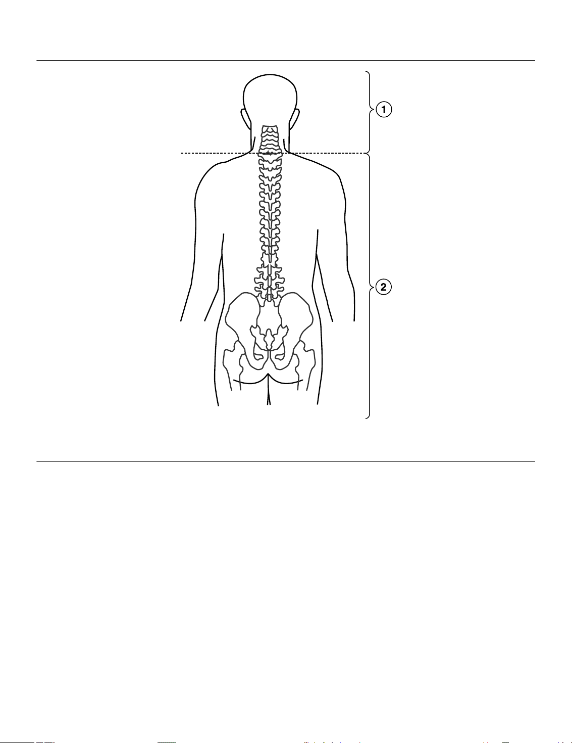
Figure 1. 3 T Scan location requirements
1 No B
2 B
1+RMS
restrictions
1+RMS
not to exceed 2.8 µT
2.3 Patient monitoring and rescue requirements
Continuous patient monitoring is required during the MRI scan.
In the event that patient rescue is required, an external defibrillator must be immediately available.
2.4 Training requirements
●
A health professional who has completed cardiology SureScan training must be present during the
programming of the MRI SureScan feature.
●
A health professional who has completed radiology SureScan training must be present during the MRI scan.
6

3 MRI warnings and precautions
Warnings:
●
Do not scan a patient without first programming the MRI SureScan mode to On. Scanning the patient without
programming the MRI SureScan mode to On may result in patient harm or damage to the SureScan CRT-P
system.
●
Do not scan patients who do not have a complete SureScan CRT-P system, which includes the following
components:
– A Percepta MRI, Serena MRI, or Solara MRI implanted device
– A SureScan right atrial pacing lead or a Model 6725 pin plug for the right atrial port
– A SureScan left ventricular pacing lead
– A SureScan right ventricular pacing lead
Any other combination may result in a hazard to the patient during an MRI scan.
●
Do not scan patients with broken, abandoned, or intermittent leads. Lead fractures or other damage to the
leads may cause changes in the electrical properties of the SureScan CRT-P system that will make the system
unsafe for an MRI scan. Patients with damaged leads may be harmed if an MRI scan is performed.
●
Do not scan patients with a SureScan CRT-P system implanted in sites other than the left and right pectoral
region. Safety and effectiveness have been assessed for left and right pectoral implant locations only.
Scanning of patients with devices implanted in other locations could lead to increased pacing capture
threshold or unintended cardiac capture.
●
Do not leave the device in MRI SureScan mode after the scan is complete. While the MRI SureScan mode is
programmed to On, the patient receives no CRT support. This lack of CRT support might cause dizziness or
shortness of breath. Be sure to program the MRI SureScan mode to Off as soon as the scan is complete.
Cautions:
●
Do not scan patients in a 1.5 T magnetic field with a whole body averaged SAR level > 2.0 W/kg. A scan above
2.0 W/kg may increase the risk of myocardial tissue damage due to lead tip heating, resulting in an increase
in the pacing capture threshold.
●
Do not scan patients in a 3 T magnetic field with a B
bore) is inferior to the C7 vertebra. A scan above 2.8 µT may increase the risk of myocardial tissue damage due
to lead tip heating, resulting in an increase in the pacing capture threshold.
●
For pacemaker-dependent patients, it is not recommended to perform an MRI scan if the right ventricular (RV)
lead pacing capture threshold is greater than 2.0 V at a pulse width of 0.4 ms. A higher pacing capture
threshold may indicate an issue with the implanted lead.
●
Do not scan patients whose device will be programmed to an asynchronous pacing mode when MRI SureScan
mode is on, and who have diaphragmatic stimulation at a pacing output of 5.0 V and at a pulse width of 1.0 ms.
It may be difficult for the patient to remain still in order to obtain a quality MRI scan.
Note: The LV lead is not paced during SureScan operation so the presence of diaphragmatic stimulation on
the LV lead at a pacing output of 5.0 V and a pulse width of 1.0 ms does not need to be considered.
●
Do not scan patients with lead extenders or lead adaptors. Lead extenders and lead adaptors may increase
the risk of MRI-related hazards, including myocardial tissue damage due to lead tip heating.
●
It is not recommended to perform MRI scans during the lead maturation period (approximately 6 weeks after
implant) because MRI scans during this period have not been prospectively studied by Medtronic.
●
Scanning patients who have multiple MR Conditional devices present is acceptable as long as the MR labeling
conditions for all implants can be satisfied.
●
Do not bring the Medtronic programmer, Patient Assistant, or patient monitor into Zone 4 (MRI magnet room),
as defined by the American College of Radiology. They are MR Unsafe.
value > 2.8 µT when the isocenter (center of the MRI
1+RMS
7

4 Potential adverse events
The SureScan CRT-P system is designed to minimize the potential adverse events that may cause patient harm.
The following potential adverse events may occur in the MRI environment:
●
lead electrode heating and tissue damage resulting in loss of sensing or capture or both
●
device heating resulting in tissue damage in the implant pocket or patient discomfort or both
●
MR-induced stimulation on leads resulting in continuous capture, VT/VF, hemodynamic collapse, or all three
●
damage to the device or leads causing the system to fail to detect or treat irregular heartbeats or causing the
system to treat the patient’s condition incorrectly
●
damage to the functionality or mechanical integrity of the device resulting in the inability of the device to
communicate with the programmer
●
movement or vibration of the device or leads resulting in dislodgment
●
potential for VT/VF induction when the patient is programmed to an asynchronous pacing mode during MRI
SureScan mode
5 Patient monitoring requirements
Proper patient monitoring must be provided during the MRI scan and includes both of the following actions:
●
maintaining continuous visual and verbal contact with the patient
●
continuous monitoring of the patient’s heart rate using instrumentation such as pulse oximetry
(plethysmography) or electrocardiography
Preparation for patient rescue – In the event that patient rescue is required, an external defibrillator must be
immediately available.
Notes:
●
If the patient’s hemodynamic function is compromised during the MRI scan, discontinue the scan, remove the
patient from the magnet room, and take the proper measures to restore the patient’s hemodynamic function.
●
While the MRI SureScan mode is programmed to On, the patient receives no CRT support. This lack of CRT
support might cause dizziness or shortness of breath. Therefore, proper patient monitoring is required during
the entire time when the MRI SureScan mode is programmed to On.
6 Cardiology-specific considerations
Lead maturation – MRI scans during the lead maturation period (approximately 6 weeks after implant) have not
been prospectively studied by Medtronic and are not recommended.
Competitive pacing – If an asynchronous MRI SureScan pacing mode is selected, be aware that some patients
may be susceptible to cardiac arrhythmia induced by competitive pacing. For these patients, it is important to first
select an MRI SureScan pacing rate that avoids competitive pacing and then minimize the duration of the
asynchronous pacing operation. For more information, contact a Medtronic representative.
Note: If the patient does not need pacing support, select a nonpacing mode (ODO).
System information and records – All pertinent information about the components of the implanted SureScan
CRT-P system such as model names, model numbers, and serial numbers should be recorded in the patient record
and on the Patient Information screen on the programmer. This information will help with system identification in
the future.
Patient ID card – Reference materials, such as an ID card, should be provided to all patients with an implanted
SureScan CRT-P system. These reference materials should indicate that the patient has a SureScan CRT-P
device and SureScan leads.
8

Note: Be sure to advise the patient to notify medical personnel that they have a CRT-P before entering the MR
environment and to present their patient ID card.
7 Radiology-specific considerations
7.1 MRI considerations
3 T whole-body transmit coil RF excitations – 3 T MRI systems using two transmit channels (or fewer) may
operate in the following RF excitations: two transmit channels (known as Multichannel-2 (MC-2)) or Circularly
Polarized (CP). Systems that use more than two transmit channels have not been studied, but such systems could
be operated in CP or MC-2, if available.
Use of transmit/receive and receive-only coils – There are no restrictions on the use of local transmit/receive
coils for MRI scanning of the head or of the extremities, and there are no restrictions on the placement of
receive-only coils.
Image artifact and distortion – SureScan leads have demonstrated minimal MRI scan distortion for areas
surrounding the implanted leads when the device is out of the field of view. Significant MRI scan distortion will result
from the presence of the device within the field of view. MRI scan artifacts and distortion resulting from the
presence of the device and the leads within the field of view must be considered when selecting the field of view
and MRI scanning parameters. These factors must also be considered when interpreting the MRI scans.
Patient sensation during MRI – The device has been evaluated to ensure no risk of tissue damage. However, the
patient may feel sensations of warmth or vibration in the implant site during the MRI scan. Tolerable levels of these
sensations do not indicate that patient safety has been compromised.
8 Pre-MRI scan operations
The steps in the following sections are required before performing an MRI scan.
8.1 Identification of SureScan CRT-P system components
Use the following methods to verify that a patient has a SureScan CRT-P system:
●
Patient records or patient ID card (if applicable): Patient records and the patient ID card, if applicable, are
the most reliable record of the medical devices that have been implanted in the patient. These records are
available to clinicians other than the device clinician and can be accessed without the presence of the patient
or the use of a programmer. These records must be complete and accurate if they are to be used to determine
whether the patient has a SureScan CRT-P system.
●
Patient information on the programmer: The programmer Patient Information feature is intended to be
used by the implanting clinician to document the components of the patient’s SureScan system. If the
implanting clinician has entered the needed information completely and accurately, you can use the Patient
Information feature to determine whether the patient has a SureScan CRT-P system. The patient may have
other implanted devices that are not approved for use in the MRI environment, but not noted in the patient
information on the programmer.
1. Tap Patient > Patient Information > MRI SureScan System/Other Hardware….
The MRI SureScan System/Other Hardware window appears.
2. View the MRI SureScan System fields for information about the patient’s leads and whether or not they
are MR conditional.
3. View the Other Hardware fields for information about other lead extenders, lead adaptors, and
abandoned leads.
9

8.2 Required patient care
Before programming the MRI SureScan mode to On, perform the following actions to help ensure patient safety:
Evaluate the patient to determine whether or not pacing support is needed while the MRI SureScan
mode is programmed to On. – For patients who require pacing support, set the MRI SureScan pacing mode to
DOO, AOO, or VOO when programming the MRI SureScan mode to On. For patients who do not require pacing
support, set the MRI SureScan pacing mode to ODO when programming the MRI SureScan mode to On.
Asynchronous pacing may increase the risk of arrhythmia. For pacemaker-dependent patients, it is not
recommended to perform an MRI scan if the right ventricular (RV) lead pacing capture threshold is greater than
2.0 V at a pulse width of 0.4 ms.
If the patient will require pacing support, ascertain an appropriate pacing rate. – An appropriate pacing
rate is one that will help avoid competitive pacing while the MRI SureScan mode is programmed to On.
9 Performing an MRI scan
Warnings:
●
Do not scan a patient without first programming the MRI SureScan mode to On. Scanning the patient without
programming the MRI SureScan mode to On may result in patient harm or damage to the SureScan CRT-P
system.
●
Do not leave the device in MRI SureScan mode after the scan is complete. While the MRI SureScan parameter
is programmed to On, the patient receives no CRT support. This lack of CRT support might cause dizziness
or shortness of breath. Be sure to program the MRI SureScan mode to Off as soon as the scan is complete.
Note: The system automatically programs the MRI SureScan mode to Off 6 hours after it is programmed to On.
Before you program the MRI SureScan mode to On, ensure that the MRI scan will be completed before this 6-hour
timeout occurs. Refer to the MRI SureScan Parameters report or the Quick Look II report for information about
when the MRI SureScan mode was programmed to On.
Caution: Do not bring the Medtronic programmer, the Patient Assistant, or the patient monitor into Zone 4 (MRI
magnet room), as defined by the American College of Radiology. They are MR Unsafe.
When programming the MRI SureScan mode to On, you must select parameters that are appropriate for the
patient. Pacing mode and rate (if applicable) are to be programmed per the physician’s discretion. Based on
whether or not the patient needs pacing support, an asynchronous pacing mode (DOO, AOO, or VOO) or sensing
only mode can be programmed. Sensed events will be ignored by the device when the MRI SureScan mode is
programmed to On, regardless of the programmed mode. The device maintains the selected parameters until the
MRI SureScan mode is programmed to Off after the MRI scan has been completed. After the MRI SureScan mode
is programmed to Off, the permanent device parameters are restored.
9.1 SureScan CRT-P system integrity verification
The SureScan CRT-P system provides automatic verification that no device or lead issues that may compromise
patient safety during an MRI scan are detected. Before allowing the user to initiate the MRI SureScan feature, the
SureScan device application software checks for the following 2 situations:
Lead impedance is out of range – If a lead impedance measurement value is < 200 Ω or > 3000 Ω, or
unavailable, the software prevents the MRI SureScan feature from being initiated. For multiple point pacing (MPP)
vectors, the impedance range applies to each individual vector. For example, when LV Pace Polarity is LV1 to LV2
and 2nd LV Pace Polarity is LV4 to Can, both LV1 to LV2 and LV4 to Can must be in the 200 Ω - 3000 Ω range.
Note: If Atrial Sensitivity is programmed to Off, the software prevents the atrial lead impedance check so that
patients without an atrial lead implanted can undergo an MRI scan.
10

Insufficient battery longevity – If the device is at Recommended Replacement Time (RRT), Elective
Replacement Indicator (ERI), or End of Service (EOS), the software prevents the MRI SureScan feature from being
initiated.
9.2 Programming the MRI SureScan feature to On
When programmed to On, the MRI SureScan feature allows patients to be safely scanned by an MRI machine.
Before MRI SureScan mode can be programmed to On, pace polarity must be set to bipolar.
Perform the following steps to program the MRI SureScan feature to On.
1. Tap Params > Additional Features… > MRI SureScan…
The MRI SureScan Checklist screen appears.
2. Review the MRI SureScan Checklist and select the check box if all items are satisfied for the patient.
Note: Tap Print… to print a copy of the MRI SureScan Checklist if desired.
3. Tap OK.
The MRI SureScan screen appears.
4. Tap the MRI SureScan field.
The MRI SureScan feature settings become available.
5. Tap the Mode field to select an appropriate MRI SureScan pacing mode as described in Table 1 and tap the
Lower Rate field to enter an appropriate MRI SureScan pacing rate.
Notes:
●
To avoid competitive pacing during the operation of asynchronous pacing, select an appropriate MRI
SureScan pacing rate.
●
If the patient’s device is programmed to the non-pacing (ODO or OVO) mode, the MRI SureScan pacing
rate (Lower Rate) is not available for programming.
Table 1. MRI SureScan pacing modes
Situation Mode
Patients with a dual chamber device that require pacing support Asynchronous modes:
DOO
AOO
VOO
Patients with a single chamber device that require pacing support Asynchronous mode:
VOO
Patients with a dual chamber device that do not require pacing
support
Patients with a single chamber device that do not require pacing
support
Non-pacing mode:
ODO
Non-pacing mode:
OVO
6. Tap PROGRAM.
The implanted device is now ready for the MRI scan. The implanted device begins pacing at the selected MRI
SureScan pacing mode and MRI SureScan pacing rate.
Notes:
●
After the device is programmed for an MRI scan, available options are Print…, End Session…, and
Emergency. The MRI SureScan parameter can also be programmed to Off.
●
Selecting the Emergency button while in MRI SureScan mode programs the MRI SureScan parameter
to Off.
●
The status of the MRI SureScan mode and the programmed parameters may be confirmed by printing the
MRI SureScan Parameters report. The MRI SureScan Parameters report may be printed by tapping
Print….
11

9.3 Device considerations
Suspension of diagnostic data – When the MRI SureScan mode is programmed to On, all device diagnostic
measurements and collection are suspended.
Suspension of Magnet Mode – When MRI SureScan mode is programmed to On, the device does not initiate
asynchronous, fixed-rate bradycardia pacing in the presence of a magnet.
Suspension of wireless telemetry and Medtronic CareAlert Monitoring – Wireless telemetry and the
Medtronic CareAlert Monitoring feature are disabled when MRI SureScan mode is programmed to On.
Automatic amplitude and pulse width selection for MRI SureScan pacing modes – When MRI SureScan
mode is programmed to On and the pacing mode is DOO, VOO, or AOO, the device may automatically set the
amplitude and pulse width values.
If the permanently programmed A. Amplitude or RV Amplitude is less than 5.0 V, the amplitude is set to 5.0 V. If the
permanently programmed A. Pulse Width or RV Pulse Width is less than 1.00 ms, the pulse width is set to 1.00 ms.
Automatic canceling of the MRI SureScan mode with Emergency programming – If you deliver emergency
therapy when the MRI SureScan mode is programmed to On, the MRI SureScan mode is automatically
programmed to Off. After Emergency VVI pacing is programmed, the MRI SureScan mode must be programmed
to On again before the patient can be scanned safely.
Suspension of PVC detection – When MRI SureScan mode is programmed to On, the device does not detect
PVCs.
Suspension of tachyarrhythmia detection – When MRI SureScan mode is programmed to On, the device does
not detect atrial or ventricular tachyarrhythmias.
Suspension of tachyarrhythmia therapies – When MRI SureScan mode is programmed to On in a dual
chamber device capable of providing tachyarrhythmia therapies, the device does not deliver these therapies.
However, bradyarrhythmia pacing therapy is provided when an asynchronous pacing mode is selected for MRI
SureScan operation.
Automatic PAV selection for DOO mode – If the DOO mode is selected when MRI SureScan mode is
programmed to On, the device automatically sets the PAV to either the permanently programmed PAV interval or
110 ms, whichever is less. However, if the permanently programmed PAV is less than 50 ms, the device
automatically sets the PAV to 50 ms when MRI SureScan mode is programmed to On.
10 Following the MRI scan
Warning: Do not leave the device in MRI SureScan mode after the scan is complete. While the MRI SureScan
mode is programmed to On, the patient receives no CRT support. This lack of CRT support might cause dizziness
or shortness of breath. Be sure to program the MRI SureScan mode to Off as soon as the scan is complete.
Six-hour timeout period – The system automatically programs the MRI SureScan mode to Off 6 hours after it was
programmed to On. This six-hour timeout period is provided to minimize the amount of time during which the
patient receives no CRT support.
Check the pacing capture threshold – Check the pacing capture threshold after the scan is complete, and be
sure that the pacing parameters are programmed adequately for the patient based on the threshold. There is a very
slight risk that the MRI will cause lead tip heating, leading to increased pacing capture threshold and loss of
capture.
Note: The Pacing Threshold Test measures capture thresholds in 0.25 V increments. The actual capture threshold
change associated with a 0.25 V change is between 0.0 V and 0.5 V. For example, actual thresholds of 1.49 V and
1.51 V correspond to measured thresholds of 1.5 V and 1.75 V, respectively. In this case, an actual change of
0.02 V results in a measured change of 0.25 V. Similarly, actual thresholds of 1.01 V and 2.00 V correspond to
measured thresholds of 1.25 V and 2.00 V. In this situation, an actual change of 0.99 V results in a measured
change of 0.75 V.
12

Atrial, LV, and RV Capture Management measures capture thresholds in 0.125 V increments.
10.1 Returning the device to the pre-MRI configuration
After the MRI scan is complete, the MRI SureScan mode must be programmed to Off using the Medtronic
programmer. Programming the MRI SureScan mode to Off restores the device parameter values to the pre-MRI
SureScan mode configuration.
The device maintains the parameters that were set while initiating MRI SureScan operation until the MRI SureScan
mode is programmed to Off after the MRI scan.
Perform the following steps to program the MRI SureScan mode to Off:
1. Tap the MRI SureScan field of the MRI SureScan screen, changing the value to Off.
2. Tap PROGRAM.
3. Tap Close.
The MRI SureScan screen closes and the programmer returns to the Parameters screen. The device
parameter values are now restored to the pre-MRI SureScan configuration.
Note: During each interrogation, the device is monitored for possible electrical reset conditions and disabled
therapies. If a condition is detected that requires attention, the programmer displays a Device Status Indicator
warning in a pop-up window and on the Quick Look II screen.
11 Medtronic warranty information
Please see the literature enclosed with the products for information regarding the product warranty or disclaimer
of warranty as applicable.
12 Explanation of MRI symbols
The following symbols are related to the magnetic resonance (MR) environment and are used to indicate the safety
of devices and components in the MR environment.
SureScan symbol
MR Conditional symbol. The Medtronic SureScan CRT-P system is MR Conditional and,
as such, is designed to allow implanted patients the ability to undergo an MRI scan under
the specified MRI conditions for use.
13 Service
Medtronic employs highly trained representatives and engineers located throughout the world to serve you and,
upon request, to provide training to qualified hospital personnel in the use of Medtronic products. Medtronic also
maintains a professional staff to provide technical consultation to product users. For more information, contact
your local Medtronic representative, or call or write Medtronic at the appropriate telephone number or address
listed on the back cover.
13


Medtronic, Inc.
*M967072A001*
710 Medtronic Parkway
Minneapolis, MN 55432
USA
www.medtronic.com
+1 763 514 4000
Medtronic USA, Inc.
Toll-free in the USA (24-hour technical
consultation for physicians and medical
professionals)
Bradycardia: +1 800 505 4636
Tachycardia: +1 800 723 4636
Europe/Middle East/Africa
Medtronic International Trading Sàrl
Route du Molliau 31
Case Postale 84
CH-1131 Tolochenaz
Switzerland
+41 21 802 7000
Technical manuals
www.medtronic.com/manuals
© 2016 Medtronic
M967072A001 A
2016-11-11
 Loading...