

Page 1
Leica FL400
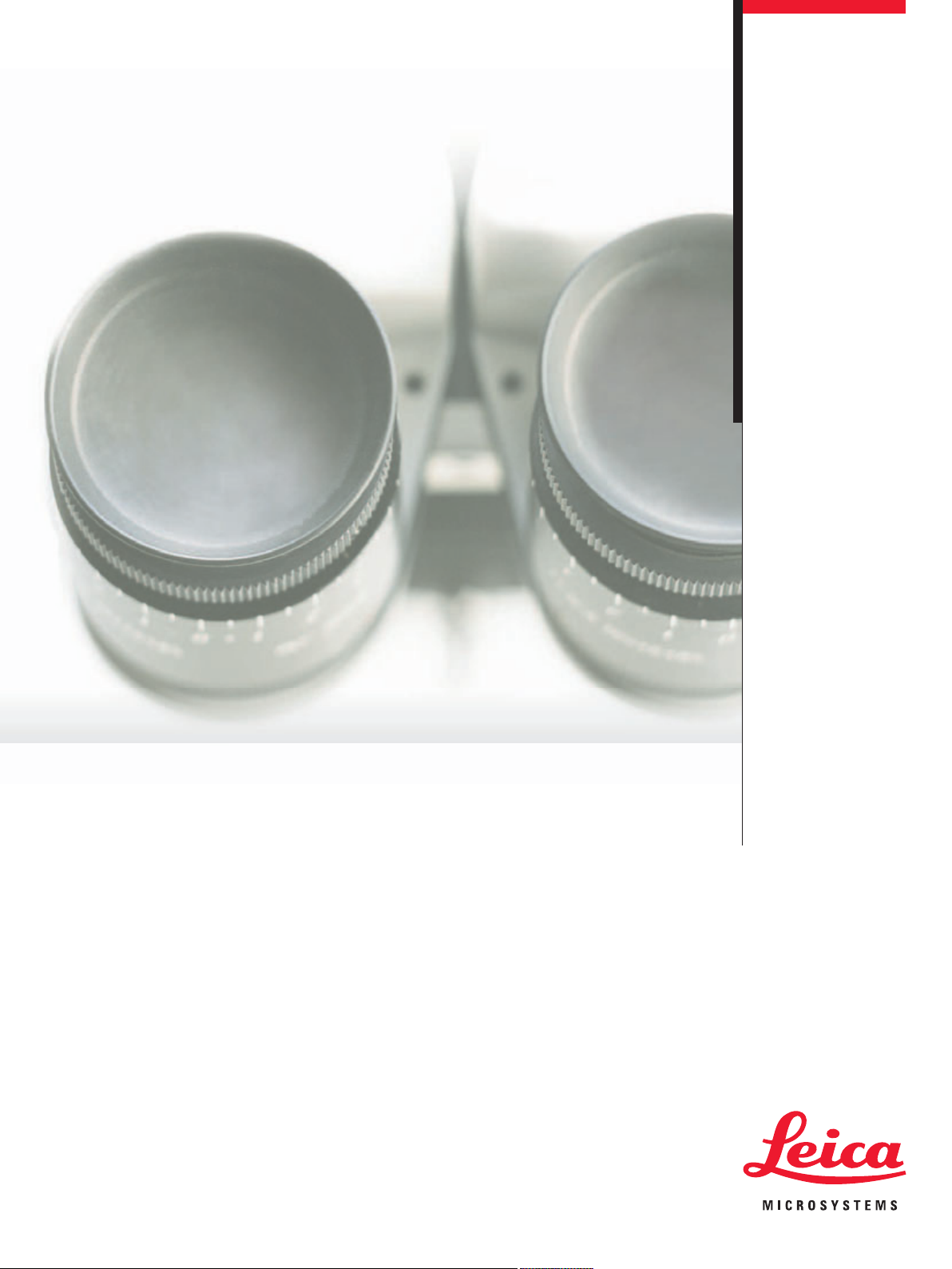
Invisible Becomes Visible
Living up to Life
Page 2

Pioneering Fluorescence Innovation
The development of fluorescence microscopy has a long tradition
at Leica Microsystems, dating back to the beginning of the 20th
century. An important contribution to biological research, fluores-
cence science now integrates with the technology of the surgical
microscope to provide state-of-the-art oncological fluorescence.
The Leica FL400 enables fluorescence guided tumor resection. In
addition to the visible white light image, the Leica FL400 provides
the surgeon with information from fluorescence imaging and
the infrared spectrum, previously not visible to the human eye.
The ability to view fluorescence images intraoperatively through
the microscope’s eyepieces during surgery can benefit surgical
* The Leica FL400 is not approved
for sale in the USA, Japan,
and some other countries.
Please check the status of approval
with your local Leica representative.
2
outcomes.
Page 3
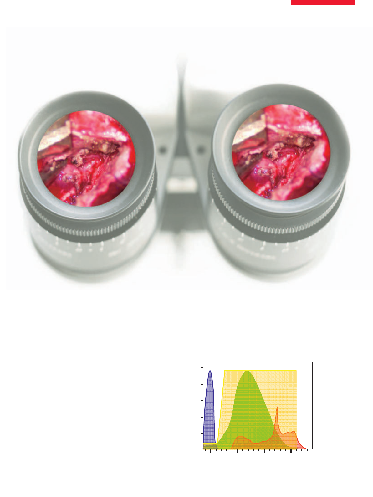
Oncological Fluorescence
*
Photo Dynamic Imaging (PDI) during surgery combines a tumor-
selective photosensitizer, excitation light of an appropriate wave-
length, and a well-adjusted observation spectrum. Leica Micro-
systems provides the ideal combination of filters and crisp, clear
optics to provide good orientation in the resection area. The vital
tumor shines with red sensitizer fluorescence in good contrast to
normal tissue under blue light illumination.
Observation modes
Changing observation modes is easy. To switch from white light
mode to fluorescence mode and back requires only the click of
a button on the handle or the foot pedal. The Leica CAN bus con-
trols the type of illumination, the observation filters for different
fluorescence applications, and an optional mode-controlled video
camera specifically aligned for fluorescence.
View: White light mode
5-ALA Spectras
0.1
8.0
6.0
4.0
2.0
excitation spectrum
retlifnoitavresbo
murtcepsytivitisneseye
noissime
murtceps
007006005004
λ/ mn
3
Page 4

IntenseBlue
Using two light sources (Leica M525) or a high
efficient 400W light source (Leica M720),
the Leica FL400 offers the best excitation and
bright fluorescence of the protoporpherin.
Furthermore, the system supports the surgeon
with longer resection cycles in the blue light
mode, without the eyestrain of frequent
changes to white light.
TM
Leica M720 FL400 illumination module
Page 5

The PDI Application
Surgeons familiar with Photo Dynamic Imaging (PDI) comment
that it is very easy to use. The patient orally receives the 5-ALA
dissolved in water, thus placing little strain on the patient, and has
almost no side effects.
Tum or resection can be performed not only in white light mode but
also in blue light mode. Blue light mode requires sufficient bright-
ness in order to resect the residuals of the tumor, which always
appear red or pink under blue light.
The Leica FL400 used for PDI reveals many new possibilities – its
potential is not yet fully realized. Overall, it represents an out-
standing new, straightforward method of intraoperative tumor
resectioning.
Malignant glioma,
white light mode
Malignant glioma,
blue light mode
Pictures taken
with Leica FL400
5-ALA Operation
5
Page 6

The Leica FL400 System
The intensified and homogeneous excitation light of the Leica M525 OH4 with the
Karl Storz D light C or the Leica M720 OH5 with 400W Xenon power offers a bright and
clear fluorescence view.
A patented long pass filter switched into the observation beampathes provides a
well-adjusted observation and a perfect photo dynamic signal.
6
Page 7

The Best Possible Image
The fluorescence technique
The Leica M720 OH5 and Leica M525 OH4 can be easily upgraded to use blue light
fluorescence. The illumination systems, the observation filters and an optional blue
mode-optimized video camera interact automatically with a simple push of a
button found on the pistol grip of the surgical microscope. This offers bright blue
illumination and an easy and ergonomic change of the observation modes.
The Leica M720 and Leica M525 surgical microscopes
The Leica M720 OH5 and Leica M525 OH4 with integrated Leica FL400 5-ALA
fluorescence module offer the easiest movement, best ergonomics and highest
quality optics of any surgical microscope on the market today. Leica’s premium
OptiChromeTMOptics provides outstanding contrast, sharpness, resolution and
color fidelity. The “light touch” maneuverability of the Leica floorstands OH5 and
OH4 give the surgeon the perfect blend of stability and ease-of-use for micro-
surgical procedure.
7
Page 8

www.leica-microsystems.com
“With the user, for the user”
Leica Microsystems. We have developed fi ve
4HE3URGICAL$IVISIONWITHIN,EICA-ICROSYSTEMS3CHWEIZ!'HOLDSTHEMANAGEMENTSYSTEM
CERTIFICATESFORTHEINTERNATIONALSTANDARDS)3/)3/AND)3/RELATINGTOQUALITY
MANAGEMENTQUALITYASSURANCEANDENVIRONMENTALMANAGEMENT
Leica Microsystems
Leica Microsystems operates globally in four divi sions,
where we rank with the market leaders.
Life Science Division
The Leica Microsystems Life Science Division supports the
imaging needs of the scientific community with advanced
innovation and technical expertise for the visualization,
measurement, and analysis of microstructures. Our strong
focus on understanding scientific applications puts Leica
Microsystems’ customers at the leading edge of science.
Industry Division
The Leica Microsystems Industry Division’s focus is to
support customers’ pursuit of the highest quality end result.
Leica Microsystems provide the best and most innovative
imaging systems to see, measure, and analyze the microstructures in routine and research industrial applications,
materials science, quality control, forensic science investigation, and educational applications.
Biosystems Division
The Leica Microsystems Biosystems Division brings histopathology labs and researchers the highest-quality,
most comprehensive product range. From patient to pathologist, the range includes the ideal product for each
histology step and high-productivity workflow solutions
for the entire lab. With complete histology systems featuring innovative automation and Novocastra™ reagents,
Leica Microsystems creates better patient care through
rapid turnaround, diagnostic confi dence, and close customer collaboration.
Surgical Division
The Leica Microsystems Surgical Division’s focus is to
partner with and support surgeons and their care of patients with the highest-quality, most innovative surgi cal
microscope technology today and into the future.
The statement by Ernst Leitz in 1907, “with the user, for the user,” describes the fruitful collaboration
with end users and driving force of innovation at
brand values to live up to this tradition: Pioneering, High-end Quality, Team Spirit, Dedication to
Science, and Continuous Improvement. For us, living up to these values means: Living up to Life.
Active worldwide
Australia: North Ryde Tel. +61 2 8870 3500 Fax +61 2 9878 1055
Austria: Vienna Tel. +43 1 486 80 50 0 Fax +43 1 486 80 50 30
Belgium: Groot Bijgaarden Tel. +32 2 790 98 50 Fax +32 2 790 98 68
Canada: Richmond Hill/Ontario Tel. +1 905 762 2000 Fax +1 905 762 8937
Denmark: Ballerup Tel. +45 4454 0101 Fax +45 4454 0111
France: Nanterre Cedex Tel. +33 811 000 664 Fax +33 1 56 05 23 23
Germany: Wetzlar Tel. +49 64 41 29 40 00 Fax +49 64 41 29 41 55
Italy: Milan Tel. +39 02 574 861 Fax +39 02 574 03392
Japan: Tokyo Tel. +81 3 5421 2800 Fax +81 3 5421 2896
Korea: Seoul Tel. +82 2 514 65 43 Fax +82 2 514 65 48
Netherlands: Rijswijk Tel. +31 70 4132 100 Fax +31 70 4132 109
People’s Rep. of China: Hong Kong Tel. +852 2564 6699 Fax +852 2564 4163
Portugal: Lisbon Tel. +351 21 388 9112 Fax +351 21 385 4668
Singapore Tel. +65 6779 7823 Fax +65 6773 0628
Spain: Barcelona Tel. +34 93 494 95 30 Fax +34 93 494 95 32
Sweden: Kista Tel. +46 8 625 45 45 Fax +46 8 625 45 10
Switzerland: Heerbrugg Tel. +41 71 726 34 34 Fax +41 71 726 34 44
United Kingdom: Milton Keynes Tel. +44 1908 246 246 Fax +44 1908 609 992
USA: Bannockburn/lllinois Tel. +1 847 405 0123 Fax +1 847 405 0164
and representatives in more than 100 countries
10 M1 750 0en/C • © Leica Microsystems (Schweiz) AG • CH-9435 Heerbrugg, 2010 • Printed in Switzerland – I.2010 – RDV – Illustrations, descriptions and technical data are not binding and may be changed without notice.
 Loading...