

Page 1

➔
Gebrauchsanweisung
Diagnosestation
Instructions
diagnostic station
Mode d’ emploi
Station de diagnostic
Instrucciones para el uso
Unidad de diagnóstico
нструкция по эксплуатации
иагностические станции
Istruzioni per I’ uso
Stazione diagnostica
ri-former
®
Page 2

2
7 7
8
4
6
5
3
1
2
Page 3

Inhaltsverzeichnis
1. Wichtige Informationen zur Beachtung vor Inbetriebnahme
2. Sicherheitshinweise und Elektromagnetische Verträglichkeit
3. Zweckbestimmung
4. Befestigung
5. Inbetriebnahme und Funktion
6. Ein und Ausschalten
7. Reinigung und Desinfektion
8. Technische Daten
Table of Contents
1. Important information to be observed before operation
2. Safety information and electromagnetic compatibility
3. Intended use
4. Attachment
5. Operation and function
6. Switching on and off
7. Cleaning and disinfection
8. Technical data
Sommaire
1. Informations importantes à lire attentivement avant la mise en
service
2. Consignes de sécurité et compatibilité électromagnétique
3. Usage prévu
4. Fixation
5. Mise en service et fonctionnement
6. Allumage et mise hors tension
7. Nettoyage et désinfection
8. Fiche technique
3
Page 4

Índice
1. Informaciones importantes que deben tenerse en cuenta antes
del uso
2. Indicaciones sobre la seguridad y la compatibilidad
electromagnética
3. Finalidad de uso
4. Fijación
5. Puesta en marcha y funcionamiento
6. Encendido y apagado
7. Limpieza y desinfección
8. Datos técnicos
одержание
1. ажная информация – читать перед вводом в эксплуатацию
2. нформация по безопасности и электромагнитной совместимости
3. азначение
4. репление
5. вод в эксплуатацию и работа
6. ключение и выключение
7. истка и дезинфекция
8. ехнические данные
Indice
1. Importanti avvertenze da rispettare prima dell'uso
2. Avvertenze di sicurezza e compatibilità elettromagnetica
3. Uso previsto
4. Fissaggio
5. Messa in esercizio e funzionamento
6. Accensione e spegnimento
7. Pulizia e disinfezione
8. Dati tecnici
4
Page 5

1. Important information read prior to start-up
You have purchased a high quality RIESTER wall instrument, which has
been manufactured according to the Directive 93/42 EEC and is subject
to the strictest quality controls at all times.
Read these instructions for use carefully before putting the unit into operation and keep them in a safe place.
If you should have any questions, we are available to answer queries at
all times. Our address can be found in these instructions for use.
The address of our sales partner will be given upon request.
Please note that all instruments described in these instructions for use
are only to be used by suitably trained personnel.
The perfect and safe functioning of this instrument is only guaranteed
when original parts and accessories from Riester are used.
2. Safety information and electromagnetic
compatibility
Meaning of the symbol on the model identification plate
Attention, read accompanying papers!
Instruments of protective class II
Application part type B
Protective conductor connection
The instrument satisfies the requirements for electromagnetic compatibility. Please note that under the influence of unfavourable field strengths,
e.g. during the operation of wireless telephones or radiological instruments, adverse effects on function cannot be excluded.
Attention!
There is a possible danger of inflammation of gases, if the instrument is
operated in the presence of inflammatory mixtures or mixtures of pharmaceuticals and air or oxygen or laughing gas!
Never attempt to take the instrument apart!
There is a danger of
Unplug the instrument before cleaning or when disinfecting.
life-threatening electrical shock.
3. Intended use
The wall instrument ri-former®described in these instructions for use was
manufactured for use with various instrument heads and modular components for non-invasive diagnostics.
9
Page 6

4. Attachment
a.) Drilling instructions/drilling plan
The drilling instructions and the drilling plan are enclosed separately.
Follow the drilling instructions in order to drill the holes in the wall.
b.) Attaching the wall mounting plates
After you have drilled the holes, take the plugs supplied and push them
into the holes as far they will go.
Take the wall mounting plate and hold it onto the wall so that the screws
can be pushed through the holes of the mounting plate into the plugs.
Now screw in the screws with a screw driver, as far as they will go.
c.) Attachment of the diagnostic station
When all screws have been screwed in tightly, take the diagnostic station and guide the screw heads through the openings (1). Then press the
diagnostic station downwards until it snaps into place.
d.) Attachment of the extension module
Connect the diagnostic station and the extension module with the help of
the connecting cable.
In order to plug in the connecting cable, remove the sliding cover (2) of
the diagnostic station.
Close the casing opening of the extension module, which is not needed,
with the sliding cover (2).
Take the extension module and guide the screw heads through the openings (1). Then press the extension module downwards.
Attention:
Take care that the connecting cable does not get caught behind the
extension module.
Push the connecting cable into the groove provided on the reverse side
of the extension module.
5. Operation and function
Putting the diagnostic station into service
with or without extension module:
Put the plug into the electrical socket. The optional clock starts to blink.
You can adjust it to local time by repeatedly pressing the keys; with the
left key marked
Move the handle (5) upwards out of the handle holder (7) and attach the
desired instrument head by placing it with the two projecting guide cams
onto the handle. Press the instrument head lightly onto the handle and
turn the handle in a clockwise direction until it stops. Removal of the
instrument head is carried out by turning in a counter-clockwise direction.
HR and the right key marked MIN.
6. Switching on and off
Switch on the instrument by using the rocker switch (3). The green control lamp (4) in the rocker switch (3) indicates that the instrument is ready
to use. Each handle (5) is automatically ready to operate as soon as it is
taken out of the handle holders (7).
By turning the ring (6) in a clockwise direction, the instrument is switched
on. By turning the ring in a counter-clockwise direction until it stops, the
instrument can be switched off.
10
Page 7

The handle (5) is automatically switched off when replaced back into the
handle holder (7).
6.1Rheostat for regulating the light intensity
It is possible to adjust the light intensity on the handle with the rheostat.
Depending on whether the fluted ring (6) is turned in a clockwise or counter-clockwise direction, the light intensity becomes stronger or weaker.
You have the possibility to adjust the light to the desired intensity and to
maintain this continuously by simply operating the handle (5) via the
automatic on/off switch in the handle holder.
Attention!
Make sure that no more than 3 handles (5) are used at the same time! If
more than 3 handles are used at the same time, the transformer in the
instrument may become overloaded and switch itself off.
7. Cleaning and disinfection
The diagnostic station ri-former(r) with extension module can be cleaned
externally with a moist cloth. Furthermore, it can also be disinfected from
the outside, with the exception of the clock glass cover (8), using the following disinfectants:
Aldehyde (formaldehyde, glutaraldeyhde, aldehyde derivatives), surfactants or alcohols.
When using these substances, the manufacturer’s instructions must be
strictly complied with.
Means for cleaning or disinfection may be a soft, possibly lint-free cloth
or Q-tips.
Attention!
We recommend unplugging the instrument before cleaning or disinfection.
Take care while cleaning and disinfecting that no liquid enters inside the
instrument!
Sterilisation
According to the current school of thought (Test Centre for Medical
Devices in Tübingen), sterilisation is only prescribed in the case of ope-
rative procedures.
8. Technical data
Model: Voltage supply
Diagnostic station ri-former®
Connection: Mains supply, see Note
Model identification plate on reverse side.
Outlet: 1 x 3,5 V
Working temperature: 0° C to + 40° C
Storage location: -5° C to + 50° C, up to 85 % relative
humidity
Dimensions
Diagnostic station: 200 x 180,5 x 75 mm
Extension module: 200 x 100 x 75 mm
Wight
Diagnostic station: 1450 g
Extension module: 490 g
11
Page 8
Gebrauchte elektrische und elektronische Geräte sollten nicht in den
normalen Hausmüll gelangen, sondern gemäß nationaler bzw. EURichtlinien separat entsorgt werden.
Used electrical and electronic products are not to be disposed as unsorted municipal waste and are to be collected separately accordingly to
national/EU regulations.
Les dispositifs électriques et électroniques usagés ne doivent pas être
éliminés avec les déchets domestiques non triés et doivent être collectés séparément conformément à la réglementation nationale/européenne en vigueur.
Los productos eléctricos y electrónicos usados no pueden eliminarse
como basura general; deberán desecharse de forma separada de acuerdo con las regulaciones nacionales/UE.
спользованные электрические и электронные изделия нельзя
утилизировать как несортированный городской мусор, их следует
собирать в отдельном месте в соответствии с национальными
правилами и правилами .
Apparecchi elettronici ed elettrici usati nom vanno smaltiti nei rifiuti casalinghi. Questi derono essere smaltiti separatamente attenendosi à le
direttive nazionali risp. direttive UE.
27
Page 9
ri-scope® Otoskop
ri-scope® Ophthalmoskop
ri-scope® Retinoskop (Skiaskop) XL 3,5 V
3.
ri-derma® Dermatoskop XL 3,5 V
3.1
28
4.
2.
3.2
Page 10

ri-scope® F.O. Lampenträger XL 3,5 V
ri-scope® F.O. Nasenspekulum XL 3,5 V
3.
2b
2a
ri-scope® F.O. Zungenspatelhalter XL 3,5 V
ri-scope® Human-Operationsotoskop XL 3,5 V
ohne Trichter
5.
3.
2.
ri-scope® Veterinär-Operationsotoskop XL 3,5 V
ohne Trichter
3.
29
2.
Page 11

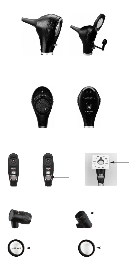
ri-scope® instrumen heads
ri-scope® L otoscope
1. Purpose
The RIESTER otoscope described in these Operating Instructions is produced for illumination and examination of the auditory canal in combination with
2 Fitting and removing ear specula
Either RIESTER disposable ear specula (blue colour) or reusable RIESTER ear specula (black colour) can be fitted to the otoscope head.
The size of the ear specula is marked at the back of the speculum.
L1 and L2 otoscopes
Screw the speculum clockwise until noticeable resistance is felt. To
remove the speculum, screw the speculum counter clockwise.
L3 otoscope
Fit the chosen speculum on the chrome-plated metal fixture of the otoscope until it locks into place.
To remove the speculum, press the blue ejection button. The speculum
is automatically ejected.
3. Swivel lens for magnification
The swivel lens is fixed to the device and can be swivelled 360°.
4. Insertion of external instruments into the ear
If you wish to insert external instruments into the ear (e.g. tweezers), you
have to rotate the swivel lens (approx. 3-fold magnification) located on
the otoscope head by 180°. Now you can use the operation lens.
5. Pneumatic test
To perform the pneumatic test (= examination of the eardrum), you require a ball, which is not included in the normal delivery package, but can
be ordered separately. The tube for the ball is attached to the connector.
Now you can carefully insert the necessary volume of air into the ear
canal.
RIESTER ear specula.
ri-scope® L ophthalmoscope
1. Purpose
The RIESTER ophthalmoscope described in these Operating
Instructions is produced for the examination of the eye and the eyeground.
2. Lens wheel with correction lens
The correction lens can be adjusted on the lens wheel. The following correction lenses are available:
L1 and L2 ophthalmoscopes
Plus: 1-10, 12, 15, 20, 40.
Minus: 1-10, 15, 20, 25, 30, 35.
L3 ophthalmoscope
Plus: 1-45 in single steps
Minus: 1-44 in single steps
The values can be read off in the illuminated field of view. Plus values
are displayed in green numbers, minus values with red numbers.
36
Page 12

3. Apertures
The following apertures can be selected with the aperture hand-wheel:
L1 ophthalmoscope
Semi-circle, small/medium/large circular aperture, fixation star, slit and
red-free filter.
L2 ophthalmoscope
Semi-circle, small/medium/large circular aperture, fixation star and slit.
L3 ophthalmoscope
Semi-circle, small/medium/large circular aperture, fixation star, slit and
grid.
Aperture function
Small circle:
Medium circle: pupils
Semi-circle:
Large circle: for normal examination results
Grid: for topographic determination of retina changes
Light slit: to determine differences in level
Fixation star: to ascertain central or eccentric fixation
4 Filters
Using the filter wheel, the following filters can be switched for each aperture:
L1 ophthalmoscope
The L1 instrument head is supplied without a filter wheel.
(the red filter is contained in the aperture wheel)
L2 ophthalmoscope
Red-free filter, blue filter and polarisation filter.
L3 ophthalmoscope
Red-free filter, blue filter and polarisation filter.
Filter function
Red-free filter: contrast enhancing to assess fine vascular
Polarisation filter: for precise assessment of tissue colours and to
Blue filter: for improved recognition of vascular abnormali-
to reduce reflection for small
changes, e.g. retinal bleeding
avoid retinal reflections
ties or bleeding, for fluorescence ophthalmology
For L2 + L3, every filter can be switched to every aperture.
5. Focussing device (only with L3)
Fast fine adjustment of the examination area to be observed is achieved
from various distances by turning the focussing wheel.
ri-scope® retinoscope (skiascope) XL 3,5 V
1. Intended use
The ri-scope®retinoscope Slit and ri-scope®retinoscope Spot described
37
Page 13

in these operating instructions (also called skiascopes) have been manufactured for examining the refraction of the eye (refractive error).
2. Function
Rotation and focusing of the slit and/or spot image may now be effected
by the knurled screw.
3. Rotation
The slit or spot image may be rotated by 360° by the control. Each angle
may be directly read from the scale on the retinoscope.
4. Fixation cards
Fixation cards are suspended and fixed on the object side of the retinoscope into the bracket for the dynamic skiascope.
5. Slit/Spot design
The slit retinoscope may be converted to a spot retinoscope by exchanging the slit lamp against a spot lamp.
ri-derma® Dermatoskop XL 3,5 V
1. Intended use
The ri-derma®dermatoscope described in these operating instructions
has been produced for early recognition of melanotic skin changes
(malign melanoma).
2. Focusing
Focus the magnifying glass by rotating the eyepiece ring.
3. Skin adapters
Two skin adapters are supplied:
1) Including a scale of 0 - 10 mm for measuring melanotic skin changes,
such as malign melanoma.
article number 10969
2) without scale
article number 10968
Both skin attachments can be removed easily and exchanged.
ri-scope® F.O. bent arm illuminator XL 3,5 V
1. Intended use
The bent arm illuminator described in these instructions for use was
manufactured for illuminating the mouth and pharynx.
ri-scope® F.O. nasal speculum XL 3,5 V
1. Intended use
The nasal speculum described in these instructions for use was manu
38
-
Page 14

factured for illumination and examination of the inside of the nose.
2. Function
Two types of operation are possible:
a) Quick retraction
Press down the adjusting screw on the instrument head with the thumb.
In this adjustment, the position of the shank of the speculum cannot be
changed.
)
Individual retraction
b
Turn the adjusting screw in a clockwise direction until you obtain the
desired speculum opening. The shanks close again when the screw is
turned in a counter-clockwise direction
3. Swivel lens
A swivel lens with an approx. 2.5-fold magnification is to be found on the
nasal speculum, which can be simply removed or replaced again in the
opening provided in the nasal speculum.
ri-scope® F.O. tougue blade holder XL 3,5 V
1. Intended use
The tongue blade holder described in these instructions for use was
manufactured for the examination of the mouth and throat in combination with commercially available wooden and plastic blades.
2. Function
Insert a commercially available wooden or plastic tongue blade through
the opening below the light outlet until it stops. After the examination, the
blade can be easily removed by pushing the ejector.
ri-scope®
Human operations otoscope XL 3,5 V
without speculum
1. Intended use
The RIESTER operation otoscope described in these instructions for use
was manufactured for the illumination and examination of the auditory
canal as well as for small operations in the auditory canal.
2. Attachment and removal of ear specula for human medi-
cine
Place the desired speculum onto the black holder of the operation otoscope so that the recess on the speculum fits into the guide of the holder.
Fix the speculum by turning it in a counter-clockwise direction.
3. Swivel lens for magnification
There is a small magnification lens which can be swivelled 360° on the
operation otoscope with approx. 2.5-fold magnification.
39
Page 15

4. Insertion of external insruments into the ear
The operation otoscope has been designed so that external instruments
can be inserted into the ear without a problem.
ri-scope®
Veterinary operation otoscope XL 3,5 V
without speculum
1. Intended use
The RIESTER operation otoscope described in these instructions for use
was manufactured solely for use in animals and thus for veterinary medicine. It can be used for illumination and examination of the auditory canal
as well as for small operations in the auditory canal.
2. Attachment and removal of ear specula for veterinary
medicine
Place the desired speculum onto the black holder of the operation otoscope so that the recess on the speculum fits into the guide of the holder.
Fix the speculum by turning it in a counter-clockwise direction.
3. Swivel lens for magnification
There is a small magnification lens which can be swivelled by 360° on
the operation otoscope with approx. 2.5-fold magnification.
13. Replacing the lamp
L1 otoscope
Remove the specula fitting from the otoscope. Screw out the lamp counter clockwise.
Screw in the new lamp clockwise and replace the specula fitting.
L2, L3 otoscopes, ri-derma®, bent-arm illuminator, nasal
speculum and blade holder
Screw the instrument head off the battery holder.
The lamp is located at the base of the instrument head.
Pull the lamp out of the instrument head with thumb and forefinger or a
suitable tool. Insert a new lamp.
Ophthalmoscopes
Remove the instrument head from the battery holder.
The lamp is located at the base of the instrument head.
Remove the lamp from the instrument head with thumb and forefinger or
a suitable tool. Insert a new lamp.
Caution: The pin on the lamp must be inserted into the guide groove on
the instrument head.
Veterinary/human operation otoscope
Screw the lamp out of the fixture in the operation otoscope and screw in
a new lamp.
Instrument heads: Retinoscope slit and spot
Remove the instrument head from the battery handle.
The lamp is located in a sleeve at the base of the instrument head.
Remove the lamp from the sleeve using the thumb and index finger or a
suitable tool. Insert the new lamp firmly into the sleeve and replace the
sleeve back into the instrument head so that the base of the lamp fits into
the slot on the instrument head.
40
Page 16

Information on care, cleaning and disinfection
All RIESTER ri-scope®instrument heads can be cleaned on the outside
with a moist cloth. Furthermore, the following disinfectants can be used:
Aldehydes (formaldehyde, glutaraldehyde, aldehyde fission products) or
tensides.
All parts of instruments with the exception of the swivel lens, magnifying
glass and the cover glass can also be disinfected with alcohols.
When using these substances it is absolutely essential to follow the
instructions of the manufacturer.
A soft and, as far as possible, lint-free cloth or cotton bud can be used
as an auxiliary aid for cleaning or disinfection.
Attention
Never place the instrument heads in liquid. Take care that no liquid
enters inside the casing.
a) Sterilisation
According to the current school of thought (Test Centre for Medical
Devices in Tübingen), sterilisation is only prescribed in the case of operative procedures.
b) Reusable ear specula
Although sterilisation is not necessary as described in a), it is nevertheless possible.
The reusable ear specula can be sterilised at 134°C and 10 minutes
retention time in a steam sterilizer.
Putting the instruments heads into operation
Place the desired instrument head onto the attachment on the handle so
that the two recesses on the lower part of the instrument head sit on top
of the two projecting guide cams of the battery handle. Press the instrument head lightly onto the handle and turn the handle in a clockwise
direction until it stops. To remove the head turn it in a counter-clockwise
direction.
Putting the anti-theft security into operation
1 Function
a
b
In order to activate the anti-theft security, turn the Allen screw (b) using
the Allen key (a) (included with the instrument head) until it stops.
The instrument head can now no longer be removed from the handle. In
order to deactivate the anti-theft security, the Allen screw (b) has to be
unscrewed again using the Allen key (a).
Place the desired instrument head onto
the attachment on the handle so that the
two recesses on the lower part of the
instrument head sit on top of the two projecting guide cams of the battery handle.
Press the instrument head lightly onto the
handle and turn the handle in a clockwise
direction until it stops.
41
Page 17

Riester bietet eine große Produktauswahl in
➔
den Bereichen
lutdruckmessgeräte l Instrumente für H.N.O.,
B
Ophthalmologische Instrumente l Dermatologische
Instrumente l Thermometer l Stethoskope l Stirnspiegel,
Stirnlampen, Untersuchungslampen l Laryngoskope l
Gynäkologische Instrumente l Perkussionshämmer l
Stimmgabeln l Produkte zur Blutstauung I
Lungendruckmessgeräte l Dynamometer
lDruckinfusionsgeräte l Veterinärmedizinische Instrumente l
Arztkoffer/ -taschen
Die detaillierten Beschreibungen der Produkte
finden Sie unter der jeweiligen Rubrik im
Gesamtkatalog
(Best. Nr. 51231-50). Oder gehen Sie online
unter www.riester.de.
Riester offers a large selection of products in
➔
the areas of
Blood pressure measuring devices I Instruments for ENT,
Ophthalmological instruments I Dermatological instruments I
Thermometers I Stethoscopes I Head mirrors, Head lights,
Examination lights I Laryngoscopes I Gynaecological instruments I Percussion hammers I Tuning forks I Products for
blood stasis I Pulmonary pressure measuring devices I
Dynamometers I Pressure infusion instruments I Veterinary
instruments I Doctor’s cases and bags
Detailed descriptions of the products can be
found in the respective sections of the omnibus
edition catalogue (Order No. 51232-50). Or
online under www.riester.de.
➔
Rudolf Riester GmbH
Postfach 35 • DE-72417 Jungingen
Deutschland
Tel.: +49 (0)74 77/92 70-0
Fax: +49 (0)74 77/92 70 70
info@riester.de • www.riester.de
ri-former
®
99203 Rev. B · Änderungen vorbehalten · Subject to alterations · Sous réserve de modifications · Sujeto a modificaciones · озможны изменения · Con riserva di apportare modifiche
 Loading...