

Page 1

FLAG® Western Detection Kit
INSTRUCTION MANUAL
Catalog #200470
Revision A
For In Vitro Use Only
200470-12
Page 2

LIMITED PRODUCT WARRANTY
This warranty limits our liability to replacement of this product. No other warranties of any kind,
express or implied, including without limitation, implied warranties of merchantability or fitness for
a particular purpose, are provided by Agilent. Agilent shall have no liability for any direct, indirect,
consequential, or incidental damages arising out of the use, the results of use, or the inability to use
this product.
ORDERING INFORMATION AND TECHNICAL SERVICES
United States and Canada
Agilent Technologies
Stratagene Products Division
11011 North Torrey Pines Road
La Jolla, CA 92037
Telephone (858) 373-6300
Order Toll Free (800) 424-5444
Technical Services
Internet
World Wide Web
Europe
Location Telephone Fax Technical Services
Austria 0800 292 499 0800 292 496 0800 292 498
(800) 894-1304
www.stratagene.com
00800 7000 7000 00800 7001 7001 00800 7400 7400 Belgium
0800 15775 0800 15740 0800 15720
00800 7000 7000 00800 7001 7001 00800 7400 7400 France
0800 919 288 0800 919 287 0800 919 289
00800 7000 7000 00800 7001 7001 00800 7400 7400 Germany
0800 182 8232 0800 182 8231 0800 182 8234
00800 7000 7000 00800 7001 7001 00800 7400 7400 Netherlands
0800 023 0446 +31 (0)20 312 5700 0800 023 0448
00800 7000 7000 00800 7001 7001 00800 7400 7400 Switzerland
0800 563 080 0800 563 082 0800 563 081
00800 7000 7000 00800 7001 7001 00800 7400 7400 United Kingdom
0800 917 3282 0800 917 3283 0800 917 3281
All Other Countries
Please contact your local distributor. A complete list of distributors is available at www.stratagene.com.
Page 3

FLAG® Western Detection Kit
CONTENTS
Materials Provided.............................................................................................................................. 1
Storage Conditions.............................................................................................................................. 1
Additional Materials Required .......................................................................................................... 2
Western Blotting and Antibody Probing of Electroblots....................................................... 2
Color Development Reaction ................................................................................................ 2
Dot Blot Assay ...................................................................................................................... 2
Notices to Purchaser ........................................................................................................................... 3
Introduction......................................................................................................................................... 4
Western Blotting and Antibody Probing of Electroblots................................................................. 4
Color Development Reaction ............................................................................................................. 6
Preparation of the Color Development Solution Containing Nitroblue Tetrazolium (NBT)
and 5-Bromo-4-chloro-3-indolyl Phosphate (BCIP) .....................................................6
Color Development on the Nitrocellulose Membrane........................................................... 6
Dot Blot Assay (Optional)................................................................................................................... 7
Troubleshooting .................................................................................................................................. 9
Western Blotting, Antibody Probing of Electroblots, and Color Development Reaction..... 9
Dot Blot Assay (Optional)..................................................................................................... 9
Preparation of Reagents ................................................................................................................... 10
Stock Solutions.................................................................................................................... 10
Standard Reagents ...............................................................................................................10
References .......................................................................................................................................... 11
Endnotes............................................................................................................................................. 11
MSDS Information............................................................................................................................ 11
Quick-Reference Protocol ................................................................................................................ 14
Page 4
FLAG® Western Detection Kit
ATERIALS PROVIDED
M
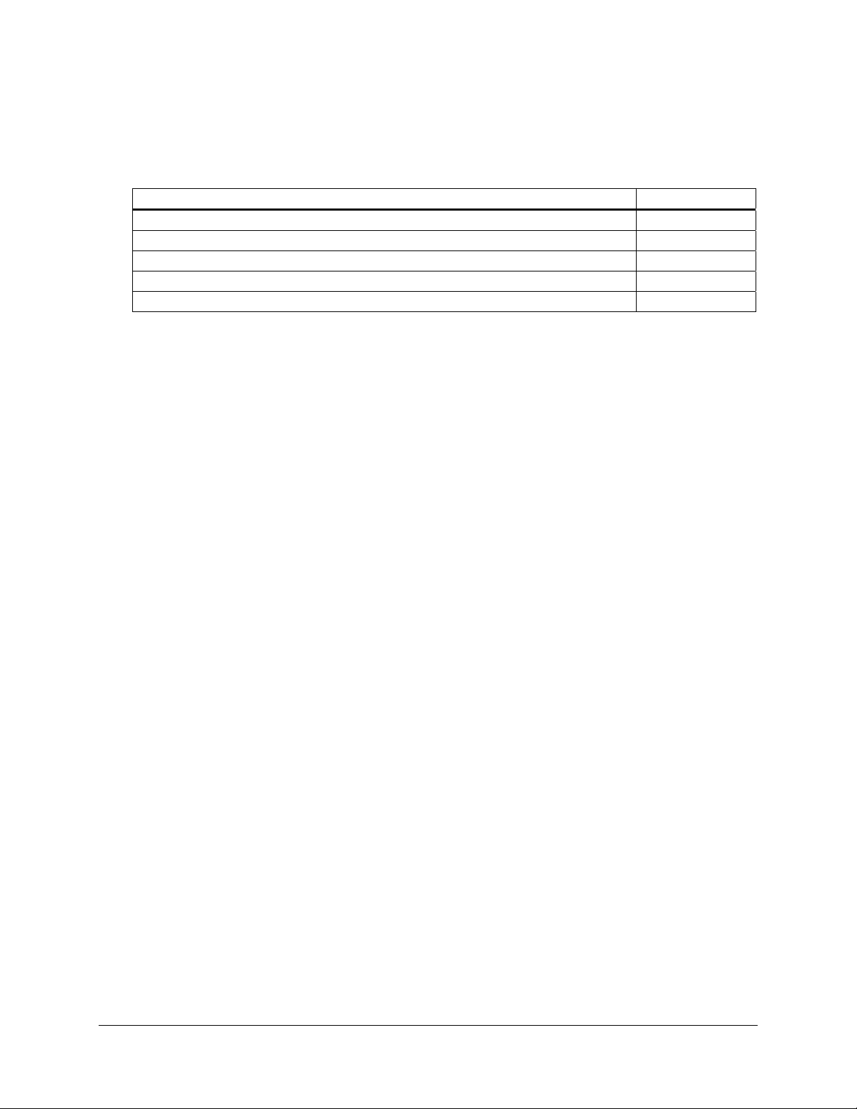
Materials provideda Quantity
Anti-FLAG M2 antibody
Goat anti-mouse antibody, AP conjugated, immunoaffinity purified, polyclonalc 100 μl
Sterile, filtered fraction V bovine serum albumin (BSA) [30% (w/v) in saline] 15 ml
Nitroblue tetrazolium (NBT)b [75 mg/ml in 70% N,N-dimethylformamide (DMF)] 2 ml
5-Bromo-4-chloro-3-indolyl phosphate (BCIP)b (50 mg/ml in 100% N,N-DMF) 1.5 ml
a
Sufficient materials are provided for ten 100-mm nitrocellulose membranes.
b
Also available separately [Anti-FLAG M2 antibody, Stratagene Catalog #200471 (200 μg), #200472 (1 mg);
BCIP (5-bromo-4-chloro-3-indolyl phosphate, Stratagene Catalog #300045 (1.5 ml);
NBT (nitroblue tetrazolium), Stratagene Catalog #300130 (2 ml)].
c
Please see the Certificate of Analysis for the concentration.
d
Immediately on receipt, store the anti-FLAG M2 antibody at –20°C and the goat anti-mouse antibody at 4°C. Avoid
multiple freeze–thaw cycles. Aliquot and fast freeze the primary and secondary antibodies for long-term storage.
b–d
200 μg
STORAGE CONDITIONS
BSA: –20°C
Anti-FLAG
®
M2 Antibody: –20°C
Note For best results, store the anti-FLAG M2 antibody at –20°C.
Goat Anti-Mouse Antibody: 4°C
NBT: 4°C
BCIP: 4°C
Revision A © Agilent Technologies, Inc. 2009.
FLAG® Western Detection Kit
1
Page 5

ADDITIONAL MATERIALS REQUIRED
Western Blotting and Antibody Probing of Electroblots
Laemmli sample buffer (1×)§
Sodium dodecyl sulfate–polyacrylamide gel electrophoresis (SDS–PAGE) system
Nitrocellulose membranes (available from Stratagene, Catalog #420106, #420107, and #420108)
Transfer buffer (1×)§
Blocking solution
Tris-buffered saline (TBS)§
Color Development Reaction
Notes Do not prepare the color development solution containing the NBT and BCIP until
Color development solution
Tris-buffered saline (TBS)
Stop solution
®
Whatman
3MM paper
Dot Blot Assay
Strip of nitrocellulose membrane (1 × 6 cm)
Blocking solution or
Tris-buffered saline (TBS)
Color development solution
®
Whatman
3MM paper
Stop solution
§
See Preparation of Reagents.
§
or bovine lacto transfer technique optimizer (BLOTTO)
§
just prior to use in the color development reaction [see Preparation of the Color
Development Solution Containing Nitroblue Tetrazolium (NBT) and 5-Bromo4-chloro-3-indolyl Phosphate (BCIP) subsection of Color Development Reaction].
§
§
BLOTTO
2
FLAG® Western Detection Kit
Page 6

NOTICES TO PURCHASER
FLAG® License Agreement
The enclosed DNA expression vector and/or antibody are specifically adpated for a method of
producing selected protein molecules covered by one or more of the following patents owned by
Sigma-Aldrich Co.: U.S. Patent Nos. (5,011912, 4,703,004, 4,782,137 and 4,851,341;EP Patent No.
150,126 (Austria, Belgium, Switzerland, France, United Kingdom, Italy, Netherlands and Sweden);
EP Patent No. 335,899 (Belgium, Switzerland, Germany, France, United Kingdom, Italy,
Luxembourg and Sweden); German Patent No. P3584260.1; Canadian Patent No. 1,307,752; and
Japanese Patent Nos. 1,983,150 and 2,665,359. Your payment includes a limited license under these
patents to make only the following uses of these products:
A. Vector License: You may use the enclosed vector to transform cells to produce proteins
containing the amino acid sequence DYKDDDDK for research purposes provided, however, such
research purposes do not include binding an unlicensed antibody to any portion of this amino acid
sequence nor using such proteins for the preparation of antibodies having an affinity for any
portion of this amino acid sequence.
B. Antibody License: You may only use the enclosed antibody for research purposes to perform
a method of producing a protein in which the protein is expressed in a host cell and purified by
use of the antibody in accordance with a claim in one of the above patents in force in a country
where the use actually occurs so long as: (1) you perform such method with a DNA expression
vector licensed from Sigma-Aldrich Co.; and (2) you do not bind (or allow others to bind) an
unlicensed antibody to any DYKDDDDK epitope of any fusion protein that is produced by use of
the method.
This license does not include any rights under any other patents. You are not licensed to use the
vector and/or antibody in any manner or for any purposed not recited above. As used above, the term
“unlicensed antibody” means any antibody which Sigma-Aldrich Co. has not expressly licensed
pursuant to Paragraph B, above. Sigma-Aldrich Co. hereby expressly retains all rights in the above
listed patents not expressly licensed hereunder.
If the terms and conditions of this License Agreement are acceptable to you, then you may open the
vessel(s) containing the vector and/or antibody and, through such act of opening a vessel, will have
shown your acceptance to these terms and conditions.
If the terms and conditions of this License Agreement are not acceptable to you, then please return
the vessel(s) unopened to Agilent for a complete refund of your payment.
For additional licensing information or to receive a copy of any of the above patents, please contact
the Sigma-Aldrich Co. licensing department at telephone number 314-771-5765.
FLAG® Western Detection Kit
3
Page 7

INTRODUCTION
Epitope tagging technology is a powerful method for the analysis of protein
®
structure and function both in vivo and in vitro, and the FLAG
(DYKDDDDK)
1
is one of the most widely used epitope tags in the field of
molecular biology, cell biology, and genetics for protein labeling.
Stratagene FLAG
®
western detection kit includes an anti-FLAG® M2
peptide
1,2,3,4
The
antibody that can detect recombinant fusion proteins with the FLAG epitope
located at either the N- or C-terminal, as well as recombinant fusion proteins
with the FLAG epitope located internally. The anti-FLAG M2 antibody can
detect as little as 5 ng of FLAG epitope-tagged recombinant fusion protein.
The anti-FLAG M2 antibody is highly specific, with low cross-reactivity
with prokaryotic and eukaryotic proteins.
3,4
The FLAG western detection kit utilizes the anti-FLAG M2 antibody as the
primary antibody and an alkaline phosphatase (AP)-conjugated goat antimouse antibody as the secondary antibody. The signal is detected by a color
development solution containing nitroblue tetrazolium (NBT) and 5-bromo4-chloro-3-indolyl phosphate (BCIP). The FLAG western detection kit can
be used to monitor expression of FLAG-tagged proteins in prokaryotic and
eukaryotic cells, to follow FLAG epitope-tagged protein during protein
purification steps, and to identify proteins following immunoprecipitation.
In addition, the FLAG western detection kit provides a rapid and sensitive
way to detect proteins expressed in the ESP yeast protein expression and
purification system (Stratagene Catalog #251600).
WESTERN BLOTTING
5,6
AND ANTIBODY PROBING OF ELECTROBLOTS
Note Unless otherwise indicated, conduct all procedures at room
temperature on a shaker.
1. Boil the antigenic protein samples in 1× Laemmli sample buffer for 2
minutes and load the samples and a molecular-weight marker into
separate lanes of an appropriate percentage SDS–polyacrylamide gel
for analysis. Resolve the gel by SDS–PAGE.
2. Equilibrate a nitrocellulose membrane in 1× transfer buffer by soaking
the membrane for 20 minutes.
Note Never touch the nitrocellulose membranes with bare hands;
always use gloves or forceps. Also, do not allow the
nitrocellulose membranes to dry during any step.
4
FLAG® Western Detection Kit
Page 8

3. Electroblot the antigenic protein samples onto the nitrocellulose
membrane either at 30 V and ~200 mA or at the voltage and
approximate current specified by the manufacturer of the transfer
apparatus for 1 hour in 1× transfer buffer.
4. Following electrophoretic transfer of the proteins onto the
nitrocellulose membrane, block the membrane by incubating the
nitrocellulose membrane in blocking solution, using at least 25 ml of
blocking solution for an 8- × 6-cm membrane, with gentle rocking at
room temperature for a minimum of 1 hour.
Notes Blocking may also be performed overnight at 4°C.
BLOTTO may be substituted for the blocking solution
throughout the western blotting, antibody probing, and the
optional dot blot assay procedures when blocking the
membrane. Do not use
BLOTTO to dilute the anti-FLAG M2
antibody or the goat anti-mouse antibody.
5. Thoroughly wash the nitrocellulose membrane twice with a generous
amount of TBS for a minimum of 2 minutes/wash.
Notes If nitrocellulose membranes are combined during the wash
steps, less of the wash solution can be used. Separate the
nitrocellulose membranes with forceps at each change to
ensure adequate exposure of each membrane.
6. For an 8- × 6-cm membrane, add 20 μg of the anti-FLAG M2 antibody
to at least 10 ml of TBS and incubate the membrane in this antibody
dilution at room temperature for a minimum of 1 hour.
7. Thoroughly wash the nitrocellulose membrane twice with a generous
amount of TBS for a minimum of 2 minutes/wash.
8. Dilute the goat anti-mouse antibody 1:5000 in blocking solution.
9. Gently rock the nitrocellulose membrane in the goat anti-mouse
antibody dilution, using at least 10 ml for an 8- × 6-cm nitrocellulose
membrane, at room temperature for a minimum of 1 hour.
10. Thoroughly wash the nitrocellulose membrane three times with a
generous amount of TBS for a minimum of 5 minutes/wash and then
proceed to the Color Development Reaction.
FLAG® Western Detection Kit
5
Page 9

COLOR DEVELOPMENT REACTION
Preparation of the Color Development Solution Containing Nitroblue
Tetrazolium (NBT) and 5-Bromo-4-chloro-3-indolyl Phosphate (BCIP)
Prepare the color development solution containing NBT and BCIP just prior
to use as outlined in the following steps.
1. Dilute 40 μl of NBT (75 mg/ml) in 10 ml of color development
solution to a final concentration of 0.3 mg/ml for a standard 8- × 6-cm
nitrocellulose membrane.
2. Add 30 μl of BCIP (50 mg/ml) dropwise to a final concentration of
0.15 mg/ml.
Note If a precipitate forms, warm the color development solution
containing the NBT and BCIP to 37°C and pass this solution
through a 0.2-
3. Protect the color development solution containing the NBT and BCIP
from light and leave the solution at room temperature until ready for
use in step 2 of the following section.
Color Development on the Nitrocellulose Membrane
μ
m filter.
Note Allow the color development reaction to proceed in the dark.
1. Remove the nitrocellulose membrane from the final TBS wash (see
step 11 of Western Blotting and Antibody Probing of Electroblots) and
blot off any excess moisture with Whatman
2. Immerse the nitrocellulose membrane in the color development
solution containing NBT and BCIP. Allow the color development
reaction to proceed in the dark until the positive reactions are clearly
visible (usually within minutes).
Notes Extended times for color development can result in high
backgrounds. If extended times (1–5 hours) are required for
the color development reaction, store the nitrocellulose
membranes in the dark and check the membranes
periodically to minimize the background.
If a visible precipitate forms during the color development
reaction that is not associated with specific dots or bands,
replace the color development solution containing NBT and
BCIP with a freshly prepared solution and allow the color
development reaction to continue.
3. Remove the color development solution containing NBT and BCIP
from the nitrocellulose membrane and rinse the membrane with TBS.
®
3MM paper.
6
FLAG® Western Detection Kit
Page 10

4. Terminate the color development reaction by immersing the
nitrocellulose membrane in stop solution.
5. Following completion of the color development reaction, air dry the
nitrocellulose membrane and store the membrane protected from light.
If desired, the optional Dot Blot Assay may be performed to quickly and
simply determine if the recombinant fusion protein has been produced.
DOT BLOT ASSAY (OPTIONAL)
The dot blot assay assesses the ability of the anti-FLAG M2 antibody
dilution and the goat anti-mouse antibody dilution to detect different levels
of recombinant fusion protein. The dot blot assay also verifies the reaction
of the NBT and BCIP with the AP-conjugated goat anti-mouse antibody.
1. Cut out a 1- × 6-cm strip of nitrocellulose membrane. Using a soft
pencil, mark the strip into six equal squares measuring ~1 cm
2. Prepare the nitrocellulose membrane with serial protein dilutions,
anti-FLAG M2 antibody, and goat anti-mouse antibody as follows:
a. Spot 1 μl of each of the following serial protein dilutions onto
separate squares of the strip: 100 ng/μl, 10 ng/μl, 5 ng/μl, and
1 ng/μl.
b. Spot 1 μl of the anti-FLAG M2 antibody diluted in TBS to a
concentration of 10 ng/μl onto a separate square of the strip.
c. Spot 1 μl of the goat anti-mouse antibody diluted 1:5000 in
blocking solution onto a separate square of the strip.
3. Air dry the nitrocellulose membrane for 5 minutes.
4. Immerse the strip in blocking solution for 1 hour at room temperature
to block nonspecific protein-binding sites.
5. Thoroughly wash the nitrocellulose membrane two to three times for
2 minutes each in a generous amount of TBS.
2
.
6. Prepare 10 ml of the anti-FLAG M2 antibody diluted in TBS to a
concentration of 2 μg/ml and incubate the strip in the anti-FLAG M2
antibody dilution at room temperature for 1 hour.
FLAG® Western Detection Kit
7
Page 11

7. Thoroughly wash the nitrocellulose membrane two to three times for
2 minutes each in a generous amount of TBS.
8. Prepare 10 ml of the goat anti-mouse antibody diluted 1:5000 in
blocking solution and gently rock the strip in the goat anti-mouse
antibody dilution at room temperature for a minimum of 1 hour.
9. Thoroughly wash the nitrocellulose membrane three times with a
generous amount of TBS for a minimum of 5 minutes/wash.
10. Prepare the color development solution containing NBT and BCIP as
outlined in the following steps:
a. Dilute 40 μl of NBT (75 mg/ml) in 10 ml of color development
solution to a final concentration of 0.3 mg/ml.
b. Add 30 μl of BCIP (50 mg/ml) dropwise to a final concentration
of 0.15 mg/ml.
Note If a precipitate forms, warm the color development
solution containing the NBT and BCIP to 37°C and pass
this solution through a 0.2-
μ
m filter.
c. Protect the color development solution containing the NBT and
BCIP from light and leave the solution at room temperature until
ready for use in step 12 of this section.
11. Remove the strip from the final TBS wash and blot off any excess
moisture with Whatman 3
MM paper.
12. Immerse the nitrocellulose membrane in color development solution
containing NBT and BCIP. Allow the color development reaction to
proceed in the dark until the positive reactions are clearly visible.
Check the membrane every minute for a positive reaction.
13. Rinse the residual precipitated dye from the strip with a generous
amount of TBS.
14. Terminate the color development reaction by immersing the
nitrocellulose membrane in stop solution.
15. Air dry the nitrocellulose membrane and store the membrane protected
from light.
8
FLAG® Western Detection Kit
Page 12
TROUBLESHOOTING
Western Blotting, Antibody Probing of Electroblots, and Color Development
Reaction
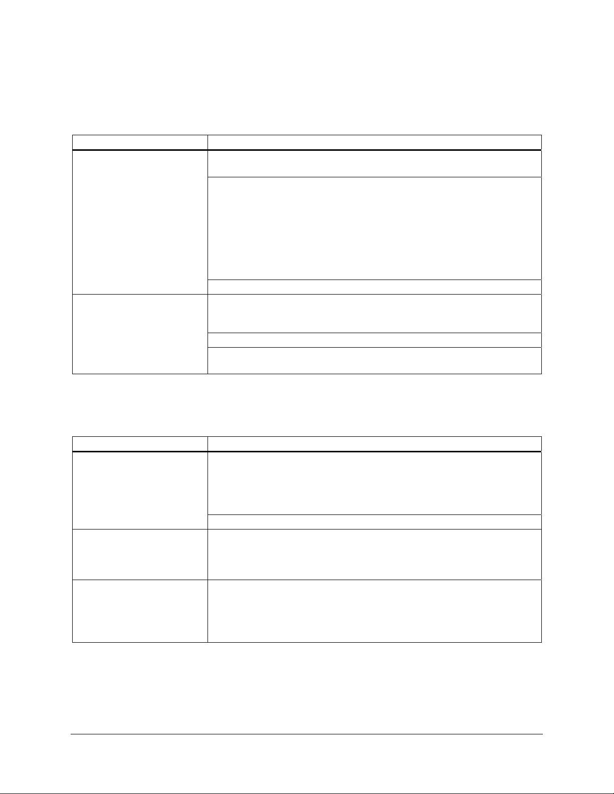
Observations Suggestions
The signal is not visible on the
nitrocellulose membrane
Excessive background on the
nitrocellulose membrane is
present
Ensure that the goat anti-mouse antibody or the color development solution is fresh
and/or properly prepared
To test the activity of the working dilution of the goat anti-mouse antibody, add 1 ml
of the color development solution containing NBT and BCIP with 1 ml of the goat
anti-mouse antibody diluted in TBS as indicated on the Certificate of Analysis; if no
color precipitates within 30 minutes, repeat this test with a fresh dilution of the goat
anti-mouse antibody
If color does appear, proceed to the Dot Blot Assay to check the protein, the antiFLAG M2 antibody, and the goat anti-mouse antibody
Verify the FLAG sequence in the protein
If the nitrocellulose membrane shows excessive background, check that the blocking
solution or BLOTTO is properly prepared and ensure that the nitrocellulose membrane
is thoroughly washed
Check to ensure that that the anti-FLAG M2 antibody is properly diluted in TBS
Check to ensure that that the goat anti-mouse antibody is properly diluted in the
blocking solution
Dot Blot Assay (Optional)
Note The observations in this section are addressed by performing the optional Dot Blot Assay.
Observations Suggestions
The protein is not visible on the
dot blot strip, while the antiFLAG M2 antibody probe is
visible
The anti-FLAG M2 antibody
probe is not visible on the dot
blot strip, while the goat antimouse antibody is visible
The goat anti-mouse antibody is
not visible on the dot blot strip
Because the protein is folded, the FLAG epitope tag, which is located in the interior of
the protein molecule, is not making contact with the anti-FLAG M2 antibody probe;
perform western blotting noting that the denaturing characteristics of the gel should
cause the protein to unfold, making the FLAG epitope tag available for detection by
the anti-FLAG M2 antibody
Verify the FLAG sequence in the protein
Use a freshly prepared dilution of the anti-FLAG M2 antibody probe or replace the
probe completely with a new anti-FLAG M2 antibody probe
To test the activity of the working dilution of the goat anti-mouse antibody, add 1 ml
of the color development solution containing NBT and BCIP with 1 ml of the goat
anti-mouse antibody diluted in TBS as indicated on the Certificate of Analysis; if no
color precipitates within 30 minutes, repeat this test with a fresh dilution of the goat
anti-mouse antibody
FLAG® Western Detection Kit
9
Page 13
PREPARATION OF REAGENTS
Stock Solutions
Use sterile concentrated stock solutions prepared with deionized, double-distilled water (ddH2O) or
the equivalent. The stock solutions necessary for all reagent preparations in the FLAG western
detection kit are as follows:
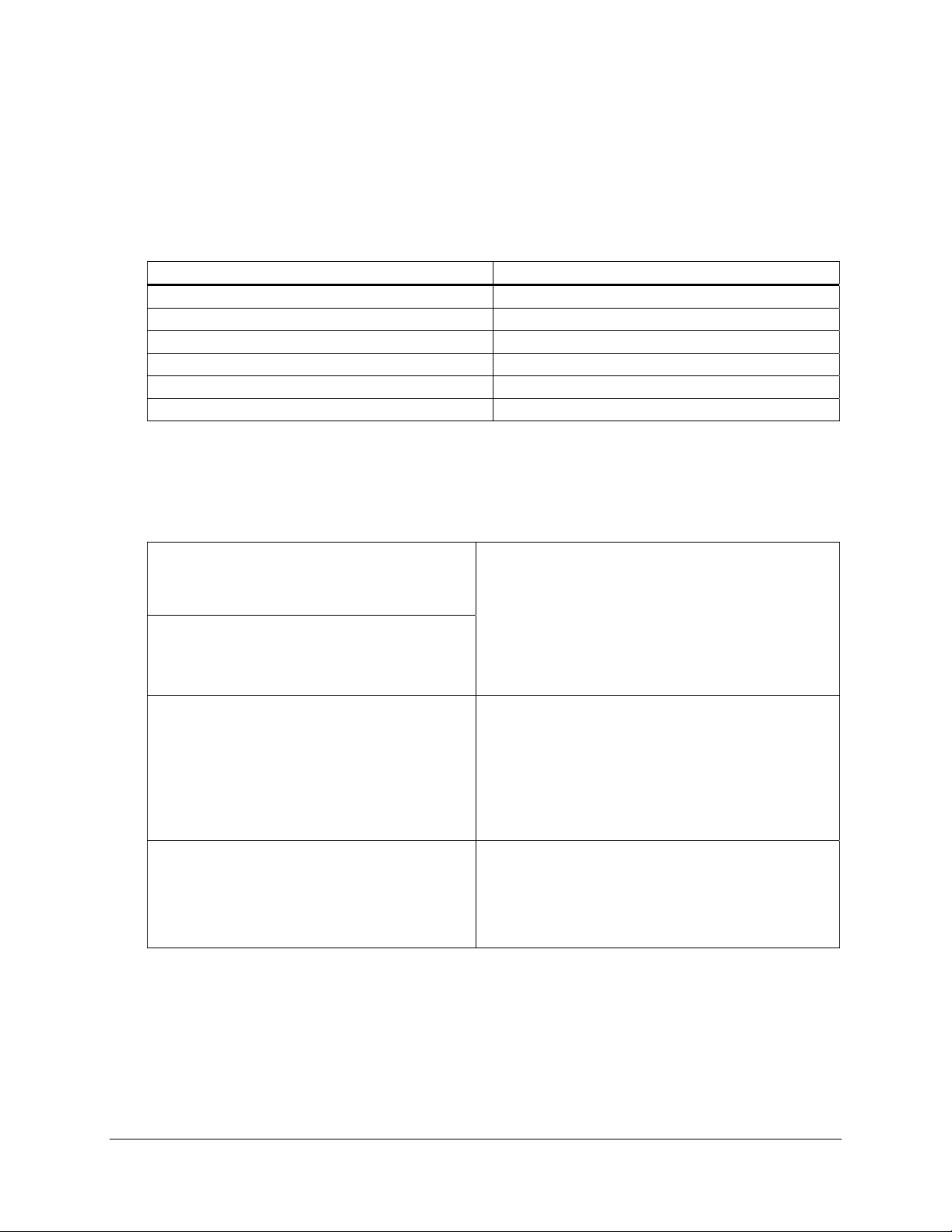
Stock solutions Sterilization method
1 M Tris-HCl (pH 7.5) Autoclave
1 M Tris-HCl (pH 9.5) Autoclave
1 M Tris-HCl (pH 2.9) Autoclave
5 M NaCl Autoclave
1 M CaCl2 Autoclave
1 M MgCl2 Filter sterilize
Standard Reagents
Note Use freshly prepared reagents. The suggested working volume is 10 ml for each 8- × 6-cm
nitrocellulose membrane.
Blocking Solution
1% (w/v) BSA in TBS
TBS
50 mM Tris-HCl (pH 7.5)
150 mM NaCl
Color Development Solution
100 mM Tris-HCl (pH 9.5)
100 mM NaCl
5 mM MgCl
2
Stop Solution
20 mM Tris-HCl (pH 2.9)
1 mM CaCl
2
BLOTTO
5 g of nonfat dry milk
1 ml of 1% (w/v) thimerosal–H
2
O
(Sigma Catalog #T 5125)
Adjust the volume to 100 ml with TBS
Store at 4°C for 7–10 days
Laemmli Sample Buffer (2×)
250 mM Tris-HCl (pH 6.8)
8% (w/v) SDS
40% (v/v) glycerol
0.01% (w/v) bromphenol blue dye
200 mM β-mercaptoethanol
Transfer Buffer (1×)
12 mM Tris-HCl (pH 8.3)
96 mM glycine
20% (v/v) methanol
10
FLAG® Western Detection Kit
Page 14

REFERENCES
ENDNOTES
1. Hopp, T., Prickett, K., Price, V., Libby, R., March, C. et al. (1988) BioTechnology
6:1204-1210.
2. Chiang, C. M. and Roeder, R. G. (1993) Pept Res 6(2):62-4.
3. Su, X., Prestwood, A. K. and McGraw, R. A. (1992) Biotechniques 13(5):756-62.
4. Smit, M. J., Timmerman, H., Alewijnse, A. E., Punin, M., van den Nieuwenhof, I. et al.
(1995) Biochem Biophys Res Commun 214(3):1138-45.
5. Harlow, E. and Lane, D. (Eds.). (1988). Antibodies: A Laboratory Manual. Cold Spring
Harbor Laboratory Press, Plainview, NY.
6. Towbin, H., Staehelin, T. and Gordon, J. (1979) Proc Natl Acad Sci U S A
76(9):4350-4.
FLAG® and Anti-FLAG® are registered trademarks Sigma-Aldrich Co.
Whatman
®
is a registered trademark of Whatman Ltd.
MSDS INFORMATION
The Material Safety Data Sheet (MSDS) information for Stratagene products is provided on the web at
http://www.stratagene.com/MSDS/. Simply enter the catalog number to retrieve any associated MSDS’s
in a print-ready format. MSDS documents are not included with product shipments.
FLAG® Western Detection Kit
11
Page 15

FLAG® Western Detection Kit
Catalog #200470
QUICK-REFERENCE PROTOCOL
Western Blotting and Antibody Probing of Electroblots
♦ Boil the protein samples in 1× Laemmli sample buffer for 2 minutes, load the samples and a
molecular-weight marker into separate lanes of a SDS–polyacrylamide gel, and resolve by
SDS–PAGE
♦ Equilibrate a nitrocellulose membrane in 1× transfer buffer for 20 minutes
♦ Electroblot the protein samples onto the nitrocellulose membrane either at 30 V and
~200 mA or at the voltage and approximate current specified by the manufacturer of the
apparatus for 1 hour in 1× transfer buffer
♦ Block the nitrocellulose membrane in blocking solution with gentle rocking at room
temperature for a minimum of 1 hour
♦ Wash the membrane twice with TBS for a minimum of 2 minutes/wash
♦ Add 20 μg of the anti-FLAG M2 antibody to at least 10 ml of TBS and incubate the
membrane in this antibody dilution at room temperature for a minimum of 1 hour
♦ Wash the membrane twice with TBS for a minimum of 2 minutes/wash
♦ Dilute the goat anti-mouse antibody 1:5000 in blocking solution
♦ Gently rock the nitrocellulose membrane in the goat anti-mouse antibody dilution at room
temperature for a minimum of 1 hour
♦ Wash the membrane three times with TBS for a minimum of 5 minutes/wash
Color Development Reaction
♦ Prepare the color development solution containing NBT and BCIP
♦ Blot off any excess moisture and immerse the nitrocellulose membrane in color development
solution containing NBT and BCIP until the positive reactions are clearly visible
♦ Rinse the membrane with TBS
♦ Immerse the nitrocellulose membrane in stop solution
♦ Air dry the membrane and store protected from light
14
 Loading...