


DiaTecne
PulsePen - Quick Reference Guide 1.0
This Quick Reference Guide contains information about the use of the PulsePen system in synthetic form, aiming at providing
some tips to obtain good signals in a simple way.
This Guide, together with the user Manual and the software on-line Help, provide comprehensive information regarding the
use of PulsePen system.
We highly recommend the users to read it.
Content:
•
General Notes
•
Operator Tips
•
Patient Tips
•
Noisy Signals
•
Good Signal
• Traffic Lights (software)
Indications:
Recommended / Correct
Wrong / Unsatisfactory
1

General Notes
DiaTecne
PulsePen - Quick Reference Guide 1.0
Press the on/off button until the
beep sound (about 1 sec).
The above images show how to
insert and remove battery for the
tonometric probe.
PulsePen includes an “auto-freeze” function that works in
association with the tonometric probe - sensor 2 (blue curve)
during the signal capture mode. When pulsatile signal is detected
(left), the computer screen is continuously updated with the
incoming curves; last captured curves are “frozen” when the probe
is removed (right), waiting for storage.
2
Operator Tips
DiaTecne
PulsePen - Quick Reference Guide 1.0
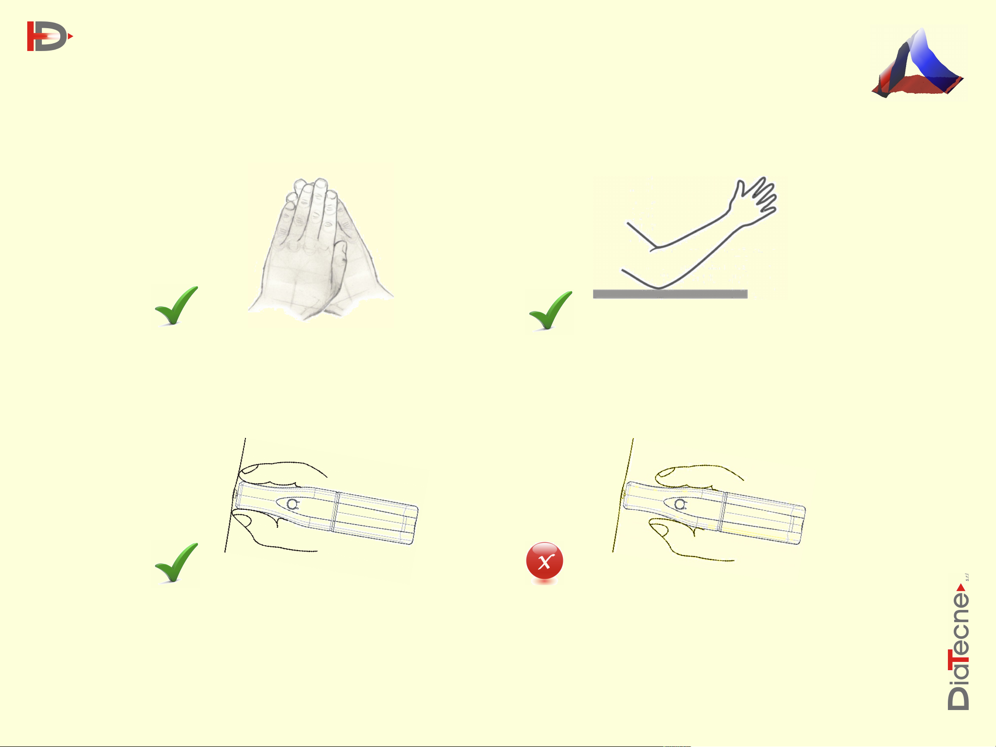
When searching for Femoral Artery, overlapping
fingertips is very helpful to find pulsation.
The tonometric probe must be kept perpendicular
to the application surface with the operator’s
Keep the operating elbow lying firmly on a
stable surface (not “floating”).
WRONG way to hold the tonometric
probe.
fingers touching the patient’s skin.
3

Patient Tips
DiaTecne
PulsePen - Quick Reference Guide 1.0
In order to obtain good quality signals, the patient
must be relaxed, in supine position on horizontal
surface.
Signals on the femoral artery can be captured also
through tights, pantyhose, leggings, or thin pants.
A small pillow (orange in the image) on the
opposite side of the target carotid artery, is
useful to keep the patient’s head stable.
4
Noisy Signals
DiaTecne
PulsePen - Quick Reference Guide 1.0
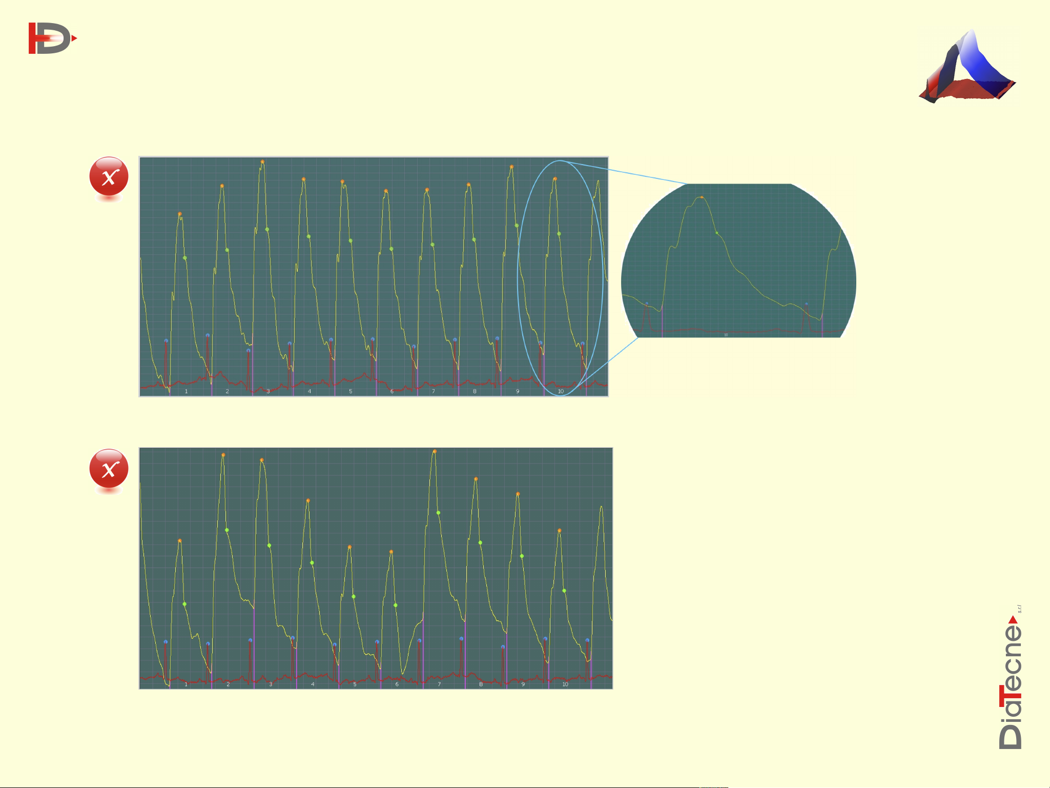
Example 1: Noisy signal superimposed by operator’s tremor- cycle 10 expanded on the right.
Important!
Bad signals could produce mistakes in
determination of the pressure wave
markers and parameters.
For remedies, please follow
Operator Tips and Patient Tips
Example 2: Noisy signal superimposed by operator’s tremor and movements
5
Good Signal
DiaTecne
PulsePen - Quick Reference Guide 1.0
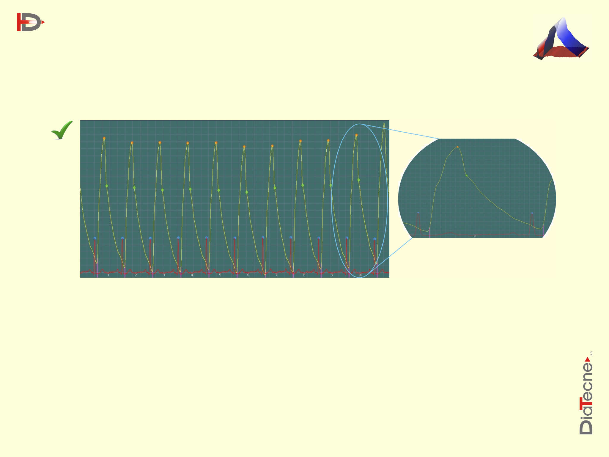
Example of clean signal - cycle 10 expanded on the right
Slight breathing fluctuations are physiological and do not cause any problem.
6
Possible causes
Remedy
Ecg + Tonometry
The ECG amplitude is too low
Re-position one or both the ECG electrodes in order to
obtain signals with higher amplitude.
The R wave is reversed (downwards)
Exchange the Ecg cables or electrodes.
The R wave has pathological shape
Proceed with the capture for at least 10 seconds, save
the signals and manually add markers on the R waves.
2 x Tonometry
Tonometer1 (red curve) is off and/or no signal is
applied
Switch on the Tonometer1 and position it on the artery
under investigation (Carotid).
Captured signal’s amplitude on Tonometer1 is too low
Re-position the Tonometer1 in order to obtain signals
with higher amplitude.
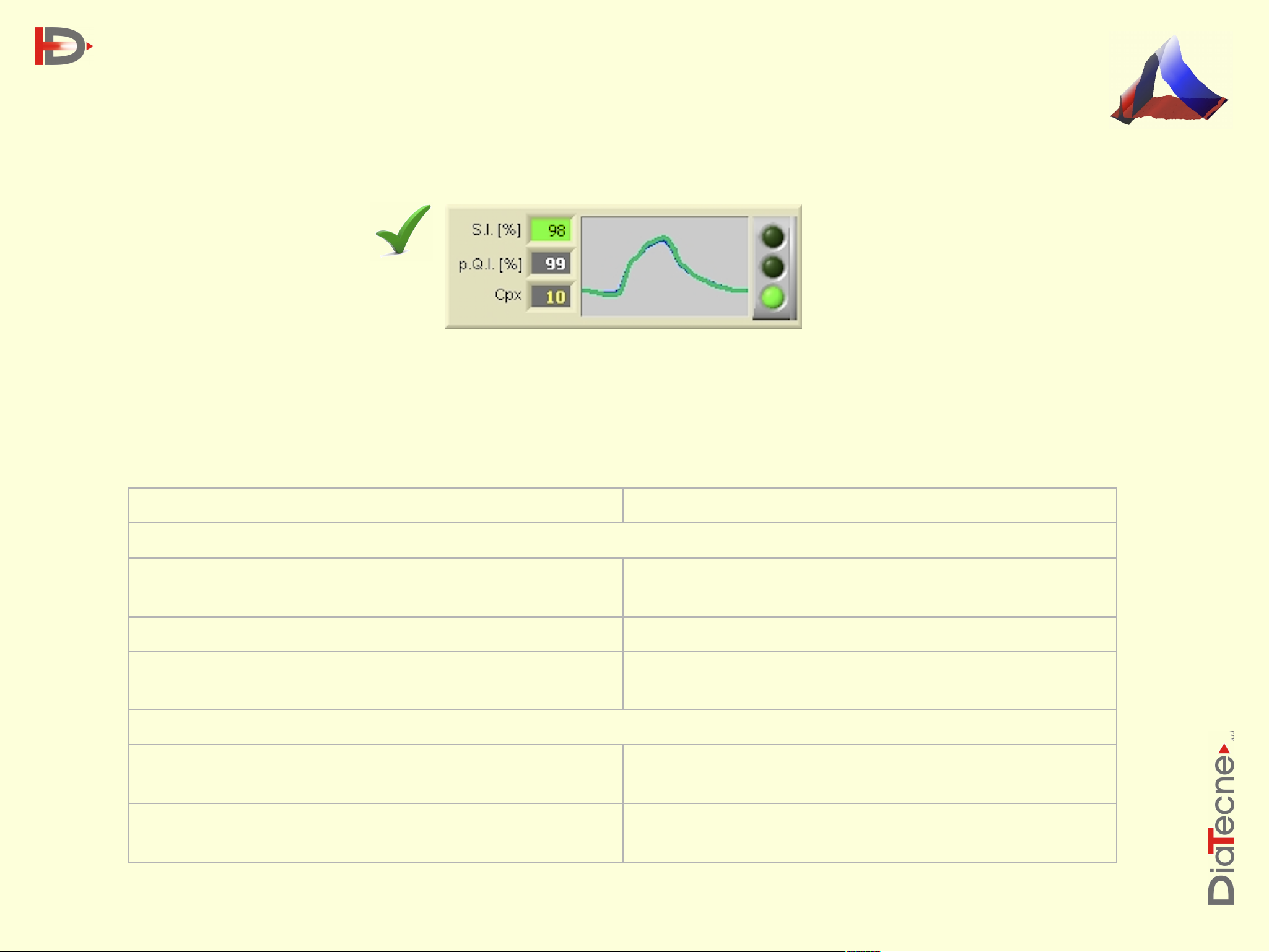
Traffic Lights (software)
The small traffic lights graph, at the top of the computer screen during the signal capture, is updated each
cardiac cycle. If it doesn’t occur, it means:
•
The R wave of the ECG is not detected (in the case of Ecg + Tonometry).
DiaTecne
PulsePen - Quick Reference Guide 1.0
•
The foot of Tonometer - sensor1 curve is not detected (in the case of 2 x Tonometry, ETT model only).
7
 Loading...