

Page 1
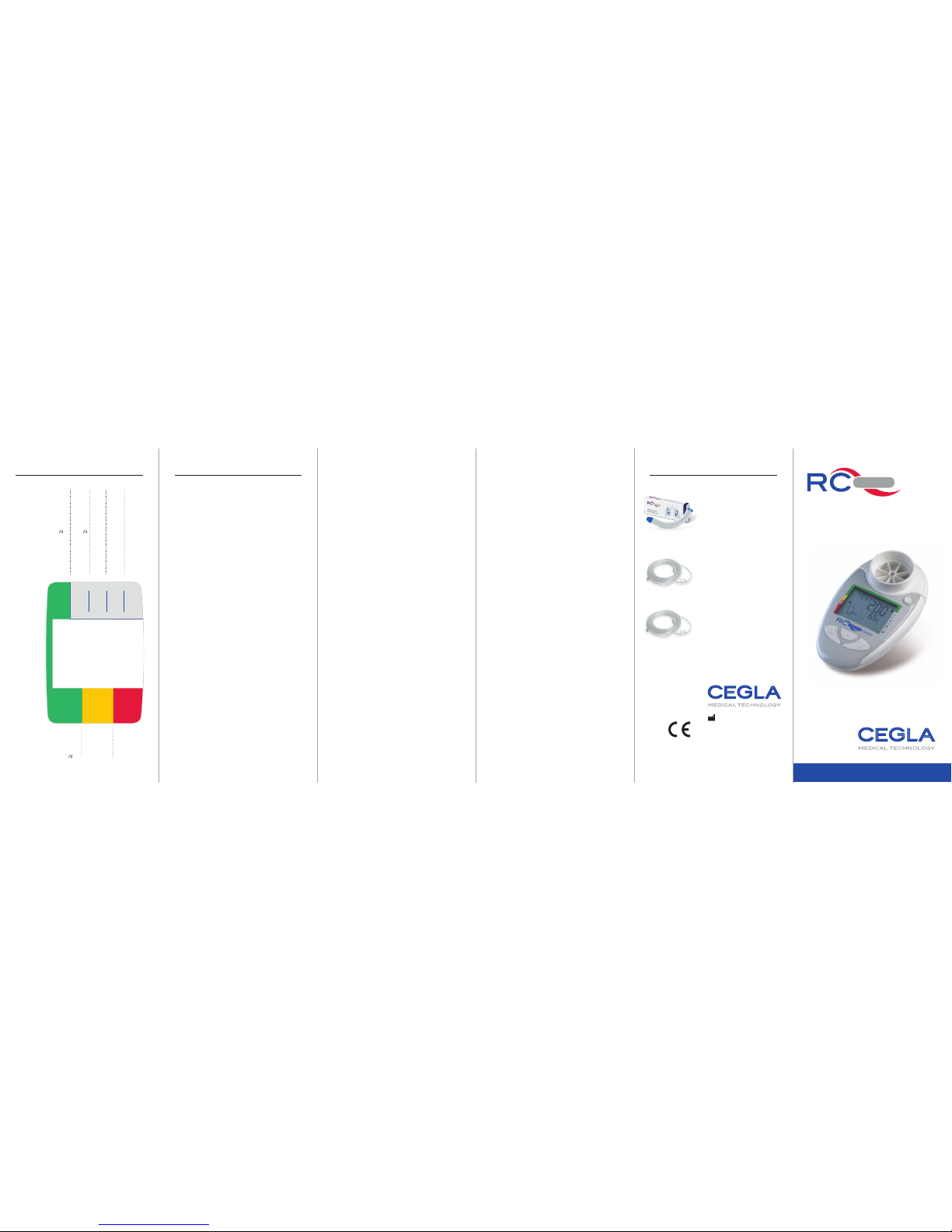
The Digital Pulmonary Function
Test for COPD and Pulmonary
Emphysema
COP D
TesT
PHYSIOLOGICAL BACKGROUND
The physiological background of the
pulmonary parameters.
The lungs are made up of airways,
bronchi, bronchiole and millions of
alveoli. The diameter of the smallest
bronchiole is very small compared to
the large bronchi and the airways.
The one second capacity (FEV1)
measures in the first second the maximum exhaled lung volume. This
measurement predominantly mirrors
a narrowing of the large airways. The
six second capacity in contrast is
the standard for the measurement of
changes to the small airways.
Unlike to the bronchi, the bronchiole are not stabilized by cartilage, but
rather, held open by elastic draws in
the lung tissue. When inhaling deeply
these draws cause the bronchiole to
expand. When exhaling the draw of
the lung tissue lets up due to the shrinking lung volume which narrows the
bronchiole even more.
The Six Second Capacity (FEV6)
measures the “rest” breathing volume
after 6 seconds of exhaling. That is, at
a point in time at which the retraction
power of the lung volume, as described above, is significantly diminished.
COPD is an illness which besides the
larger airways (mucous buildup, narrowing) primarily effects the smallest
airways and the pulmonary alveoli.
This results in dissolution of the setting
of the smallest airways and thereby
causing a diminished holding open of
these bronchial parts during exhaling.
The FEV6 mirrors therefore the disturbances in the pulmonary periphery significantly better than the FEV1. From
the ratio FEV1/FEV6 the narrowing of
the smallest airways can be determined.
With illnesses such as pulmonary fibrosis (an airway restricting illness)
the fibrosis affected lung tissue of the
bronchiole is pulled apart, so that with
a generally diminished FEV1 (due to
diminished lung volume with pulmona-
ry fibrosis) the FEV6 is in comparison
significantly increased.
With COPD we observe disturbances,
which can be (partially) rectified with
medicine and other changes which are
caused by physical disturbances of the
lung tissue and thereby cannot respond
to medicine.
Here the physical therapy, for example
with the RC-Cornet (basic cornet), can
help. This respiratory therapy device
loosens mucous, stabilizes and expands
the small bronchi and reduces shortness
of breath of the patient.
Detailed instructions for use are available at www.cegla.de.
07/2015
Article-no. 160EN
PPN 1107 1177 1670
The related products as well as accessories for
the RC-Test are available in pharmacies, medical
supply stores or at www.cegla-shop.com.
RC-Cornet
®
Respiratory Therapy Device
Loosens mucous, reduces shortness of breath and coughing.
Article-no. 210EN
PPN 1108 4186 6756
PIP 392-5542
Oxygen cannula craton
adults
Comfortable to wear due to
the soft kraton nose tips.
Article-no. 710EN
PPN 1103 3673 0429
Oxygen cannula
phthalate-free adults
With soft kraton nose tips,
free of phthalate (plasticizers).
Article-no. 760EN
PPN 1110 3980 1541
RELATED PRODUCTS
INSTRUCTIONS FOR USE
0
1
2
3
Normal
I
II
III
IV
Obstruction
Index
COPD-
Classifi-
cation
FEV1/ FEV6-
ratio and
FEV1 value in % of
the standard value (SV)
FEV1 /FEV6 0,70
FEV1 /FEV6 < 0,70 &
FEV1
80 % of the NW
FEV1 /FEV6 < 0,70 &
FEV1 < 80 % of the NW
FEV1 /FEV6 < 0,70 &
FEV1 < 50 % of the NW
FEV1 /FEV6 < 0,70 &
FEV1 < 30 % of the NW
FEV1-Value
iin % of
the standard
value
80 %
< 80 %
< 50 %
< 30 %
DISPLAY EXPLAINATION
Please read the instructions for use
of the RC-Test COPD carefully before
first use.
Manufacturer:
R. Cegla GmbH & Co. KG
Horresser Berg 1
56410 Montabaur
Germany
www.cegla.d e
Page 2
RC-TEST PREPARATION APPLICATION DESCRIPTION
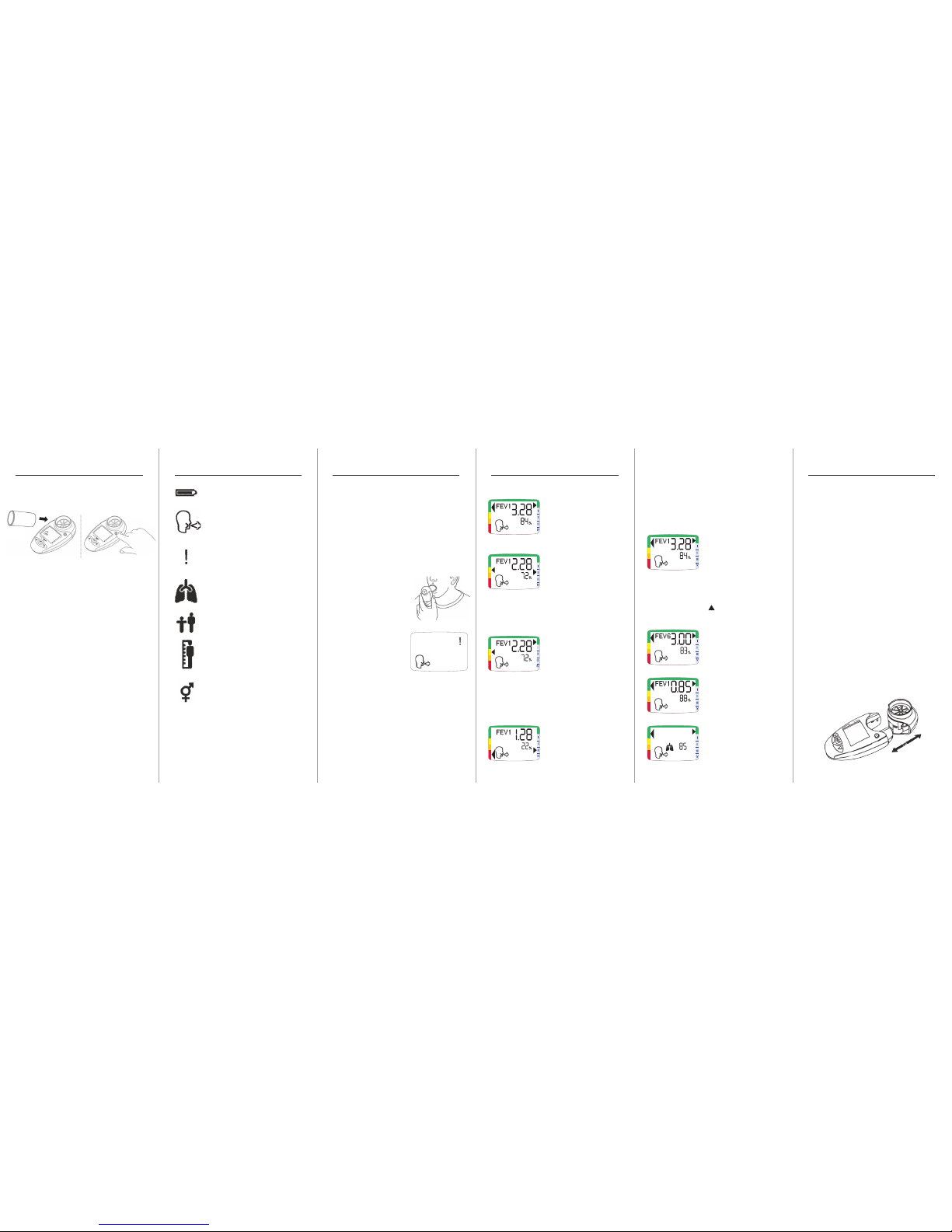
Normal
Notice that both arrows
point to the green area!
Possible COPD or Emphysema Result
A possible COPD indication results when the left
arrow points to the yellow
area and the right arrow
indicates a number I to IV. Recommendation:
Spirometer Examination
Possible Restriction Result
Indications of a restrictive
pulmonary illness, for
example fibrosis, result
when the left arrow points
to the yellow area and the right arrow to the
green area. COPD indication is unlikely.
Recommendation: Spirometer Examination
Indication of severe COPD
When the left arrow
points to the red and
the right arrow indicates
a number III to IV, then
that is a sure indication
of severe COPD. Recommendation:
Spirometer examinationCOPD.
Display of the pulmonary function
parameters
1. The display shows
the FEV1 (absolute) and
the FEV1 in % of the
standard value. Each
value of the breathing maneuver is displayed until the enter key is pushed.
2. By pushing the
-key you can check
the following displays one after the other:
a) a. FEV6 (absolute)
and FEV6 in % of the
standard value
b) a. FEV1/FEV6 ratio
and FEV1/FEV6 in %
of the standard value
c) a. Estimated
biological age of the
lung
TEST ASSESSMENT CLEANING AND DISINFECTION
Battery charge condition
Now blow
Bad maneuver
(delayed start or cough)
Biological lung age
Age
Size
Sex
DEVICE SYMBOLS
Device Head
Device Body
Before first use, please put in the batteries.
1. Fasten the mouth
piece* onto the device.
3. After the battery indicator goes out
then the device is ready for use.
4. Enter the age of the patient and
confirm the entry with the enter key.
5. Enter the size of the patient and
confirm the entry with the enter key.
6. Enter the sex of the patient and
confirm the entry with the enter key.
All surfaces should be cleaned weekly
(wiped down), more often when needed. We recommend for cleaning the
use of disinfectant disposable wipes.
Special attention should be paid to the
device head.
To clean or disinfect please remove
the device head with a light tug from
the device body. Now the device head
can be soaked in a soap water bath
or disinfectant solution for 15 minutes.
Afterwards rinse with warm water to
accelerate the drying process. The
device body itself may not come into
contact with water.
* With the RC-Test COPD can be used the safety
mouthpiece (per patient | no to be cleaned |
article-no. 460EN or the reusable mouthpiece
(for one patient | article-no. 180EN). Mouthpieces are available separately in pharmacies or at
www.cegla-shop.com
The patient must position the device in
the hand so that the backside opening is
not covered. The patient stands upright,
inhales deeply and encloses the mouthpiece tightly with his/her lips. Afterwards
he/she exhales as fast and as powerful
as possible for 6 seconds.
After 6 seconds a beep will sound.
Afterwards the exhaling
maneuver should be
carried out another two
times.
The device recognizes
a bad or an insufficient
exhale maneuver and
shows this as a ! on the
display.
The best test can be displayed with the
enter key.
If the device is not used for two minutes
then it automatically turns itself off.
To turn the device off please hold down
the power button for three seconds.
2. Turn on
the device.
 Loading...