

Page 1
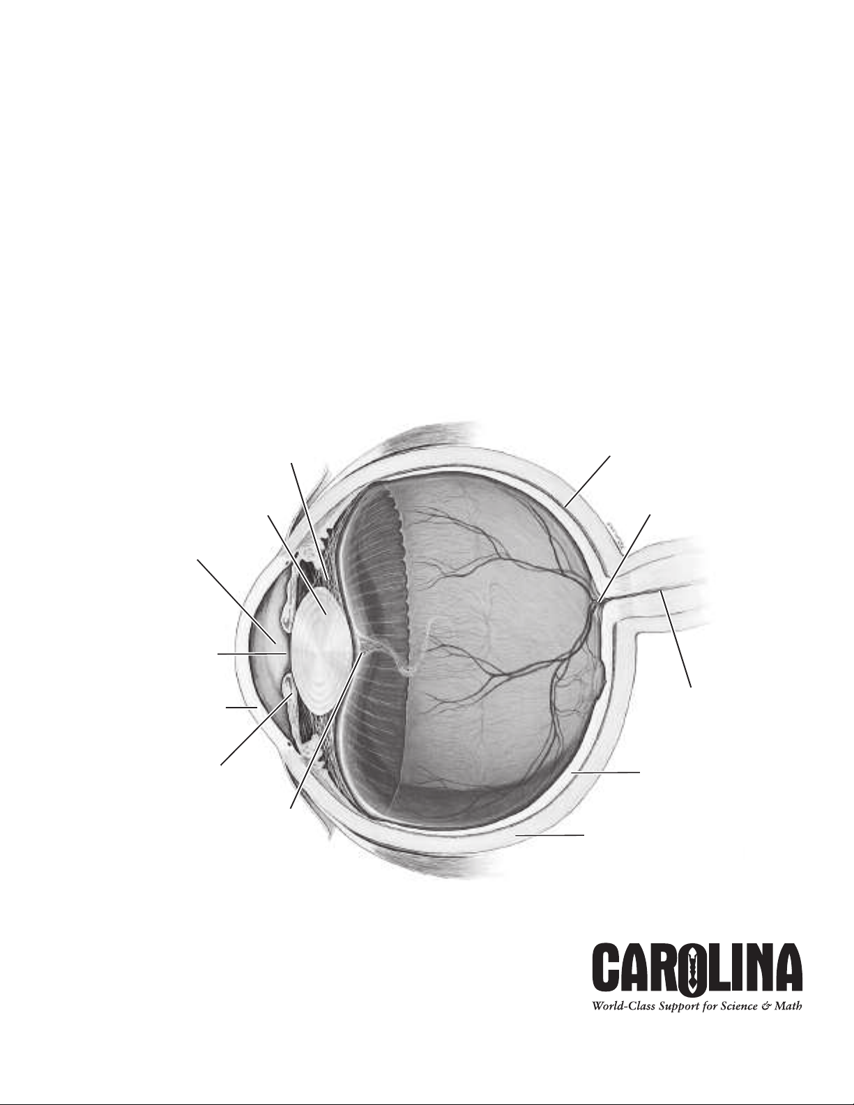
Sclera
Retina
Optic nerve
Blind spot
Choroid
Zonula ciliaris
Lens
Aqueous humor
Pupil
Cornea
Iris
Hyaloid fossa
Vitreous
humor
CarolinaTMMammal Ey
e
Dissection Guide
C80130
Page 2
CarolinaTMMammal Eye Dissection Guide
Overview
The Carolina Mammal Eye Dissection Guide is a general set of instructions for dissecting mammal eyes. With
each type of eye, there will be differences in the size of the structures and in the thickness and coloration of
the tissue, but the general structures and their relative location are the same.
Safety
Follow safe laboratory practices when performing any dissection. Wear splashproof safety goggles, gloves, and
lab aprons at all times. Be careful when using sharp tools, such as scalpels, forceps, teasing needles, and
scissors. Perform dissections on a dissecting tray to contain specimens and fluids.
Procedure
Review the glossary provided at the end of this dissection guide. Refer to the diagram of the eye as a general
reference as you observe and identify external and internal structures.
1. Observe the outer structure of the eye. Identify the following: optic nerve, sclera, cornea.
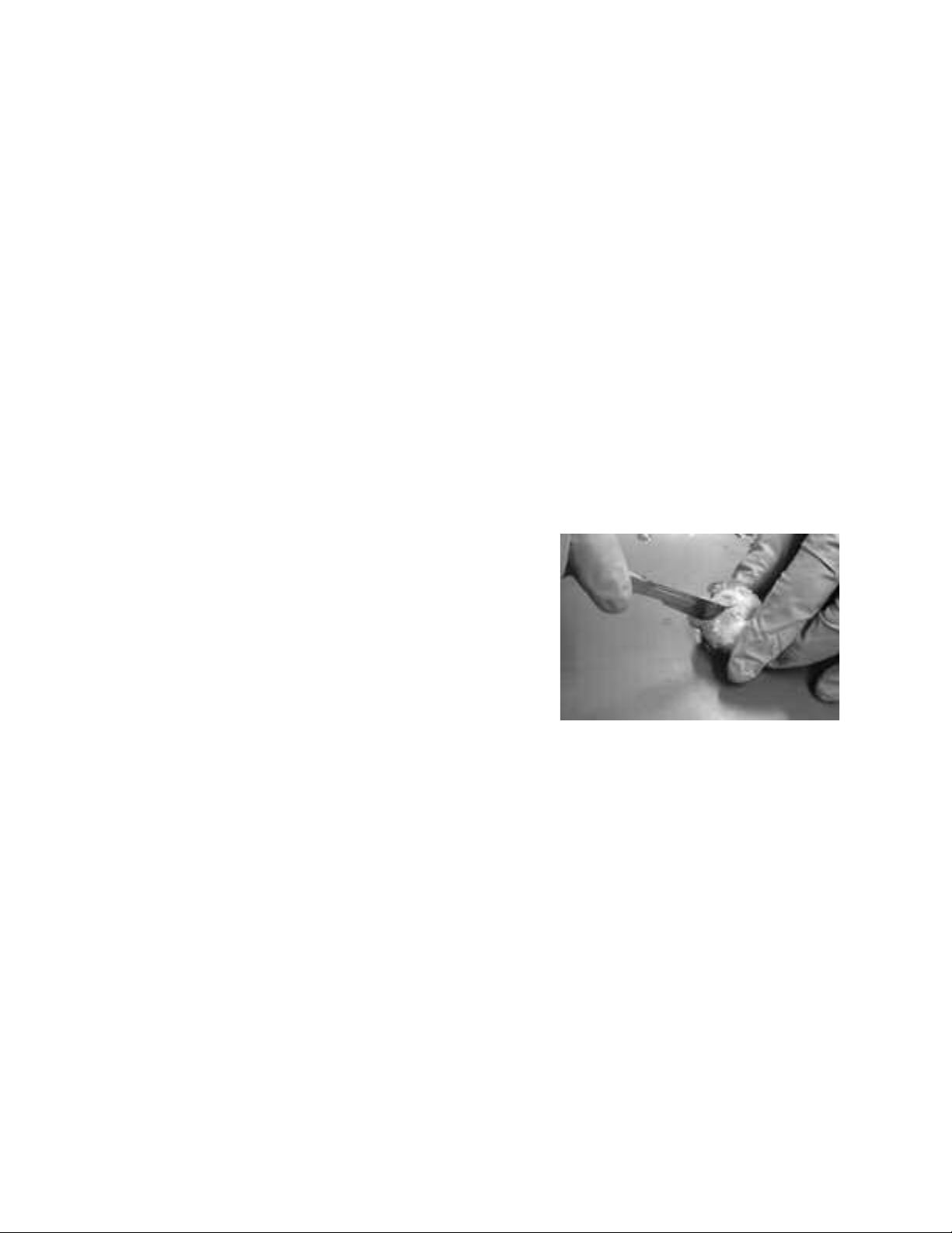
2. Trim away excess tissue surrounding the eyeball on the sclera.
3. Hold the eyeball gently with your thumb and forefinger at the
cornea and near the optic nerve.
4. Begin a cross-section of the eye by making an incision slightly
behind the middle of the eyeball through the sclera. Do not
cut deep into the eyeball or squeeze it too tightly
damage the interior structures. You may begin the cut with a
scalpel and finish with small scissors, or you may use a scalpel
for the entire cut. If some of the vitreous humor begins to
come out of the eyeball as you cut through the sclera, let it come out slowly.
5. Once you have made a cut around the eyeball, separate the eye into halves. Let the vitreous humor—
gelatinous, transparent material found inside the eye behind the lens—and any associated structures slowly
slide out of the eye. You may need to tease the vitreous humor gently away from the lining of the eye.
6. Look at the inside front portion of the eyeball. The lens may still be suspended in the middle of the pupil.
ip the front portion over and let the lens and associated structures fall out. Again, depending on the
T
viscosity of the vitreous humor, you may need to tease the material loose from the inside of the eye.
7. Observe the vitreous body, lens, and associated structures. The hyaloid fossa is an indention in the center
of the vitreous body that supports the lens. Surrounding the hyaloid fossa is the zonula ciliaris, made up of
suspensory ligaments that suspend the lens and stretch it to focus. You will also notice dark lines around
the hyaloid fossa. These lines are pigment from the iris.
8. Pick up the lens with a pair of forceps. Pat it dry with a paper towel. Note: You may want to place the
lens on some printed text on paper and observe its ability to magnify. This works best if you allow the lens
to dry overnight.
9. Turn the front half of the eyeball over so that you are looking at the cornea. Cut the front of the eye
around the outside of the cornea (where the cornea meets the sclera) to remove the cornea. This cut
works best if you mak
e the initial incision with a scalpel but then use scissors to finish.
, so as not to
©2005 Carolina Biological Supply Company
Printed in USA
Page 3

10. Place the cornea on your dissecting tray and cut it in half to observe its thickness.
Sclera
Retina
Optic nerve
Blind spot
Choroid
Zonula ciliaris
Lens
Aqueous humor
Pupil
Cornea
I
ris
Hyaloid fossa
Vitreous
humor
11. Insert the forceps through the opening created and carefully separate the edge of the iris from the inner
surface of the eye. You may be able to
remove the iris intact.
12. Pick up the back half of the eyeball and
observe the structures on the inside. Identify
the retina, which contains the cone and rod
receptor cells. Follow this mass of nerve cells
to their convergence at the back of the eye,
where the optic nerve begins. This is called
the blind spot.
13. Turn the back half of the eyeball over and
observe the optic nerve on the outside of the
eye. Pinch the nerve with your forceps to see
the separate fibers of the nerve.
14. Look at the interior of the back portion of
the eyeball again. Move the retina so that
you can see the dark, metallic-looking tissue
at the back of the eye. This is the choroid, a thin layer that lies between the retina and the sclera. The
portion of the choroid that appears iridescent blue and green with shades of yellow is called the tapetum.
15. Once you have observed all the structures of the eye, dispose of the specimen in accordance with local
guidelines and your teacher’s instructions.
Glossary
Aqueous humor - clear fluid filling the area between the lens and the cornea, composed mostly of water;
helps maintain the shape of the eyeball.
Blind spot - area of the retina where the receptor cells converge to form the optic nerve.
Choroid - thin, dark sheet of tissue between the retina and the sclera.
Cones - receptor cells of the retina that are responsible for perceiving color
Cornea - transparent covering that allows light to enter the eye; on a preserved specimen, the cornea is cloudy
Hyaloid fossa - indention in the center of the vitreous body that supports the lens.
Iris - diaphragm that regulates the size of the pupil.
Lens - biconve
x transparent structure that focuses the light coming in through the cornea and pupil.
Optic nerve - bundle of nerve cells that send signals from the eye to the brain.
Pupil - opening through which light enters the eye.
Retina - light-sensitive portion of the eye composed of receptor cells called cones and rods.
Rods - receptor cells of the retina that are responsible for perceiving difference in light intensity.
Sclera - outer covering of the eyeball; a tough, opaque sheet of connective tissue that protects inner
structures of the eyeball and helps maintain rigidity
.
Tapetum - iridescent portion of the choroid tissue.
Vitreous body - the cavity between the retina and the back of the lens.
Vitreous humor - viscous fluid that fills the vitreous body; helps maintain the shape of the eyeball.
Zonula ciliaris - ligaments that suspend the lens and stretch it to focus vision.
.
Teacher’s Manual 3
.
Page 4

Carolina’s Perfect Solution
®
Classrooms and labs using Carolina’s Perfect Solution specimens require no special ventilation and no special
disposal methods (check with local officials, as procedures may vary.) Independent, certified laboratory analyses of
specimens fixed in Carolina’s Perfect Solution show less than 0.002% formaldehyde, and OSHA’s safety standards
for off-gassing are far surpassed. Furthermore, these laboratory tests found all volatile and semivolatile chemical
levels are so low they are undetectable in the most sophisticated and demanding analysis. Additional laboratory
tests of Carolina’s Perfect Solution found no toxicity.
Carolina’s Perfect Solution®Specimens Available From Carolina Biological Supply Company
Carolina’s Perfect Solution®Cow Eye RN-22-8903
®
Carolina’s Perfect Solution
Carolina’s Perfect Solution
Carolina
’s Perfect Solution
Carolina’s Perfect Solution
Unless otherwise indicated, preserved specimens are shipped in Carosafe “odorless” shipping and holding fluid.
Carosafe is not a fixative; it is a preservative designed to prevent mold and deterioration after tissue has been
fixed properly in formalin or alcohol. Carosafe is an effective substitute for the standard formalin preservative and
minimizes the unpleasant odor of formaldehyde. Specimens in Carosafe are shipped in vials or pails.
Sheep Eye RN-22-8763
®
Sheep Brain RN-22-8703
®
Pig Heart
®
Pig Kidney RN-22-8573
RN-22-8563
Carolina Carosafe
M
T
Preservative
Caropak®Packaging
You can order preserved animals “damp-packed.” Our trade name for this improved method of packaging is
Caropak
effective fixation and preservation techniques allow. They are packaged in vacuum-sealed, double-layered, plastic
barrier bags. Specimens may be packaged one specimen per pack or many per pack.
Additional Specimens
Pig Eye (Caropak®Bulk) RN-22-8552
Cow Eye (Caropak
Cow Eye (Caropak
Sheep Eye (Carosafe
Sheep Eye (Caropak
Sheep Eye (Caropak
Carolina’s preserved organisms comply with all OSHA requirements for formaldehyde fumes.
®
. Preserved animals shipped in Caropaks have been processed with Carosafe and are as “odorless” as
®
Single) RN-22-8901
®
Bulk) RN-22-8902
TM
Bulk) RN-22-8760
®
Single) RN-22-8761
®
Bulk) RN-22-8762
Disposal
Because local regulations may vary from federal and state regulations, we recommend that you discuss disposal of
preserved specimens with your institution
’s or system’s environmental representative.
Carolina Biological Supply Company
2700 York Road, Burlington, North Carolina 27215
Phone: 800.334.5551 • Fax: 800.222.7112
Technical Support: 800.227.1150 • www.carolina.com
CB280950507
 Loading...