

Page 1
CarolinaTMMammal Brain
Dissection Guide
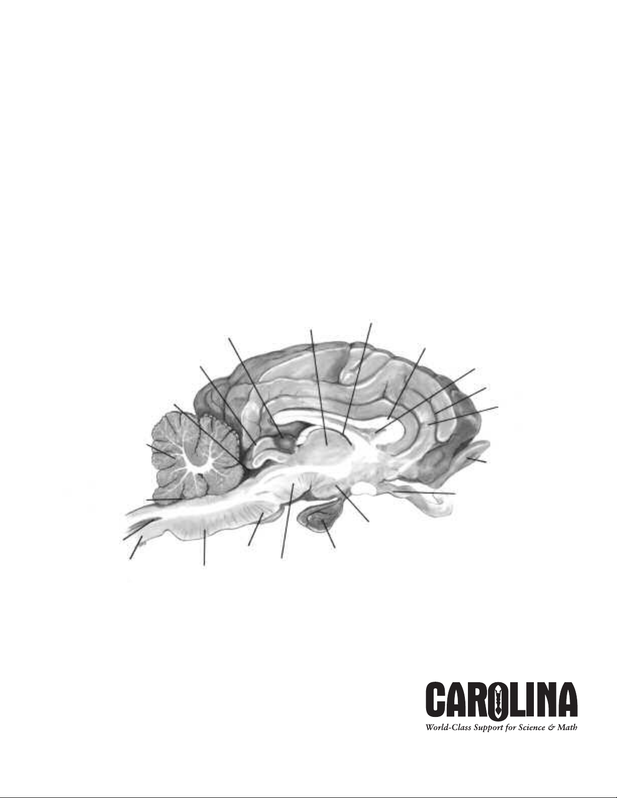
Cerebral aqueduct
Cerebellum
Fourth ventricle
Central canal
Spinal cord
Pineal body
Superior colliculus
Medulla
Pons
Thalamus
Midbrain
Third ventricle
Corpus callosum
Septum pellucidum
Sulcus
Gyrus
Olfactory bulb
Optic nerve
Hypothalamus
Hypophysis
C80135
Page 2

©2005 Carolina Biological Supply Company
Printed in USA
Page 3

CarolinaTMMammal Brain Dissection Guide
Overview
The Carolina™ Mammal Brain Dissection Guide is a general set of instructions for dissecting mammal brains.
With each type of brain, there will be differences in the size of the structures and brain regions, but the
general structures and their relative location will be the same or very similar.
Safety
Follow safe laboratory practices when performing any dissection. Wear safety glasses or goggles, gloves, and lab
aprons when dissecting. Perform dissections on a dissecting tray or pan to contain specimens and fluids. Be
careful when using sharp instruments such as scalpels, forceps, teasing needles, and scissors.
Procedure
1. Review the glossary provided at the end of the dissection guide. Refer to the Mammal Brain Section
diagram to help you observe and identify external and internal structures.
2. Place the brain on a dissecting tray
3. Observe the dura mater, or outer meninges, if they are present and were not removed prior to
preservation. The two remaining meninges, pia and arachnoid, form a thin covering which adheres to the
surface of the cerebrum. Use forceps to gently remove these layers.
4. Identify the cerebrum. On the cerebral surface, observe the grooves known as sulci. Also observe the
ridges called gyri. Identify the medial longitudinal fissure, which separates the right and left hemispheres
of the cerebral cortex.
5. Locate the four lobes of the cerebrum. At the anterior portion of the brain is the frontal lobe, which
controls motor functions. Dorsal to this lobe is the parietal lobe, which receives and processes somatic
sensory information. Inferior to the parietal lobe are the temporal lobes. The temporal lobes receive and
process auditory sensations. The dorsal portion of the cerebrum mak
receives and processes sensations from the eyes.
6. Locate the cerebellum, which is inferior to the occipital lobe of the cerebrum. The cerebellum has an
outer cortex and is folded. It is incompletely divided by a dorsal central ridge called the vermis. The
cerebellum controls muscle coordination.
7. Place the brain on the dissecting tray
brain stem, and spinal cord.
8. The cranial nerves and pituitary were cut when the brain was removed from the skull. You should be able
to identify the olfactory bulb, which lies below the frontal lobe of the cerebrum. Identify the optic
chiasma. This x-shaped structure is formed by the crossover of the right and left optic nerves. The optic
nerves have been removed, but portions of the optic chiasma are visible.
, dorsal side up.
es up the occipital lobe, which
, ventral surface up. Locate the following structures: medulla, pons,
9. Place the brain on a dissecting tray
longitudinal fissure. Insert a scalpel into the fissure and cut through the corpus callosum connecting the
two cerebral hemispheres. Continue to cut, dividing the cerebrum, cerebellum, and brain stem into two
longitudinal halves.
10. Each hemisphere contains a lateral ventricle, referred to as the first and second ventricles. The lateral
ventricles can be located by removing the septum pellucidum. The septum pellucidum is a thin,
transparent membrane located inferior to the corpus callosum on each hemisphere.
, dorsal side up. Using your fingers, gently widen the medial
Teacher’s Manual 3
Page 4

11. Locate the third and fourth ventricles. The fourth ventricle connects to the central canal of the spinal
cord. It is also connected to the third ventricle by a cerebral aqueduct. Examine each ventricle and try to
identify the choriod plexus, which produces cerebrospinal fluid.
12. With the cut side facing up, locate the following parts: thalamus, hypothalamus, pineal body, pons, and
medulla.
13. Observe the cut surface of the cerebellum. In medial section, the white matter of the cerebellum forms a
branched, treelike pattern called the arbor vitae. Try to identify this pattern.
14. Locate the midbrain region, located inferiorly between the thalamus and pons. This area contains
important nerve tracts. Dorsal areas of the midbrain are concerned with responses to visual and auditory
stimuli.
15. Make a cross section through a cerebral hemisphere just anterior to the thalamus. Examine the cross
section and identify the inner white matter and outer gray matter.
16. Remove the cerebellum and the remainder of the cerebral hemisphere by dissecting away everything
dorsal to the floor of the lateral ventricle. This will expose an infolding of the cerebral cortex, called the
hippocampus. The hippocampus is involved with emotions and memory.
17. Remove the hippocampus to locate the remainder of the thalamus.
18. Once you have observed all the structures of the brain, dispose of the specimen in accordance with local
guidelines and your teacher’s instructions.
Cerebral aqueduct
Cerebellum
Fourth ventricle
Central canal
Spinal cord
Pineal body
Superior colliculus
Medulla
Mammal Brain Section
Thalamus
Pons
Midbrain
Third ventricle
Hypothalamus
Hypophysis
Corpus callosum
Septum pellucidum
Optic ner
Sulcus
Gyrus
Olfactory bulb
ve
4
Page 5

Glossary
Arachnoid mater - middle of three layers (meninges) surrounding the brain and spinal cord.
Cerebellum - part of the brain that controls balance and muscle coordination; located inferior to the
cerebrum.
Cerebral aqueduct - channel connecting the third and fourth ventricles and containing cerebrospinal fluid.
Cerebrum - two hemispheres divided by the medial longitudinal fissure; largest portion of the mammalian
brain.
Choroids plexus - network of capillaries located in the roof of ventricles; contributes to production of
cerebrospinal fluid.
Corpus callosum - large band of nervous tissue that connects the two cerebral hemispheres.
Cortex - outer portion of the cerebrum.
Cranial nerves - twelve pairs of nerves that leave the brain.
Diencephalon - region of the brain made up of the thalamus and hypothalamus.
Dura mater - tough connective tissue layer that serves as the outer layer of the meninges.
Gray matter - areas of the brain and spinal cord containing cell bodies, dendrites, and unmyelinated axons;
found in the cerebral cortex of the brain and inner portion of the spinal cord.
Gyri - the folds of the cerebral cortex (singular = gyrus).
Hippocampus - a region below the lateral ventricles; involved with emotional states and converting short-
term memory to long-term memory.
Hypophysis - pituitary gland; controls a number of endocrine glands.
Hypothalamus - part of the diencephalon; inferior to the thalamus and responsible for regulation and
maintenance of internal homeostasis by controlling body temperature, appetite, fluid balance, etc.
Medulla - the most inferior portion of the brain stem; contains centers for heart rate, blood pressure, and
respiration. Also contains reflex centers controlling coughing
Midbrain - the part of the brain between the pons and the diencephalon.
Olfactory bulb - contains cell bodies of neurons that synapse with neurons of the olfactory nerves.
Optic chiasma - crossing point of the optic nerves.
Pia mater - innermost of the meninges layers.
Pineal body - endocrine gland located in the roof of the third ventricle; secretes melatonin.
Pons - anterior to the medulla; contains nerve tracts that connect the cerebellum with other parts of the
brain and spinal cord.
Sulci - grooves between gyri of the brain (singular = sulcus).
Thalamus - part of the diencephalon, superior to the hypothalamus; serves as a sensory relay center. Most
sensory nerves enter it and their impulses are sent to the appropriate cerebral region.
Ventricle - one of four cavities in the brain filled with cerebrospinal fluid.
White matter - bundles of myelinated axons within the brain and spinal cord; found in the inner portions of
the cerebrum and outer regions of the spinal cord.
, sneezing, hiccupping, etc.
5
Page 6

Carolina’s Perfect Solution
Independent, certified laboratory analyses of specimens fixed in Carolina’s Perfect Solution®have found it to be
nontoxic and free of dangerous off-gassing. This means that, for safety purposes, classrooms and labs using
Carolina’s Perfect Solution specimens do not require specialized ventilation. Carolina does recommend using
some active ventilation when working with any preserved specimens or chemicals. The safe nature of
Carolina’s Perfect Solution also means that in most localities there are no mandated disposal requirements. Be
sure to check with local sewer and landfill authorities, as local procedures may vary.
®
Carolina’s Perfect Solution®Specimens Available From Carolina Biological Supply Company
Carolina’s Perfect Solution®Cow Eye RN-22-8903
Carolina’s Perfect Solution®Sheep Eye RN-22-8763
Carolina’s Perfect Solution®Sheep Brain RN-22-8703
Carolina’s Perfect Solution®Pig Heart RN-22-8563
Carolina’s Perfect Solution®Pig Kidney RN-22-8573
M
Carolina Carosafe
Carosafe™ is a holding solution for biological specimens. It contains no formaldehyde and is not a tissue
fixative. Most specimens in
formaldehyde-free
student and educator e
Carosafe. This produces a formalin-preserved specimen that, when dissected, minimizes
Carosafe are first preserved with a formalin solution and then placed in
xposure to formaldehyde.
T
Preservative
Caropak®Packaging
Preserved animals shipped in Caropaks packaging have been processed with Carosafe and are as “odorless” as
effective fixation and preservation techniques allow. They are packaged in vacuum-sealed, double-layered
plastic barrier bags. Specimens may be pack
aged one specimen per pack or many per pack.
Additional Specimens
Sheep Brain (Caropak®Single) Dura mater removed; optic chiasma intact RN-22-8701
Sheep Brain (
Sheep Brain (
Sheep Brain (Car
Sheep Brain in Cranial Case
Sheep Half Brain (
Sheep Half Brain (
Caropak®Bulk) Dura mater removed; optic chiasma intact RN-22-8702
Caropak®Single) Dura mater intact; hypophysis and RN-22-8711
cranial nerve roots intact
®
opak
Bulk
opak
Car
Caropak®Bulk) RN-22-8732
Dura mater intact; hypophysis and RN-22-8712
)
cranial nerve roots intact
(Carosafe™) RN-22-8720
®
Single
)
RN-22-8731
Disposal
Because local regulations may vary from federal and state regulations, we recommend that you discuss
disposal of preserved specimens with your institution
’s or system
’s environmental representative.
6
Page 7

Carolina Biological Supply Company
2700 York Road, Burlington, North Carolina 27215
Phone: 800.334.5551 • Fax: 800.222.7112
Technical Support: 800.227.1150 • www.carolina.com
CB281000508
 Loading...