

Page 1
CXDI Control Software NE
CXDI Controller RF Software
Secure, intuitive, ecient workflow and comprehensive
image processing for static and dynamic imaging
Page 2
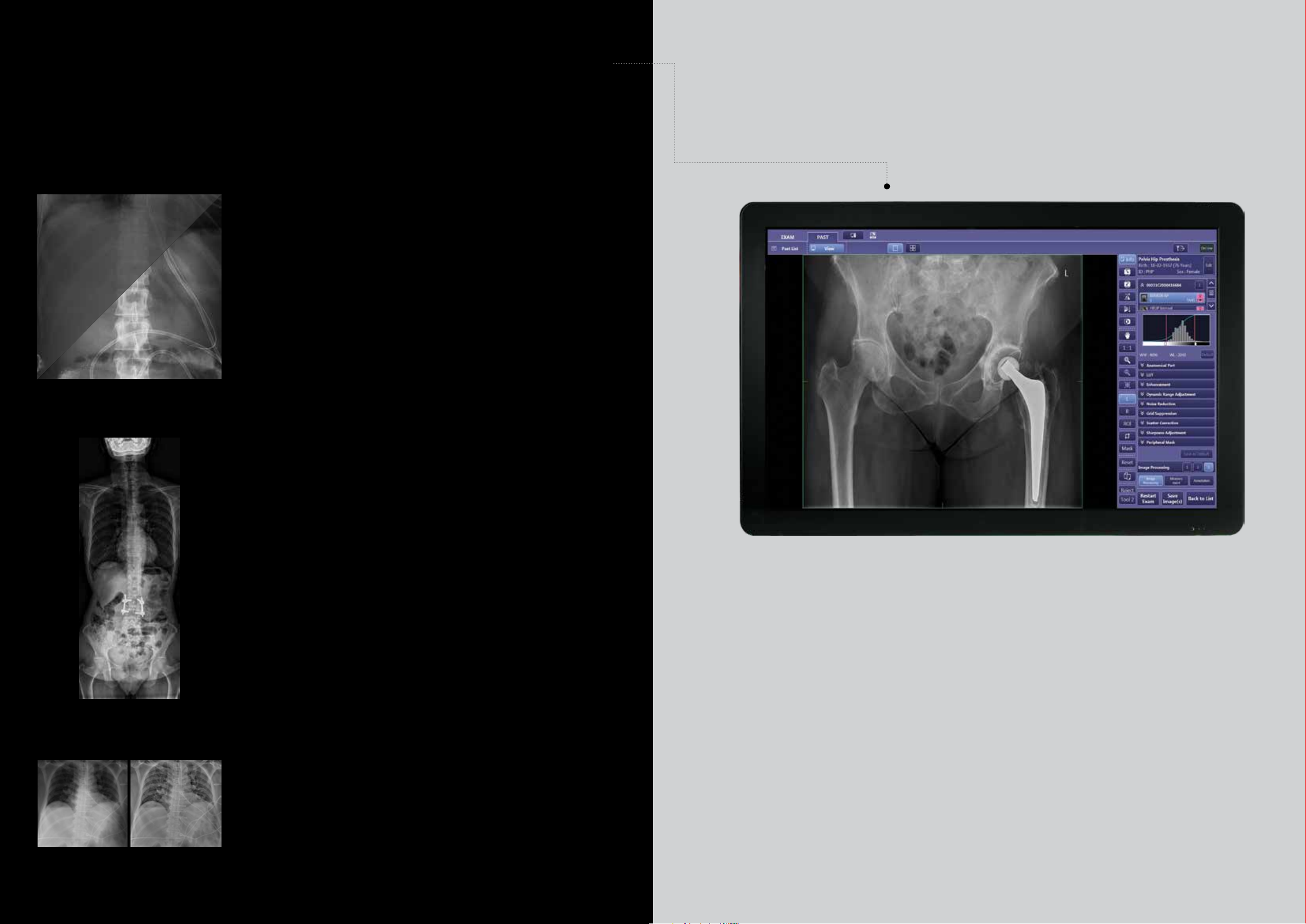
Intuitive interface
Canon’s intuitive ‘CXDI-NE/RF’ Graphical User Interface (GUI) can be used for all types of digital
radiography modality and this commonality of GUI across the entire DR product range is a major
advantage when it comes to speed of operator training, user confidence, convenience and familiarity.
Canon CXDI-NE/RF software configuration options ensure a GUI that is always right for you.
Comprehensive image processing including ‘Scatter Correction’ and ‘One Shot Long-Length’
imaging options guarantee optimised image quality with the lowest possible dose; the industry
standard DICOM 3.0 interface ensures multi-vendor and cross-platform connectivity in any situation.
CXDI Control Software NE / Controller RF
CXDI Control Software NE / Controller RF is made exclusively for use with Canon Digital Radiography
systems. This software helps to optimise workflow and reduce the steps needed to complete exams. It
provides quick image confirmation and timely network distribution, supports multiple study acquisition,
can easily be tailored to individual clinical preferences and helps provide the delivery of consistent, highresolution images with the Canon CXDI Digital Radiography systems. In addition, this proprietary software
solution is Integrating the Healthcare Enterprise (IHE) compliant and has features that can help practitioners
with their HIPAA compliance eorts.
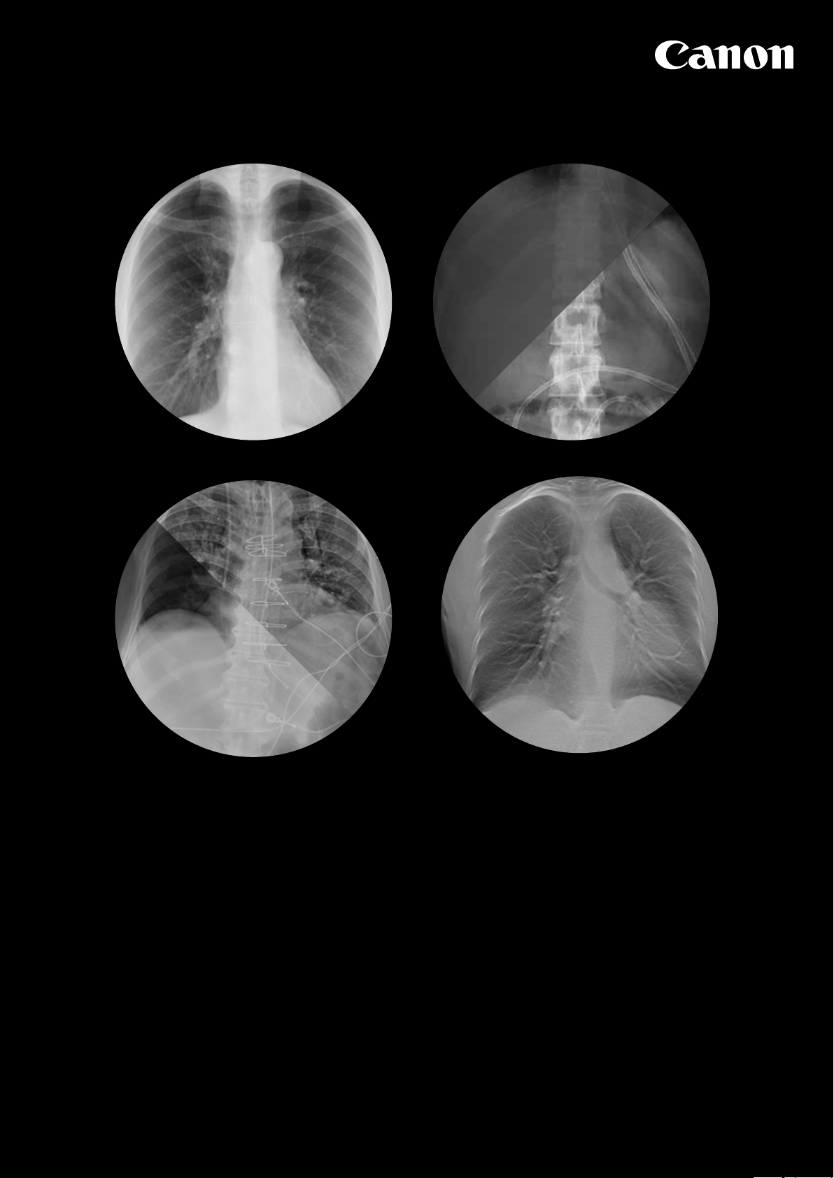
Before
After
Scatter Correction (optional)
Canon’s Scatter Correction reduces the eect of scattered radiation for nongrid bedside examinations, allowing you to obtain images with outstanding
contrast while avoiding the grid handling and improve your workflow
Benefits:
● Significantly lower X-ray dose compared to imaging with a grid*
● Superior image contrast without the need for a grid
● Improved workflow: no need to carry, fit, position and remove a grid
● Enhanced eciency: no repeat exposures due to grid misalignments
and resulting artefacts
● Potential to improve patient comfort in bed examinations as the imaging
receptor is thinner without a grid fitted
*Confirmed result after testing Canon Scatter Correction at Linköping
University Hospital, Sweden
One Shot Long-Length (optional)
One Shot Long-Length exams enhance eciency compared to conventional
stitch exams; shorter examination time, lower risk on patient movement,
reduced dose and increased image quality.
Expected benefits:
● Patient positioning stand with motorised height adjustment
● Fixed installation or mobile for convenient relocation
● Large, ergonomic grip rails for confident patient positioning
● Optional grid
● Ability to use 3 existing detectors for cost-eective one shot
Long-Length imaging
● Versatile configuration; use either 3 x CXDI-710CW or 3 x CXDI-410CW
wireless detectors
Advanced Edge Enhancement (optional for static imaging)
Improved visualization of tubes, lines and bone details. The software has
three dierent image processing algorithms (small structures, bone detail
and catheter setting):
● Automatic copy of the image acquired, allows various views on the same
acquisition
1
RF software supports only 710 series
Main features:
● Real-time viewing of high
quality images
● Large high-resolution monitor
for comfortable viewing
● Optimised workflow with less
operation steps
● Interactive GUI for intuitive
operation
● Supports various workflows to
match local requirements
● Single and Prepacked Protocols
● Emergency study capability
● Suspend Exam
● Reject Analysis
● Automatic forwarding rejected
images to a designated analysis
workstation
● Automatic Image stitching
(static FPD)
Optimises your workflow
Protocol planning with the right
sequence of the positions in the
study.
Instant display of the image taken in
high resolution within one second.
Comfortable viewing on large screen
with overview and less operation
steps.
Designed to enhance image quality
Provides wide range of the algorithm
and dynamic formatting before
saving.
Enables significant dose reductions
through optimising image processing
parameters.
Adaptive to your local standards
Is giving you the tailored preset that
you require, is adaptable to any local
language needs, preference or taste
of imaging, accommodating standard
or unique protocols such as trauma
protocol and protocols for paediatric
imaging.
Flexible and Secure
The Canon NE software is outstanding
in communication with X-ray generator
and brilliant in the non-synchronised
mode.
Page 3

Additional functionality CXDI Controller RF software
Tomosynthesis (optional for CXDI Controller RF software)
Performing high-resolution limited-angle tomography at radiation dose levels
comparable with traditional projection radiography:
● For imaging of lung nodules or lung tissue that is partly obscured by ribs,
heart or other structures
● For arthritic changes in extremities
● Extreme/ complex fracture imaging
● For localization Brachytherapy seeds
DSA (optional for CXDI Controller RF software)
Digital subtraction angiography (DSA) is a fluoroscopy technique used in
interventional radiology to clearly visualize blood vessels in a bony or dense
soft tissue environment:
● Basic functionality like road mapping is supported DSA technology on
your digital radiography modality.
Research studies on Canon Flat Panel Detectors
CONRAD Radiographic Research Centre
•
Study 1. Optimization of image quality and dose using multi-frequency software
Purpose:
Examine whether the use of multi-frequency software (MLT[S]) may allow dose
reduction without significant loss of image quality. The processing parameters
explored include brightness, contrast, edge enhancement, frequency band, noise
reduction and dynamic range (dark/bright region).
Conclusion:
By optimizing image processing parameters, a significant dose reduction of 61% is
possible without significant loss of image quality on anthropomorphic phantoms.
Reference:
Precht et al (2012) Pediatri Radiol 42: 1112-1118
•
Study 2. Large dose reduction by optimization of Multi Frequency Processing
Software in DR within follow-up examinations in pediatric femur
•
Study 3. New Developed DR Detector Performs Radiographs of Hand, Pelvic and Premature
Chest Anatomies at a Lower Radiation Dose and/or a Higher Image Quality
Purpose:
Examine whether the new detector design could increase image quality and/or
reduce dose in hand, pelvic and premature chest examinations
Conclusion:
Optimal image quality can be maintained at a lower dose and/or image quality,
and could be improved using the CXDI-70C detector for both hand, pelvic and
premature chest examinations, based on the technical and anthropomorphic
•
Study 4. Software optimization in Pediatric Pelvic DR examinations - ongoing
Purpose:
To investigate potential image quality optimization or dose savings associated with using the new multi frequency
software together with a newly developed DR detector at pediatric pelvic examinations.
Conclusion:
By using a new DR detector and optimizing image processing parameters, a significant dose reduction is possible
without significant loss of image quality in a pediatric pelvis examinations. Statistical significance was found for
processing combinations with the biggest impact from the noise reduction parameter.
Reference:
June 2014: Poster and oral presentation at “European Society for Pediatric Radiology”, Amsterdam Scientific article
will be published during 2015
0205W172
http://www.canon-europe.com/Medical
http://conradint.ucl.dk
Figure 1: Experimental images taken
of the anthropomorphic lamb femur
phantom.
(a) Reference image for the VGA
study processed with MLT(M)
at 16 mAs and 523 REX,
(b) and (c) are optimized with
MLT(S),
(b) was produced with 6.3 mAs
and 208 REX;
(c) with 2 mAs and 45 REX.
A B C
Purpose:
Figure 2: Experimental images taken
Examine whether the use of MFP processing could
of the anthropomorphic lamb femur
allow for a dose reduction based on dierentiated image
phantom.
quality while still maintaining an acceptable diagnostic
The images (A), (B) and (C) were
image quality in pediatric femur follow-up DR examinations.
MLT(S) optimized image taken at
(A) 8 mAs [244 REX],
Conclusion:
(B) 3.2 mAs [107 REX],
By optimizing image processing parameters and to adapt
(C) 0.5 mAs approved for diagnosis
the image quality depending on the requested pathology a
[18 REX],
significant dose reduction of 92% was shown possible while
(D) MLT(M) optimized image
still maintaining sucient diagnostic image quality.
(previous non frequency software
version)
Reference:
taken at 8 mAs [241 REX]. The femur
Precht et al (2014) Pediatri Radiol 44: 239-240
fraction is indicated by an arrow.
phantom results. The performance of the CXDI-70C detector in terms of IQFInv
values was on average 45 % better than the CXDI-55C detector, depending on
anatomy, kVp and mAs levels. According to the VGA results, depending on anatomy
and kVp levels, optimal image quality was maintained for the CXDI-70C detector at
an estimated dose reduction of 30 % on average
Reference:
Precht et al (2014) J Digit Imaging 1: 68-76
Enabling significant dose reductions
Canon’s NE / RF Control Software enables significant dose reductions.
Through a wide range of algorithms for dynamic formatting before saving,
it optimises the images with intelligent image processing parameters,
as confirmed by various clinical evaluation studies conducted in Europe.
CONRAD Radiographic Research Center in Denmark, proved with our static
FPD that by optimising image processing parameters and adapting the image
quality depending on the requested pathology a significant dose reduction has
been achieved while still maintaining sucient diagnostic image quality. Read
the conclusions of the specific reports in the dedicated leaflet.
For further information about the Canon Medical Components Business Group and details of local
distributors please visit: www.canon-europe.com/medical
Canon Europa N.V.
Bovenkerkerweg 59 • 1185 XB Amstelveen • The Netherlands
NERFY20VA
www.canon-europe.com/medical
 Loading...