

Page 1

ACCESS SYSTEMS
An issued or revision date for these instructions
is included for the users information. In the
event two years have elapsed between this date
and product use, the user should contact Bard
Access Systems, Inc. to see if additional product
information is available.
Revision date: March, 2014
Bard, Sherlock, Sherlock 3CG, and Site~Rite are
trademarks and/or registered trademarks of C. R.
Bard, Inc. All other trademarks are the property of
their respective owners.
©2014 C. R. Bard, Inc. All rights reserved.
Manufacturer:
Bard Access Systems, Inc.
605 North 5600 West
Salt Lake City, UT 84116 U.S.A.
(801) 522-5000
Customer Service: (800) 545-0890
Technical/Clinical Support: (800) 443-3385
www.bardaccess.com
www.discoversherlock.com
0736736 1403R
Page 2

For use with
Site~Rite™ 6 Ultrasound System
Instructions for Use
Page 3

Sherlock 3CG™ Tip Confirmation System
15
o
C
38
o
C
10
o
C
38
o
C
%
Keep Dry
Manufacturer:
Consult instructions
for use
Operating Humidity Parameters
Storage Humidity Parameters
(unpackaged)
Operating Temperature
Parameters
Fragile
%
Rx Only
CF Type Equipment
Storage Humidity Parameters
(packaged)
Storage Temperature
Parameters
Do not dispose with
ordinary municipal waste
Federal (U.S.A.) law restricts
this device to sale by or on the
order of a physician
1
Page 4

- TABLE OF CONTENTS -
Section Section
Instructions For Use
1 Overview 3
1.1 Indications for Use
1.2 Post Market Clinical Trial
1.3 Sherlock 3CG™ Tip Confirmation System (TCS) Description
1.4 System Components
1.5 Warnings and Precautions
2 Assembling the Sherlock 3CG™ TCS 6
2.1 Attaching the Sherlock™ 3CG TCS Sensor
Holster to the Roll Stand
2.2 Connecting the Sherlock 3CG™ TCS Sensor
to the Site~Rite™ 6 Ultrasound System
3 Sherlock 3CG™ TCS Information 6
3.1 Switching Between Ultrasound and TCS
3.2 Sherlock 3CG™ TCS Graphical Interface
3.3 Sherlock 3CG™ TCS Controls and Indicators
3.4 Sherlock 3CG™ TCS Audio Information
3.5 Parallax
4 Sherlock 3CG™ TCS Catheter Guidance 14
4.1 Verifying Catheter Compatibility
4.2 Catheter Placement
Step 1: Prepare Device
Step 2: Position Patient and Perform Ultrasound Pre-scan
Step 3: Measure Catheter Length
Step 4: Prepare Sensor
Step 5: Position Sensor and ECG electrodes
Step 6: Evaluate External ECG waveform
Step 7: Perform Initial Magnet Tracking Calibration
Step 8: Prepare Catheter Sterile Field
Step 9: Access the Vein
Step 10: Attach Catheter Stylet to Fin Assembly
Step 11: Perform Final Magnet Navigation Calibration
Step 12: Insert Catheter
Step 13: Catheter Tip Guidance and Positioning
Step 14: Complete Catheter Placement
Step 15: Procedural Record
5 Magnetic Navigation Only Mode 17
Step 1: Prepare Device
Step 2: Position Patient and Perform Ultrasound
Step 3: Measure Catheter Length
Step 4: Prepare Sensor
Step 5: Position Sensor
Step 6: Perform Initial Magnet Navigation Calibration
Step 7: Prepare Catheter Sterile Field
Step 8: Access the Vein
Step 9: Perform Final Magnet Navigation Calibration
Step 10: Insert Catheter
Step 11: Catheter Tip Guidance and Positioning
6 Troubleshooting and Error Messages 19
6.1 Error Screens
6.2 ECG Troubleshooting
6.3 Magnetic Navigation Troubleshooting
7 Magnetic Navigation Printing 23
8 Cleaning and Disinfection 23
8.1 Cleaning Procedure
8.2 Disinfection Procedure
9 Warranty 24
10 Service and Repair 25
11 Technical Specifications 25
11.1 Sherlock 3CG™ TCS Sensor and Display
Operational and Storage Conditions
12 Disposal Information 25
2
Page 5

Sherlock 3CG™ Tip Confirmation System
1 Overview
1.1 Indications for Use
The Sherlock 3CG™ Tip Confirmation System (TCS) is indicated for guidance and positioning of Peripherally Inserted Central Catheters (PICCs). The
Sherlock 3CG™ TCS provides real-time PICC tip location information by using passive magnet navigation and the patient's cardiac electrical activity
(ECG). When relying on the patient’s ECG signal, the Sherlock 3CG™ TCS is indicated for use as an alternative method to chest X-ray and fluoroscopy
for PICC tip placement confirmation in adult patients.
Limiting but not contraindicated situations for this technique are in patients where alterations of cardiac rhythm change the presentation of the P wave as
in atrial fibrillation, atrial flutter, severe tachycardia, and pacemaker driven rhythm. In such patients, who are easily identifiable prior to catheter insertion,
the use of an additional method is required to confirm PICC tip location.
1.2 Post Market Clinical Trial
The Sherlock 3CG™ Tip Confirmation System (TCS) is Bard’s next generation, fully integrated magnetic navigation and ECG-based peripherally inserted
central catheter (PICC) tip confirmation technology, which represents the next evolution of the Sherlock™ II Tip Location System and the previouslymarketed Sapiens Tip Confirmation System. The Sapiens TCS Post-Market Clinical trial was a prospective, single arm, single center study designed to assess
the efficacy of the ECG method for correctly positioning the tip of catheters in adult patients. The primary endpoints were to assess the performance of
the Sapiens technology with respect to: (1) the accuracy of the Sapiens technology with regard to correct positioning of the catheter tip when compared
to postoperative chest x-ray, (2) safety of using an invasive intracardiac electrode, and (3) compatibility with peripherally inserted central catheters. One
hundred fourteen (114) subjects received a PICC. Final PICC tip location was confirmed at the cavoatrial junction or within +/- 1 cm in 99.1% (113/114)
of the subjects. No adverse events were reported.
1.3 Sherlock 3CG™ TCS Description
The Sherlock 3CG™ TCS is designed to aid in PICC tip positioning through magnet navigation and ECG technology. It is designed to operate with
Bard Access Systems’ catheter kits labeled [ ] and “Sherlock 3CG™ TPS Stylet”.
Note: When used in conjunction with catheter kits labeled [ ] the device provides magnetic navigation information but does not allow positioning
through ECG technology.
ECG Positioning
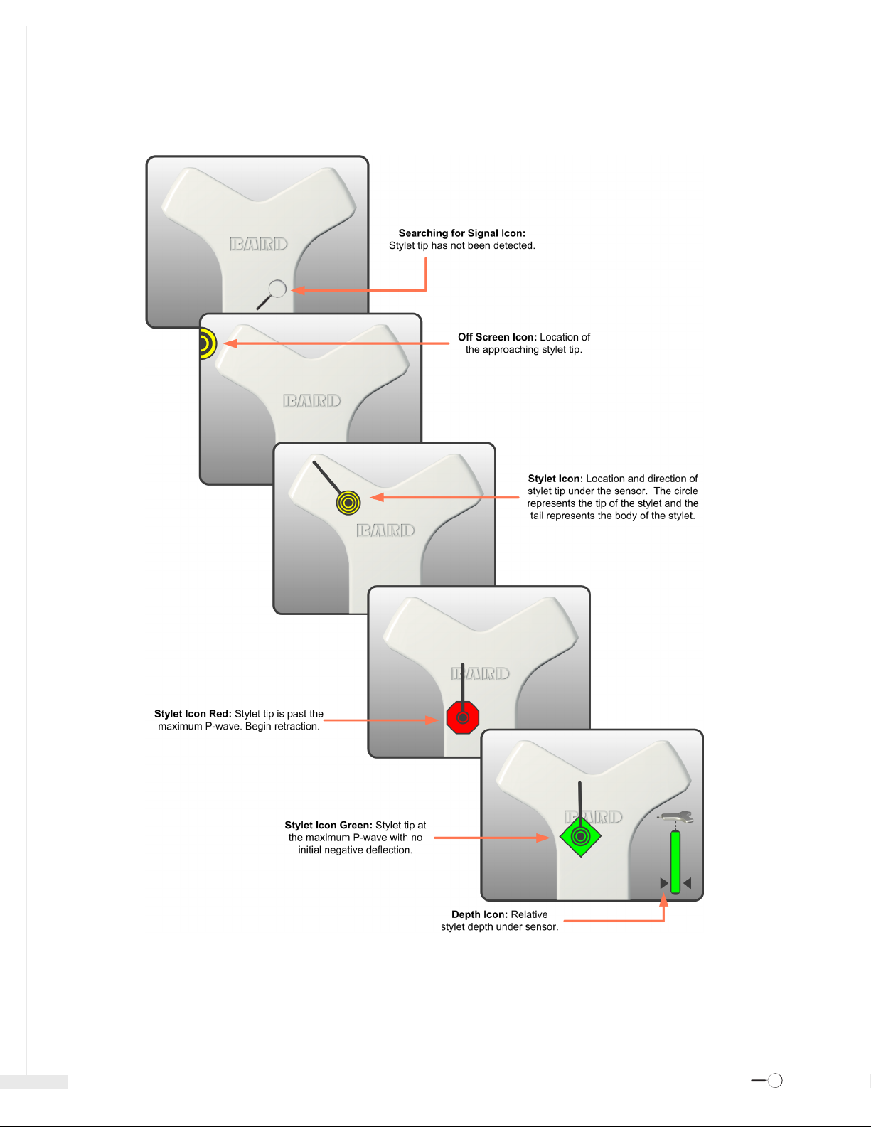
The Sherlock 3CG™ TCS displays an ECG signal detected by the intravascular and body electrodes, which can be used for catheter tip positioning.
In patients with a distinct P-wave, the P-wave will increase in amplitude as the catheter approaches the top of the cavoatrial junction. As the catheter
advances into the right atrium, the P-wave will decrease in amplitude and may be biphasic or invert. In addition to the ECG signal, the magnet tracking
stylet icon is augmented with the shape and color changes shown below.
Note: In instances where the P-wave is not present, not identifiable, or intermittent the stylet icon will remain a yellow circle.
3
Page 6
Instructions For Use
Magnetic Navigation
Permanent magnets are encapsulated within the tip of the Sherlock 3CG™ TPS Stylet. No magnetic energy is generated by the Site~Rite™ 6 Ultrasound System
or the sensor. The Sherlock 3CG™ TCS displays the relative position of the magnet-tipped stylet to the sensor. It does this in two steps:
1. Sherlock 3CG™ TCS takes a background measurement of the ambient magnetic field during the calibration cycle.
2. Sherlock 3CG™ TCS senses changes in the magnetic field. When the Sherlock 3CG™ TCS detects the stylet, it displays the stylet tip location
and orientation.
1.4 System Components
Ultrasound probe:
Ultrasound System Display Controls:
- - System controls for using Sherlock 3CG™ TCS Mode on the Site~Rite™ 6 Ultrasound System
Authorized accessories include:
- Site~Rite™ 6 Ultrasound System
- Sherlock 3CG™ TCS Sensor
- Sherlock 3CG™ TPS Stylet (included with specially marked central venous catheter kits [ ]
- Sherlock™ TLS Stylet (included with specially marked central venous catheter kits [ ]
- Fin Assembly (included with specially marked central venous catheter kits)
- Sherlock™ Sensor Holder (ordered separately or with specially marked central venous catheter kits) or .
- Bard Access Systems' supplied printer (purchased separately).
1.5 Warnings and Precautions
This section specifies warnings and precautions specific to the functionality of the Sherlock 3CG™ TCS.
- See the Bard Access Systems' catheter Instructions for Use (IFU) for possible complications associated with peripherally inserted central catheter (PICC)
placements and ECG positioning.
4
Page 7

Sherlock 3CG™ Tip Confirmation System
Warnings
Warning: This product should only be operated by qualified medical personnel.
Warning: Do not power the Sherlock 3CG™ TCS in the presence of noncontained flammable anesthetic gases. Explosion may result.
Warning: Do not attempt to sterilize the sensor. Damage to the equipment may occur.
Warning: The following actions void the warranty of the Sherlock 3CG™ TCS and may result in injury or equipment damage.
- Opening or servicing the
- Removing system labels by anyone other than Bard Access Systems’ authorized service personnel.
- Connecting the sensor or applied patient components to any unauthorized system or accessory. Refer to Section 1.4 for complete components.
- Installation of unauthorized software.
- Modification of system software settings without authorization by Bard Access Systems.
Warning: If the
Warning: Do not submerge the sensor in liquid or allow fluid to enter the connectors. Damage to the equipment may occur.
Warning: Sherlock 3CG™ TCS is not intended to diagnose or treat disease.
Warning: Only Bard Access Systems’ authorized service personnel should attempt to service this equipment.
Warning: Do not rely on ECG signal detection for catheter tip positioning when interpretation of the external or intravascular ECG P-wave is difficult.
- P-wave is not present
- P-wave is not identifiable
- P-wave is intermittent
These conditions may be a result of heart rhythm abnormalities, atrial fibrillation, atrial flutter, severe tachycardia or presence of cardiac rhythm
Warning: Do not rely on ECG signal detection for catheter tip positioning when there are no observable changes in the intravascular P-wave. In this case,
Warning: Do not place and/or use the Sherlock 3CG™ TCS in the presence of strong magnetic fields such as Magnetic Resonance Imaging (MRI) devices.
Warning: Do not remove Sherlock 3CG™ TCS enclosures. To avoid electrical shock, use only the power cord supplied with the system and connect only to
Warning: Ensure all connecting cables and connections are electrically insulated and do not come into contact with other electrical cables or metal surfaces.
Warning: Ensure that the patient does not directly or indirectly contact non-insulated metal surfaces.
Warning: Place skin electrodes carefully at locations indicated in these Instructions for Use and ensure good skin-electrode contact. Failure to do so may cause
authorized service personnel.
Site~Rite™ 6 Ultrasound System with Sherlock 3CG™ TCS is visibly damaged, discontinue use immediately. Use of the damaged system
may result in injury or equipment damage.
sensitive components and circuits. Failure to observe proper static control procedures may result in damage to the system.
For example, when:
devices. In these cases, rely on magnetic navigation and external measurement for tip positioning and use chest x-ray or fluoroscopy to confirm
catheter tip location, as indicated by institutional guidelines and clinical judgment.
rely on magnetic navigation and external measurement for tip positioning and use chest X-ray or fluoroscopy to confirm catheter tip location, as
indicated by institutional guidelines and clinical judgment.
The high magnetic fields created by an MRI device will attract the equipment with a force sufficient to cause death or serious injury to persons
between the equipment and the MRI device. This magnetic attraction may also damage the equipment. The magnetic and the RF fields associated
with the MRI environment may interfere with the display of ECG waveforms. Consult the MRI manufacturer for more information.
properly grounded wall outlets. Only Bard Access Systems qualified personnel should service the system
unstable ECG waveforms and/or ECG waveforms that are not described in these Instructions for Use. In such a case, rely on magnetic navigation
and external measurement for tip positioning and use chest X-ray or fluoroscopy to confirm catheter tip location, as indicated by the institutional
guidelines and clinical judgment.
Site~Rite™ 6 Ultrasound System with Sherlock 3CG™ TCS by anyone other than Bard Access Systems’
Site~Rite™ 6 Ultrasound System contains static
Precautions
Caution:
Federal (U.S.A) law restricts this device to sale by or on the order of a physician.
Caution: Do not pull the cables to disconnect from the system. Pulling the cable may damage the cable or cable connection.
Caution: Excessive twisting or bending of the sensor cable may cause system failure.
Caution: Use only Bard Access Systems’ cleaning and disinfection procedures. Failure to do so may damage the device.
Caution: Do not use excessive force when connecting or disconnecting the Fin Assembly to or from the sensor or equipment damage may occur.
Caution: When the sensor is not in use, store in the holster, roll stand basket or other secure location to avoid damage.
Caution: Do not allow any ferromagnetic objects, e.g., wired undergarments, metal instruments, watches, jewelry, electronics, metal bedrails, etc. to be
within 12 in (30 cm) of the sensor once the calibration process is complete. These items may interfere with the sensor's ability to accurately locate
the Sherlock
™ Stylet tip.
Caution: Equipment operating in close proximity may emit strong electromagnetic or radio frequency interference which could affect the performance
of this device. Avoid operating the device near pumps, cauterizers, diathermy equipment, cellular phones, or other portable and mobile radio
frequency communications equipment. Maintain equipment separation of at least 5 ft (1.5 m).
Caution: Electrodes should be applied only to intact, clean skin (e.g., not over open wounds, lesions, infected or inflamed areas).
5
Page 8
Caution: Placement of red electrode outside of the correct region may result in reduced ECG performance. See Section 4.
Caution: Discontinue electrode use immediately, if skin irritation occurs.
Caution: To avoid damage to the device operating system, shutdown the device through the Power Control button from the Application Toolbar.
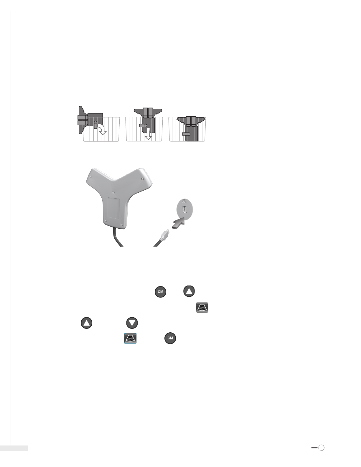
2 Assembling the Sherlock 3CG™ TCS
2.1 Attaching the Sherlock 3CG™ TCS Sensor Holster to the Roll Stand
The Sherlock 3CG™ TCS Sensor can be placed in the holster when not in use. To attach the sensor holster to the roll stand see illustrations below.
Caution: When the sensor is not in use, store in the holster, roll stand basket or other secure location to avoid damage.
2.2 Connecting the Sherlock 3CG™ TCS Sensor to the Site~Rite™ 6 Ultrasound System
Instructions For Use
The Sherlock 3CG™ TCS Sensor connects to the USB port on the display.
3 Sherlock 3CG™ TCS Information
3.1 Switching Between Ultrasound and TCS
To switch from Ultrasound mode to TCS mode the buttons [ ] and [ ] need to be pressed simultaneously.
To switch from TCS Mode to Ultrasound Mode, select the ULTRASOUND icon [ ] by:
1. Selecting the UP [ ] and DOWN [ ] buttons on the Ultrasound Probe, or on the Ultrasound Machine, to navigate to the Ultrasound icon.
2. Once the Ultrasound icon is highlighted [ ], select the [ ] button to switch back to Ultrasound Mode.
6
Page 9
Sherlock 3CG™ Tip Confirmation System
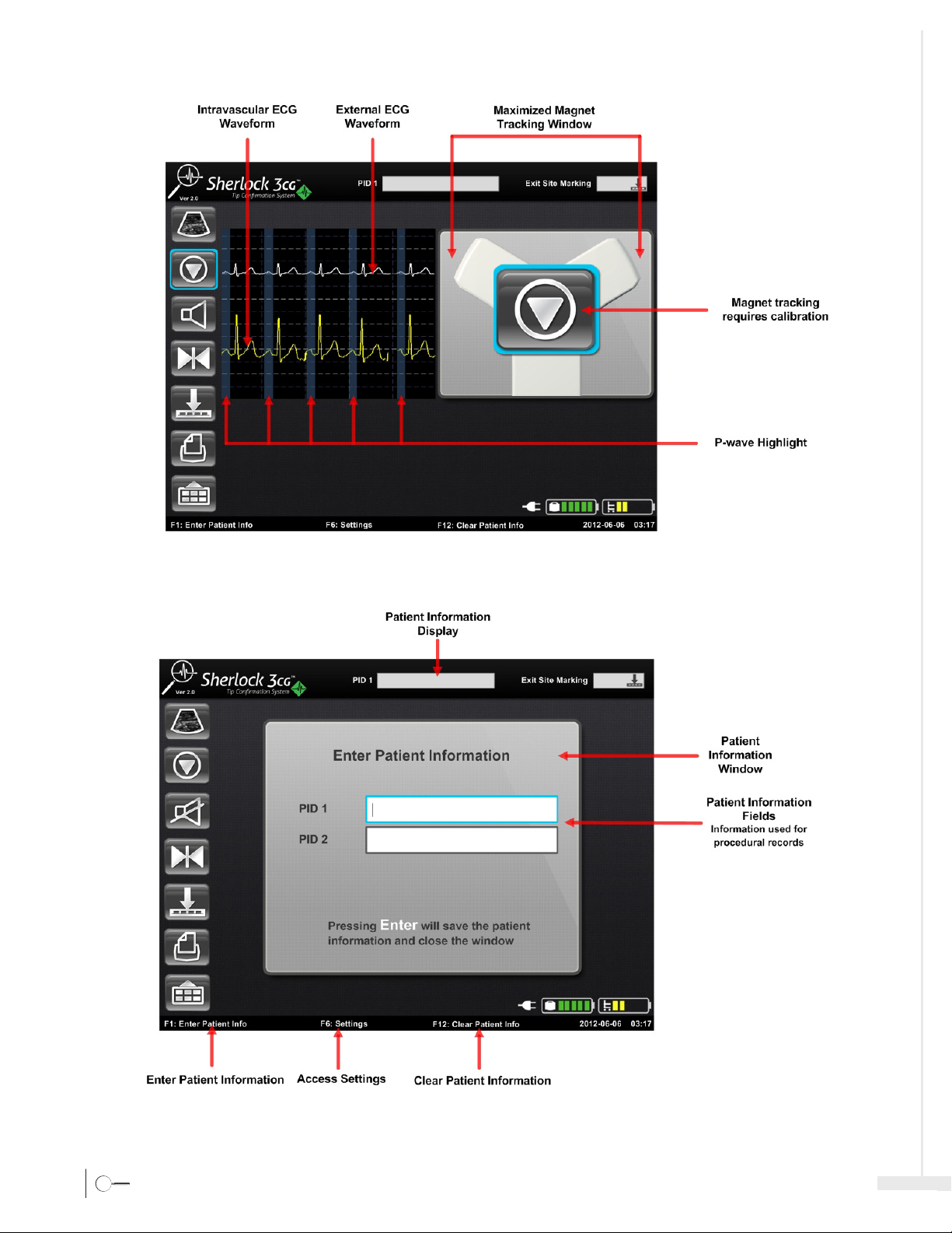
3.2 Sherlock 3CG™ TCS Graphical Interface
7
Page 10
Instructions For Use
8
Page 11

Sherlock 3CG™ Tip Confirmation System
9
Page 12

Instructions For Use
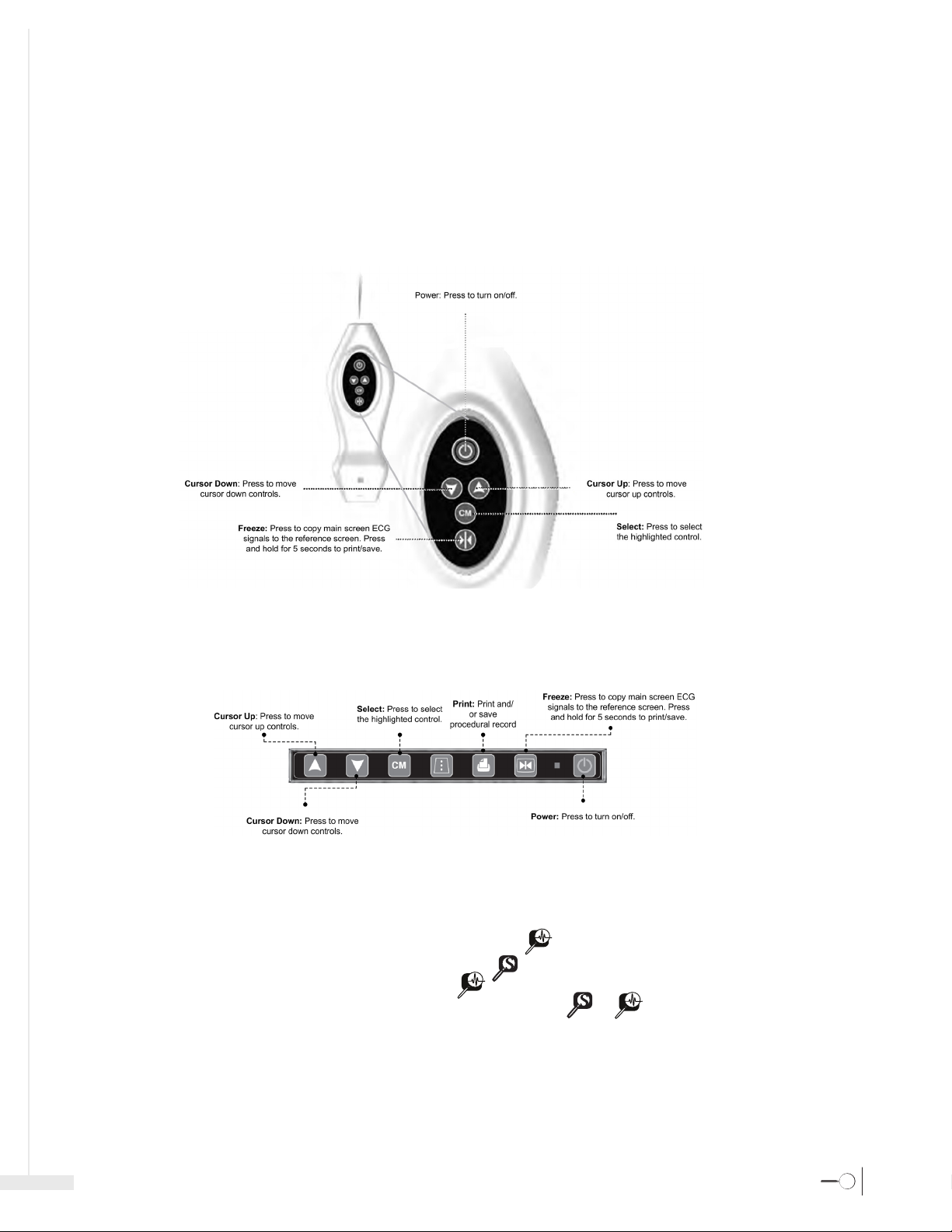
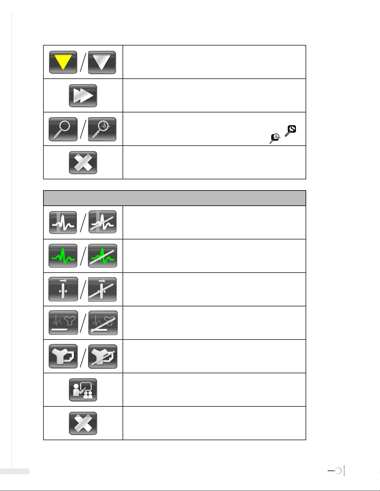
3.3 Sherlock 3CG™ TCS Controls and Indicators
Ultrasound Mode: Select to exit the TCS Mode and enter Ultrasound Mode.
Calibrate Button: Select to calibrate magnet navigation.
Audio On: Select to mute the magnet navigation audio.
Audio Mute: Select to turn the magnet navigation audio on.
Freeze: Select to copy the current ECG waveforms from the Main Screen to the Reference
Screen.
Main Controls
Exit Site Marking: Select to open the Exit Site Marking Window.
Print: Select to Print and Save the ECG and magnet navigation data. Data will be printed to
paper and saved to a file on an external USB storage device.
10
Page 13

Sherlock 3CG™ Tip Confirmation System
Menu: Select to open the Menu Window.
F1
F2
F3
F6
F12
esc
Select the ‘F1’ key on the keyboard to open the Patient Information window.
Select the ‘F2’ key on the keyboard to open the Menu Window
Select the ‘F3’ key on the keyboard to open the Exit Site Marking Window.
Select the ‘F6’ key on the keyboard to open the Settings Window.
Select the ‘F12’ key on the keyboard to clear the patient information.
Select the ‘esc’ key on the keyboard to close the Exit Site Marking Window, Menu Window,
Patient Information Window, Settings Window, or quit Demonstration Mode.
Exit Site Marking Controls
Increase: Select to cycle through the exit site marking options in a positive progression.
Decrease: Select to cycle through the exit site marking options in a negative progression.
Exit: Select to close the Exit Site Marking Window.
Menu Controls
Channel Select: Select either the external (white) or intravascular (yellow) ECG waves for
manipulation.
Scale: Select to adjust the ECG signal amplitude of the external (white) or intravascular
(yellow) ECG waveforms on the Main Screen.
Level Up: Select to increase the vertical position of the external (white) or intravascular (yellow)
ECG waveforms on the Main Screen.
11
Page 14
Instructions For Use
Level Down: Select to decrease the vertical position of the external (white) or intravascular
(yellow) ECG waveforms on the Main Screen.
Speed: Select to adjust the speed of the ECG waveforms on the Main Screen.
Magnet Navigation Toggle: Select to toggle between ECG & Magnet and Magnet Only
Navigation . Magnet Navigation Only mode is compatible with PICCs labeled with [
ECG and Magnet Navigation mode is compatible with PICCs labeled with [
Exit: Select to close the Menu Window.
] .
].
Settings Controls
P-wave Highlighting: Select to enable or disable highlighting the P-wave on the ECG signals.
ECG Color: Select to enable or disable the intravascular (yellow) ECG waveforms changing
color to green and red in sync with the stylet icon.
Depth Gauge: Select to enable or disable the depth gauge.
Confirmation Statement Printing: Select to enable or disable the confirmation statement
printing on the ECG printout.
Magnet Tracking Printout: Select to enable or disable the magnet navigation printout.
Demonstration: Select to initiate demonstration mode.
For training, contact your Bard Access Systems’ Sales Representatives.
Exit: Select to close the Menu Window.
12
Page 15

Sherlock 3CG™ Tip Confirmation System
3.4 Sherlock 3CG™ TCS Audio Information
When the audio is on, there are four possible tones:
- Tone 1: The stylet tip is detected and the tip is not under the sensor.
- Tone 2: The stylet tip is under the sensor and is at or above the Bard logo.
- Tone 3: The stylet is under the sensor and below the Bard logo.
- Tone 4: The stylet is past max P-wave location.
1
2
3
4
Note: Tone 1 – Tone 3 audio signals are only applicable to the magnetic navigation capability of the device. Tone 4 is applicable to ECG in patients
where there are no alterations to the cardiac rhythm that change the presentation of the P-wave.
3.5 Parallax
When a patient's chest is not flat, the sensor will rest at an angle, causing an effect known as parallax. Parallax is the difference in the apparent location
of an object from two different points of view. The difference between the point of view of the sensor and the user can be several centimeters.
13
Note: Parallax should only be considered in relation to magnetic navigation.
Page 16

4 Sherlock 3CG™ TCS Catheter Guidance
4.1 Verifying Catheter Compatibility
The Sherlock 3CG™ TCS on Site~Rite™ 6 Ultrasound System is compatible with the following PICCs as described below:
Instructions For Use
- The PICCS indicated with [
- The PICCs indicated with [
] are compat ble for ECG and Magnet Navigation [ ].
] are only compatible for Magnet Navigation [ ].
4.2 Catheter Placement
Step 1: Prepare Device
- Connect the sensor to the Site~Rite™ 6 Ultrasound System via USB cable.
- Verify that the ECG flatline signal is scrolling.
- Verify the battery charge is sufficient for the procedure.
- Enter Patient Information as needed.
Step 2: Position Patient and Perform Ultrasound Pre-scan
- Refer to the Bard Access Systems’ Site~Rite™ 6 Ultrasound System Instructions for Use.
Step 3: Measure Catheter Length
- Refer to Bard Access Systems’ Catheter Instructions for Use.
Step 4: Prepare Sensor
- Slide the Fin Assembly on to the sensor until fully seated.
- Place sensor in the sensor holder with the fin and ECG electrodes remaining outside the holder and tighten the cinch ring.
[1]
[3]
[2]
[4]
14
Page 17

Sherlock 3CG™ Tip Confirmation System
Step 5: Position Sensor and ECG electrodes
- Remove the adhesive backing from the sensor holder and lace the sensor directly on the patient’ skin with the adhesive side down. Place the sensor as flat as
possible for best results.
Note: The sensor should be positioned the same for left or right side placements.
- Prepare and attach external ECG electrodes per the following steps:
• Ensure electrode locations are oil-free and completely dry.
Caution: Electrodes should be applied only to intact, clean skin (e.g., not over open wounds, lesions, infected or inflamed areas).
- Attach electrodes to all lead wires.
- Remove backing and press electrodes firmly onto skin at the specified locations.
• Place black electrode on patient’s lower right shoulder.
• Place red lead on patient’s lower left side, inferior to the umbilicus and laterally along the mid-axillary line.
Caution: Placement of red electrode outside of this region may result in reduced ECG performance.
15
Warning: Place skin electrodes carefully at locations indicated in these Instructions for Use and ensure good skin-electrode contact. Failure to do so may cause
unstable ECG waveforms and/or ECG waveforms that are not described in these Instructions for Use. In such a case, rely on magnetic navigation and external
measurement for tip positioning and use chest X-ray or fluoroscopy to confirm catheter tip location, as indicated by the institutional guidelines and clinical
judgment.
Caution: Discontinue electrode use immediately, if skin irritation occurs.
Tips:
- Prior to securing the sensor holder to the patient, it may be necessary to clean the skin and remove excess hair.
- Do not move the sensor after it is secure. Best results will be achieved if the patient remains still and the sensor is not placed on open wounds, over bandages,
drapes, gowns or other coverings.
Page 18

Step 6: Evaluate External ECG waveform
- Refer to the Bard Access Systems’ catheter Instructions for Use.
Step 7: Perform Initial Magnet Navigation Calibration
- Calibrate the Sherlock 3CG™ TCS by selecting CALIBRATE [ ] prior to setting up the sterile field to ensure there is no environmental interference.
Tip: If calibration fails, remove any items that may be causing magnetic interference (e.g., active motor driven equipment, monitor leads, cell phones, name
tags, jewelry, etc.).
Step 8: Prepare Catheter Sterile Field
- Refer to the Bard Access Systems’ catheter Instructions for Use.
Step 9: Access the Vein
- Refer to the Bard Access Systems’ Site~Rite™ 6 Ultrasound System Instructions and Bard Access Systems’ catheter Instructions for Use.
Step 10: Attach Catheter Stylet to Fin Assembly
- Refer to Bard Access Systems’ catheter Instructions for Use.
Step 11: Perform Final Magnet Navigation Calibration
- Ensure the catheter tip is at least 12 inches (30 cm) away from the sensor before calibrating.
- Select CALIBRATE [ ] immediately prior to catheter insertion.
- Once calibration is complete, ask the patient to remain still and do not reposition the patient.
- Refer to Bard Access Systems' catheter instructions for use for catheter insertion.
Step 12: Insert Catheter
- Refer to Bard Access Systems’ catheter Instructions for Use for catheter insertion.
Step 13: Catheter Tip Guidance and Positioning
- Refer to Bard Access Systems’ catheter Instructions for Use for catheter insertion.
- Initially a searching magnifying glass will indicate that the stylet tip is outside the sensor range.
- Use a slow steady motion while advancing the catheter.
Instructions For Use
Magnetic Navigation and Catheter Tip Navigation
- As the stylet tip approaches the sensor, an icon appears at the edge of the screen indicating the approach of the stylet tip.
- When the stylet is under the sensor, the stylet and depth icons will display the location, orientation, and depth of the stylet in relation to the sensor.
- Advance the catheter slowly to achieve optimal performance (1 cm per second). There may be a slight delay from the time the catheter is moved until the
stylet icon moves on the display. Advancing the catheter too quickly may result in erratic movements of the stylet icon on the display.
- Insert the catheter until the magnetic navigation shows the stylet moving consistently downward.
- Continue to slowly advance catheter until the catheter is inserted to the external measurement as determined in the Bard Access Systems’ catheter
instructions for use.
- Select [ ] to minimize the magnetic navigation window and freeze / save the current ECG waveforms in the reference screen.
Note: select the minimized magnetic navigation window to return to a maximized state
ECG Positioning
- In patients with a distinct P-wave, the P-wave will increase in amplitude as the catheter approaches the cavoatrial junction. As the catheter advances into
the right atrium, the P-wave will decrease in amplitude and may become biphasic or inverted.
Note: If the intravascular ECG waveform is not displayed, flush the catheter with saline. If the problem continues, check the stylet-to-fin connection.
To freeze and compare ECG waveforms select [ ] to copy ECG waveforms in the reference screen.
Note: Pause to let the rhythm settle before freezing the reference ECG waveforms.
- For final catheter positioning, refer to Bard Access Systems' catheter instructions for use.
Step 14: Complete Catheter Placement
- Complete catheter insertion, securement and remaining procedure according to the catheter Instructions for Use and facility protocol.
16
Page 19

Sherlock 3CG™ Tip Confirmation System
Step 15: Procedural Record
- Select [ ] to print / save a procedural record.
Note: Selecting [ ] will send the procedural record to a storage device and/or an approved printer, if connected. A typical printout is shown below.
5 Magnet Navigation Only Mode
Step 1: Prepare Device
- Connect the sensor to the Site~Rite™ 6 Ultrasound System via USB cord.
- Verify the battery charge is sufficient for the procedure.
- Enter patient Information as needed.
Step 2: Position Patient and Perform Ultrasound Pre-scan
- Refer to the Bard access Systems’ Site~Rite™ 6 Ultrasound System Instructions for Use.
Step 3: Measure Catheter Length
- Refer to Bard Access Systems’ Catheter Instructions for Use.
Step 4: Prepare Sensor
- Place the Sherlock 3CG™ TCS Sensor in the sensor holder and tighten the cinch ring.
1]
3]
2]
4]
17
Page 20

Step 5: Position Sensor
- Remove the adhesive backing from the sensor holder and place the sensor directly on the patient's skin with the adhesive side down. Place the sensor as flat
as possible for best results.
Note: The sensor should be positioned the same for left or right side placements.
Step 6: Perform Initial Magnet Navigation Calibration
Instructions For Use
- Calibrate the Sherlock 3CG™ TCS by selecting CALIBRATE [ ] prior to setting up the sterile field to ensure there is no environmental interference.
Tip: If calibration fails, remove any items that may be causing magnetic interference (e.g. active motor driven equipment, monitor leads, cell phones,
name tags, jewelry, etc.).
Step 7: Prepare Catheter Sterile Field
Refer to the Bard Access Systems’ catheter Instructions for Use.
Step 8: Access the Vein
- Refer to the Bard Access Systems’ Site~Rite™ 6 Ultrasound System Instructions for Use and Bard Access Systems’ catheter Instructions for Use.
Step 9: Perform Final Magnet Navigation Calibration
- Ensure the catheter tip is at least 12 inches (30 cm) away from the sensor before calibrating.
- Select CALIBRATE [
- Once calibration is complete, ask the patient to remain still and do not reposition the patient.
- Refer to Bard Access Systems' catheter Instructions for Use for catheter insertion.
] immediately prior to catheter insertion.
Step 10: Insert Catheter
- Refer to Bard Access Systems’ catheter Instructions for Use for catheter insertion.
Step 11: Catheter Tip Guidance and Positioning
- Refer to Bard Access Systems’ catheter Instructions for Use for catheter insertion.
- Initially a searching magnifying glass will indicate that the stylet tip is outside the sensor range.
- Use a slow steady motion while advancing the catheter.
Magnetic Navigation
- As the stylet tip approaches the sensor, an icon appears at the edge of the screen indicating the approach of the stylet tip.
- When the stylet is under the sensor, the stylet and depth icons will display the location, orientation, and depth of the stylet in relation to the sensor.
- Advance the catheter slowly to achieve optimal performance (1 cm per second). There may be a slight delay from the time the catheter is moved until
the stylet icon moves on the display. Advancing the catheter too quickly may result in erratic movements of the stylet icon on the display.
- Insert the catheter until the magnetic navigation shows the stylet moving consistently downward.
- Continue to slowly advance catheter until the catheter is inserted to the external measurements as determined in the Bard Access Systems’ catheter
instructions for use.
18
Page 21

Sherlock 3CG™ Tip Confirmation System
- Below is an image of Magnetic Navigation only mode.
6 Trouble Shooting and Error Messages
6.1 Error Screens
Sensor Disconnected
Sensor Error
Cause:
Solution:
Cause:
Solution:
Cause:
Solution:
Cause:
Solution:
Cause:
Solution:
Sensor not connected to the display.
Ensure the sensor is properly connected. Disconnect and reconnect the sensor.
The display power supply is connected to a noisy electrical outlet.
Disconnect the display power supply from the electrical outlet to run the system
on battery power.
Sensor cannot be detected by the display.
Call the technical support hotline at(800) 443-3385.
Interference caused by magnetic field changes or by sensor movement.
1. Move all metal objects at least 12 inches (30 cm) away from the sensor.
2. Lower bed rails.
3. Move all active electronic devices at least 5 feet (1.5 m) away.
4. Do not move the sensor and ask the patient to remain still.
5. Calibrate.
6. If the error continues, discontinue use and call the technical support
hotline at (800) 443-3385.
Sensor malfunction.
1. Turn off and restart the system.
2. If the error continues, discontinue use and call the technical support
hotline at (800) 443-3385.
19
Page 22

Instructions For Use
Magnet Error
Battery Error
Printer Error
Cause:
Solution:
Cause:
Solution:
Cause:
Solution:
Interference caused by magnetic field changes or by sensor movement.
1. Move all metal objects at least 12 inches (30 cm) away from the sensor.
2. Lower bed rails.
3. Move all active electronic devices at least 5 feet (1.5m) away.
4. Do not move the sensor and ask the patient to remain still.
5. Ensure catheter tip is at least 12 inches (30 cm) away from the sensor.
6. Calibrate.
7. If the error continues, discontinue use and call the technical support
hotline at (800) 443-3385.
Battery malfunction.
Call the technical support hotline at (800) 443-3385.
Printer error.
1. Check and refill paper.
2. Refer to Printer Instructions for Use.
Storage Device Error
Storage Device Error
Cause:
Solution:
Cause:
Solution:
Storage device error.
Replace external USB storage device.
Storage device error.
External USB storage device has no storage space available.
1. Remove files from external USB storage device to have available storage space.
2. Replace external USB storage device.
20
Page 23

Sherlock 3CG™ Tip Confirmation System
6.2 ECG Troubleshooting
Flat-line ECG signal
Poor ECG signal
Wandering ECG baseline
ECG signal abnormal
Cause:
Solution:
Cause:
Solution:
Cause:
Solution:
Cause:
Solution:
Cause:
Solution:
Cause:
Solution:
Cause:
Solution:
Cause:
Solution:
Cause:
Solution:
Solution:
Cause:
Solution:
Cause:
Solution:
Cause:
Solution:
Cause:
Solution:
Cause:
Solution:
Cause:
Solution:
Cause:
Solution:
Cause:
Solution:
Cause:
Solution:
No fin connection.
Fully seat fin on the sensor.
No ECG electrode connection.
Ensure that the area on the skin where the ECG electrodes were placed is oil free
and completely dry.
Damaged ECG electrodes.
Ensure catheter T-lock connections are fully seated.
Replace electrodes.
Air bubble at the catheter tip.
Flush the catheter lumen containing the stylet with saline.
No stylet-to-fin connection.
Fully seat the stylet connector on the fin. Refer to the Bard Access Systems’ catheter
instructions for use.
Fin and/or stylet connectors not fully seated.
Fully seat fin on sensor. Fully seat stylet connector on fin.
Poor ECG electrode contact.
Ensure that the area on the skin where the ECG electrodes were placed is oil free
and completely dry.
Patient movement.
Ask the patient to remain still. Ensure the patient is warm and relaxed.
Extraneous noise.
Unplug the Site~Rite™ 6 Ultrasound System from AC power.
Make Sure the Ultrasound probe is not in close proximity to the sensor and/or lead wires.
Damaged ECG electrodes.
Replace electrodes.
Catheter not locked with saline.
Ensure catheter T-lock connections are fully seated. Flush the catheter lumen
containing the stylet with saline.
The External ECG signal, with black electrode on the patient’s left shoulder
(Lead III view) is weak.
Place black electrode on the patient’s right shoulder (Lead II view).
Poor ECG electrode contact.
Ensure that the area on the skin where the ECG electrodes were placed is oil
free and completely dry.
Patient movement.
Ask the patient to remain still. Ensure the patient is warm and relaxed.
Extraneous noise.
Avoid touching the stylet or electrodes.
Catheter not locked with saline.
Ensure catheter T-lock connections are fully seated. Flush the catheter lumen
containing the stylet with saline.
Body ECG leads swapped.
Place ECG leads correctly.
No distinct P-wave.
Do not rely on ECG guidance for catheter tip confirmation. Refer to
Warnings Section.
21
Page 24

Instructions For Use
Absent ECG signal
ECG Screen (main/reference screen)
is not visible
6.3 Magnetic Navigation Troubleshooting
Magnetic navigation does not
detect stylet
Cause:
Solution:
Cause:
Solution:
Cause:
Solution:
Cause:
Solution:
Cause:
Solution:
Cause:
Solution:
Cause:
Solution:
Cause:
Solution:
Cause:
Solution:
Cause:
Solution:
Cause:
Solution:
Sensor / sensor cables damaged.
Call technical hotline at (800) 443-3385 to replace sensor and/or cables.
Sensor disconnected.
Ensure the sensor is properly connected.
Sensor is not communicating with display.
Reboot the display.
Incompatible sensor.
Use appropriate sensor. Contact customer service (800) 545-0890.
Incorrect PICC compatibility is selected.
Make sure to have [
[
].
] selected from the menu when using a catheter kit labeled
Sensor incorrectly oriented.
Orient sensor correctly per procedure.
Stylet outside sensor depth range (3 to 11 cm).
- If the vein is deep, ensure that the top of the sensor on the insertion side is touching
the patient's skin (e.g. by using tape).
- If the vein is shallow, lift the sensor away from the skin (e.g., by using a folded blanket).
Too much time has passed since calibration. Magnetic fields have changed.
Pull catheter tip back at least 12 inches (30 cm) from the sensor, recalibrate and
re-advance catheter.
Stylet did not advance under the sensor.
Pull catheter back and re-advance.
Stylet not at the catheter tip.
Ensure stylet tip is within 1 cm of distal end of catheter. Adjust as necessary.
Incompatible catheter.
Verify that the Bard Access Systems’ catheter kit has a [
] icon or [ ] icon.
Note: Depth icons will
not be present.
Cause:
Solution:
Cause:
Solution:
Cause:
Solution:
Stylet magnet trimmed off.
Replace catheter with a new Bard Access Systems’ catheter from a kit labeled
[
] or [ ] .
Stylet is out of sensor's depth range (3 to 11 cm)
1. Pull catheter back until stylet icon is stable.
2. Verify catheter is moving in the correct direction and continue placement using
ECG guidance and catheter measurement.
Magnetic interference.
Pull catheter tip back at least 12 inches (30 cm) from the sensor, recalibrate and
re-advance catheter.
22
Page 25

Sherlock 3CG™ Tip Confirmation System
Cause:
Solution:
Stylet at edge of sensor’s depth range (3 to 11 cm).
1. Pull catheter back until stylet icon is stable.
2. Verify catheter is moving in the correct direction and continue placement.
Magnetic navigation Stylet icon
has erratic behavior
Cause:
Solution:
Cause:
Solution:
Cause:
Solution:
Magnetic interference.
Pull catheter tip back at least 12 inches (30 cm) from the sensor, recalibrate,
and re-advance catheter.
Too much time has passed since calibration. Magnetic fields have changed.
Pull catheter tip back at least 12 inches (30 cm) from the sensor, recalibrate
and re-advance catheter.
Stylet is out of sensor’s depth range (3 to 11 cm).
1. Pull catheter back until stylet icon is stable.
2. Verify catheter is moving in the correct direction and continue placement using
ECG guidance and catheter measurement.
Magnetic navigation Stylet icon
disappears, "sweeping magnifying glass"
displayed.
Cause:
Solution:
Cause:
Solution:
Magnetic interference.
Pull catheter tip back at least 12 inches (30 cm) from the sensor, recalibrate, and
re-advance catheter.
Too much time has passed since calibration. Magnetic fields have changed.
Pull catheter tip back at least 12 inches (30 cm) from the sensor, recalibrate and
re-advance catheter.
If problems continue, call the technical support hotline at (800) 443-3385.
7 Magnetic Navigation Printing
Select the 'F6' key on the keyboard [ ] to open Settings.
- Under the menu, navigate down until the MAGNET PRINTOUT DISABLED [
- Select the MAGNET PRINTOUT DISABLED [
- After being selected the MAGNET PRINTOUT DISABLED button will toggle to the MAGNET PRINTOUT ENABLED [
- Close Settings by selecting the EXIT [
Note: Selecting [
A typical Magnet Navigation Only printout is shown below.
] after Magnet Navigation Only Mode will send the procedural record to a storage device and/or an approved printer, if connected.
] button
] button.
] button is highlighted.
] button.
23
Page 26

8 Cleaning and Disinfection
8.1 Cleaning Procedure
To clean the Sherlock 3CG™ TCS Sensor:
1. Turn off the system.
2. Dampen a nonabrasive cloth with either warm water or isopropyl alcohol.
3. Gently wipe the dampened cloth over exterior surfaces.
8.2 Disinfection Procedure
For a list of disinfectants approved for use on the Sensor and Display, contact Bard Access Systems, Inc.
Warning: Do not submerge the sensor or allow fluid to enter the connector. Damage to the equipment may occur.
Warning: Do not attempt to sterilize the sensor. Damage to the equipment may occur.
9 Warranty
The manufacturer, Bard Access Systems, Inc., warrants to the original purchaser that this product will be free from defects in material and workmanship
for a period of one year from the date of purchase. If this product proves to be so defective, purchaser may return the same to Bard Access Systems, Inc. for
repair, replacement or credit at Bard Access Systems Inc.'s option in accordance with Bard Access Systems Inc.'s Return Goods Policy found in the current
price list. The warranty on the repaired or replaced unit continues from the purchase date of the original unit. This warranty does not cover damages resulting
from misuse, abuse, modification, or alteration of this product or the repair of this product by anyone other than an authorized Bard Access Systems, Inc.
representative.
Instructions For Use
Without limitation, the following actions void the warranty of the Sherlock 3CG™ TCS Sensor:
Warning: The following actions void the warranty of the Sherlock 3CG™ TCS Sensor and may result in injury or equipment damage.
- Opening or servicing the Sherlock 3CG™ TCS Sensor by anyone other than Bard Access Systems’
authorized service personnel.
- Removing system labels by anyone other than Bard Access Systems’ authorized service personnel.
- Connecting the sensor or applied patient components to any unauthorized system or accessory. Refer to Section 1.4 for complete components.
- Installation of unauthorized software.
- Modification of system software settings without authorization by Bard Access Systems.
TO THE EXTENT ALLOWABLE BY APPLICABLE LAW, THIS LIMITED PRODUCT WARRANTY IS IN LIEU OF ALL OTHER WARRANTIES,
WHETHER EXPRESS OR IMPLIED, INCLUDING, BUT NOT LIMITED TO, ANY IMPLIED WARRANTY OF MERCHANTABILITY OR
FITNESS FOR A PARTICULAR PURPOSE. THE LIABILITY AND REMEDY STATED IN THIS LIMITED PRODUCT WARRANTY WILL BE
THE SOLE LIABILITY OF BARD ACCESS SYSTEMS, INC. AND REMEDY AVAILABLE TO PURCHASER FOR THIS PRODUCT, WHETHER IN
CONTRACT, TORT (INCLUDING NEGLIGENCE) OR OTHERWISE.
IN NO EVENT WILL BARD ACCESS SYSTEMS, INC. BE LIABLE TO YOU FOR ANY INDIRECT, INCIDENTAL OR CONSEQUENTIAL
DAMAGES RESULTING FROM YOUR HANDLING OR USE OF THIS PRODUCT EVEN IF BARD ACCESS SYSTEMS, INC. HAS BEEN
ADVISED OF THE POSSIBILITY OR LIKELIHOOD OF SUCH DAMAGES. IN NO EVENT WILL BARD ACESS SYSTEMS, INC.'S LIABILITY
UNDER THIS WARRANTY WITH RESPECT TO THIS PRODUCT EXCEED THE PURCHASE PRICE PAID TO BARD ACCESS SYSTEMS, INC.
FOR SUCH PRODUCT.
Some states/countries do not allow an exclusion of implied warranties, incidental or consequential damages. You may be entitled to additional remedies under
the laws of your state/country.
24
Page 27

Sherlock 3CG™ Tip Confirmation System
10 Service and Repair
There is no periodic or preventative maintenance required for the Sherlock 3CG™ TCS.
For servicing information or to return your Sherlock 3CG™ TCS for repair, please contact Bard Access Systems' technical support hotline at (800) 443-3385.
Warning: Only qualified personnel should attempt to service this equipment. The Sherlock 3CG™ TCS contains static sensitive components and circuits.
Failure to observe proper static control procedures may result in damage to the system.
Warning Opening or servicing the Sherlock 3CG™ TCS Sensor by anyone other than Bard Access Systems' authorized service personnel will void the warranty
and may result in injury or equipment damage.
11 Technical Specifications
11.1 Sherlock 3CG™ TCS Sensor and Display Operational and Storage Conditions
Operating Temperature: 15°C to 38°C (59°F to 100°F)
Storage Temperature: 10°C to 38°C (50°F to 100°F)
Operating Humidity: 5% to 85% non-condensing
Storage Humidity: 5% to 95% non-condensing
12 Disposal Information
To return the Sherlock 3CG™ TCS for end of life recycling, please contact your nearest Bard sales or distributor office in the country of purchase.
25
Page 28

Instructions For Use
26
 Loading...